Drainage Procedures for Petrous Apex Lesions

Chapter 45 Drainage Procedures for Petrous Apex Lesions


image Videos corresponding to this chapter are available online at www.expertconsult.com.


With the help of cranial computed tomography (CT) and magnetic resonance imaging (MRI), petrous apex lesions can be correctly diagnosed preoperatively. Consequently, cystic lesions requiring drainage should be approached with procedures designed for drainage, not total en bloc removal. Although the transmastoid infralabyrinthine procedure has been the usual approach for these lesions, transcanal infracochlear drainage offers a more dependent drainage site. The eventual role of the various approaches for petrous apex drainage depends on the long-term follow-up of these patients.


Advances in radiologic imaging during the past 2 decades have made it possible to differentiate reliably lesions of the petrous apex preoperatively. The development of CT scanning was the first major step in imaging the temporal bone since the development of polytomography. CT scanning gives the surgeon the ability to visualize the size of the lesion and its relationship to vital structures, including the internal auditory canal (IAC), cochlea, vestibular labyrinth, carotid artery, and jugular bulb. It also helps characterize the border of the lesion as expansile or invasive, which may differentiate between benign lesions and malignant neoplasms. MRI of the temporal bone added the capability of characterizing the substance of the lesion, rather than its effect on bony interfaces, allowing the surgeon to distinguish between mucus, fat, cholesterol granuloma, cholesteatoma, and neoplasm. The combination of CT, with its superior bone imaging algorithms, and MRI, with its enhanced tissue imaging capabilities, allows the surgeon to differentiate accurately and reliably among benign cystic lesions, normal anatomic variants, and neoplastic lesions of the petrous apex.


Patients with petrous apex lesions present with various symptoms and physical findings. The most widely recognized finding is Gradenigo’s syndrome, consisting of retro-orbital pain, otorrhea, and ipsilateral sixth cranial nerve paresis. The signs and symptoms of noninflammatory or neoplastic lesions may be more subtle. Hearing loss and vestibular abnormalities are frequently associated with lesions of the petrous apex as they enlarge and compress the IAC. The facial nerve is relatively resistant to paresis from slowly expansile lesions, but may be involved early with neoplastic lesions of the petrous apex or non-neoplastic lesions compressing the IAC.


Pain may be present with benign or cystic lesions, but is more common in neoplastic lesions. Its distribution depends on the region involved. The mastoid cavity is innervated by CN IX and may radiate pain into the neck. The middle fossa and superior petrosal regions are innervated by CN V and may cause retro-orbital or facial pain. Lesions extending into the posterior fossa may cause pain along the routes of distribution of CN IX and X and the first three cervical nerves.1 Although 80% of adult mastoid bones are aerated, only 30% of petrous bones have air cells extending to the apex, and 7% may have asymmetric pneumatization of the petrous apex.2,3 The increasing use of MRI of the head and neck makes it imperative that the clinician can differentiate pathology from normal variant in the temporal bone. Table 45-1 summarizes common lesions of the petrous apex and their associated radiologic findings.


Asymmetric pneumatization is clearly seen on CT scanning, but supplementary MRI may be needed to rule out pathologic lesions in a symptomatic ear. Normal bony architecture can be seen on CT scanning, with hyperintensity on MRI T1-weighted scans and hypointensity on T2-weighted scans because of the large fat content in marrow. Retained mucus in the air cells also appears with normal bony architecture on CT scan, but is hypointense on T1-weighted MR images and hyperintense on T2-weighted MR images. Cholesteatoma is usually associated with chronic otitis media, but may arise from congenital rest cells in the petrous apex. Because of its high water content, cholesteatoma is isointense with cerebrospinal fluid on CT, and is hypointense on T1-weighted MR images and hyperintense on T2-weighted MR images. Cholesterol granuloma is isointense with brain on CT scanning and appears as a classic image on MRI with hyperintensity on T1-weighted and T2-weighted scans.


Radiologic descriptions of other, less common lesions are also summarized in Table 45-1. Differentiating between chordoma, chondroma, and chondrosarcoma of the temporal bone remains difficult, even with the scanning techniques currently available. The area of origin and the age of the patient must be considered when the pathology of destructive lesions of the petrous apex is determined.46


PATIENT SELECTION


Most surgical approaches to the petrous apex were developed in the preantibiotic era for drainage of petrous apex abscesses and cure of Gradenigo’s syndrome. With the arrival of modern antibiotics, infectious processes of the petrous apex have greatly declined in frequency, but these same approaches may be equally effective in draining cystic lesions of the petrous apex.


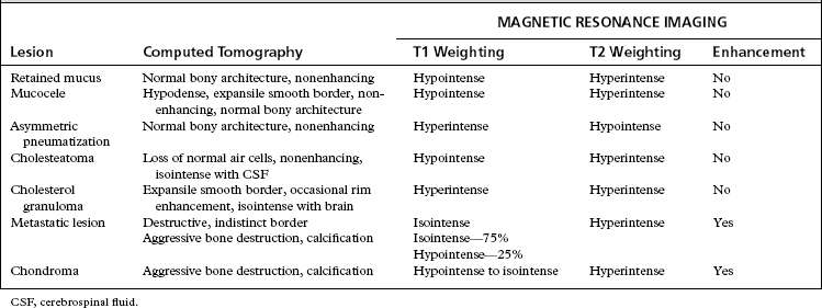
Air cell tracts extend above, below, and anterior to the otic capsule, allowing the potential of safe passage to the petrous apex. Approaches that follow superior air cell tracts include middle fossa,7 through the superior semicircular canal,8 the attic, and the root of the zygomatic arch.9 Approaches below the inner ear include the infralabyrinthine and the infracochlear.1013 Anterior approaches have been described by Ramadier,14 Eagleton,15 and Lempert,16 who used the triangle between the anterior border of the cochlea, the carotid artery, and the middle fossa dura. All these approaches are used for drainage of inflammatory disease processes that are not responsive to antibiotic therapy or simpler operations for chronic ear disease (Fig. 45-1).


Infralabyrinthine, infracochlear, and transsphenoidal approaches are most commonly chosen for drainage of cystic lesions of the petrous apex in an ear with serviceable hearing. These lesions are frequently detected at an asymptomatic stage with current imaging techniques. Because the natural history of small benign cystic lesions is not well documented, surgical drainage should be reserved for patients with larger lesions or with symptoms including pain, visual changes, diplopia, hearing loss, vertigo, or facial nerve weakness. For patients without serviceable hearing, these lesions should be drained through a translabyrinthine approach. Because other vital structures may be affected by enlargement of the cyst, delaying surgery in symptomatic patients provides no advantage.


Cholesterol granuloma is the most common cystic lesion of the petrous apex, occurring 30 times less frequently than acoustic neuroma.17 It may develop in any aerated portion of the temporal bone, but most commonly occurs in the mastoid air cells distant to a lesion that prevents normal aeration. Cholesterol granuloma of the petrous apex probably develops when a pathologic process or trauma obstructs the air cell tracts to a well-pneumatized petrous apex.


The treatment for cholesterol granuloma of the temporal bone is drainage and re-establishment of adequate aeration to the involved area. The cyst wall is composed of a fibrous connective tissue. It is free of keratinizing squamous epithelium that characterizes cholesteatoma, and complete removal of the cyst is unnecessary.


Solid tumors of the temporal bone and cholesteatoma are removed when first identified, rather than after further symptoms develop because these symptoms frequently reveal further involvement of other vital structures. Drainage procedures are inadequate treatment for these lesions, and all reasonable efforts should be made to remove them entirely. Total removal may require the sacrifice of cranial nerves and major vascular structures.


PREOPERATIVE EVALUATION AND PATIENT COUNSELING


Preoperative evaluation of patients is based on their symptoms. Patients presenting with hearing loss are evaluated initially with audiometric testing, including air, bone, and speech reception thresholds and speech discrimination scores. Electronystagmography is performed in patients who complain of imbalance or vertigo. In patients with otherwise normal results on physical examination, asymmetric hearing is next evaluated with auditory brainstem response testing. If these results are abnormal, MRI is indicated. In patients with cranial nerve involvement other than CN VIII with asymmetric hearing, auditory brainstem response testing is not performed, and the physician proceeds directly to MRI.


Patients who have normal hearing but have other cranial nerve deficits that may be referable to the petrous apex may be screened with a high-resolution, thin-section CT scan of the temporal bone or an MRI scan with gadolinium. If an abnormality is found, all patients undergo air, bone, and speech reception thresholds and speech discrimination audiometric testing before surgery to document hearing levels before a procedure that jeopardizes hearing.


Preoperatively, patients are counseled to expect resolution of pain, if present, and the possibility of improvement in cranial nerve function if it is decreased preoperatively. Cranial nerves that have been affected for shorter periods seem to have a better prognosis and fewer long-standing deficits than nerves that have been affected longer. Patients are reminded that this is a drainage procedure with the goal of decompressing the lesion and providing an aerated cavity, if possible. The goal is not the removal of the lesion, and close follow-up may be necessary. Recurrence of the lesion secondary to inadequate drainage is usually heralded by the return of preoperative symptoms. Follow-up MRI frequently reveals a cholesterol granuloma cyst that remains full of fluid, but the T1-weighted image is hypointense compared with the preoperative hyperintense image on T1-weighted views. A return of hyperintensity on the T1-weighted image suggests inadequate drainage in a symptomatic lesion.18


SURGICAL TECHNIQUES


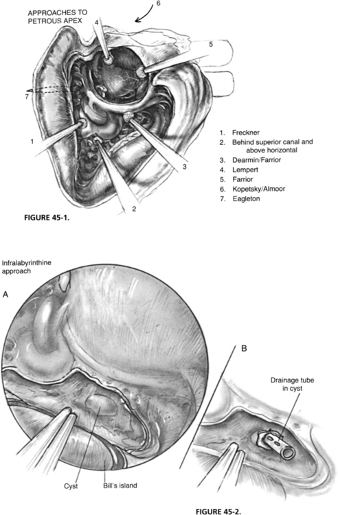
Infralabyrinthine Drainage of the Petrous Apex


In preparation for infralabyrinthine drainage of the petrous apex, the patient is prohibited from eating and drinking for at least 8 hours preoperatively. Unless an infectious process is suspected, no preoperative antibiotics are used.


The surgical ear is prepared similarly to any other chronic otitis media case operated on through a postauricular approach. Hair is shaved to one fingerbreadth above the auricle and two fingerbreadths behind the postauricular crease. A facial nerve monitor is typically used, and monitoring electrodes are placed in the standard positions. Surgical preparation is an antibacterial scrub followed by painting with antibacterial solution. Sterile Mastisol is applied around the auricle and allowed to dry. An adhesive aperture drape is placed over the ear, and sterile sheets cover the patient. Routine chronic otitis media instruments and drill are the only equipment required.


Only gold members can continue reading. Log In or Register to continue

Stay updated, free articles. Join our Telegram channel

Jun 14, 2016 | Posted by in OTOLARYNGOLOGY | Comments Off on Drainage Procedures for Petrous Apex Lesions

Full access? Get Clinical Tree

Get Clinical Tree app for offline access