Chapter 123 Diagnostic and Therapeutic Vitrectomy for Uveitis
Introduction
Uveitis is a type of intraocular inflammation that can affect vision and may lead to legal blindness. Uveitis is primarily diagnosed upon clinical manifestation rather than laboratory findings.1,2 A patient’s medical history, course of uveitis, and therapeutic response are also important in making a diagnosis. Histopathological diagnosis of biopsy specimens is not common because of the difficulty in obtaining ocular specimens. Uveitis can be classified into autoimmune, infectious, and malignant forms based on the underlying pathogenic mechanism. Regardless of cause, accurate diagnosis and appropriate pharmacotherapy are critical for positive visual outcomes. Diagnostic vitrectomy can be helpful in discriminating between different causes of uveitis.
The vitreous, which constitutes most of the ocular volume, plays an important role in the pathogenesis of many inflammatory diseases of the eye. Ocular fluid can be a valuable tool for accurate diagnosis, and considering specimen volume requirements, vitreous samples are more useful than aqueous humor samples for analysis.3 In addition, a vitrectomy can be beneficial for the management of vitreoretinal complications in uveitis patients. This chapter, which is divided into diagnostic and therapeutic vitrectomy sections, covers the surgical indications, principles, and techniques for the management of uveitis. Novel advances in laboratory techniques will also be discussed.
Diagnostic vitrectomy
Indications
Diagnostic vitrectomy is indicated in an inflamed eye when the course or appearance of uveitis is not typical for an autoimmune disease and the presence of an infectious agent or malignant process is suspected (see also Chapter 122, Infectious endophthalmitis, and Chapter 124, Vitreous, retinal, and choroidal biopsy). If the disease fails to respond to therapy in an expected manner, the physician may suspect that the original diagnosis is incorrect; in such cases, other diagnoses need to be considered. Indications for diagnostic vitrectomy are shown in Table 123.1.
Table 123.1 Indications of diagnostic vitrectomy
| Infectious uveitis | Endophthalmitis4: bacterial, fungal, parasitic or viral |
| Traumatic endophthalmitis including intraocular foreign body | |
| Postsurgical endophthalmitis | |
| Endogenous endophthalmitis | |
| Sterile endophthalmitis | |
| Vitritis | |
| Retinitis | |
| Choroiditis | |
| Retinal vasculitis | |
| Noninfectious uveitis | Autoimmune uveitis5 |
| Primary intraocular lymphoma6 | |
| Carcinoma metastasis7 including leukemia infiltration | |
| Choroidal melanoma8 |
Surgical principles and techniques
Preoperative preparation
From the technical viewpoint, cutting with a vitrector does not appear to cause more cellular degeneration compared with simple aspiration. Standard 3-port vitrectomy is typically performed under local anesthesia. Current studies have yet to determine if the use of 23- or 25-gauge (G) sutureless vitrectomy affects diagnostic yield compared with conventional 20G vitrectomy. Some authors advocate a lower cutting rate and higher duty cycle because larger and more intact pieces of the aspirated vitreous may have a better yield.9 However, these settings have not yet proven to be advantageous compared with traditional sampling techniques.10
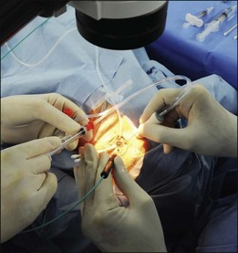
Vitreous sampling
The standard technique for performing 3-port pars plana vitrectomy (PPV) involves keeping the cutter tip within the vitreous. The undiluted vitreous humor is collected through lines using the vitreous cutter connected directly to a 3 or 5 mL syringe. This syringe is manually aspirated by an assistant while the operator compresses the eyeball with his/her fingers or a cotton swab (Fig. 123.1). When the eye visibly softens, the infusion is turned on.11 In our experience, up to 1.5 mL of undiluted vitreous can be safely obtained using this technique. Other techniques, such as using continuous air or perfluorocarbon liquid (PFCL) infusion can yield large volumes of undiluted vitreous without compressing the eyeball. Air substitutes the vitreous removed from the eyeball, and this can yield vitreous samples ranging in volume from 0.6 to 1.5 mL.9,12 Perfluorocarbon-perfused vitrectomy, in which balanced salt solution is replaced with perfluorocarbon liquid during vitreous aspiration, can be a good option for preventing complications of hypotony such as suprachoroidal hemorrhage.13 Using this technique, an average of 2.24 mL of undiluted vitreous could be obtained. However, a major disadvantage, in addition to the high cost of perfluorocarbon, is that samples obtained using this technique need to be frozen to completely remove the perfluorocarbon.
Handling and preparation of vitreous samples
The overall yield of diagnostic vitrectomies varies considerably in different studies. Generally, the number of tests that can be carried out on a vitreous specimen depends on the amount of material retrieved. In addition, patient selection, surgical technique, and vitreous specimen analysis are factors that may contribute to this variation. Ideally, cytological analysis and microbiologic cultures should be performed with undiluted vitreous specimen when first obtained. The vitreous wash fluid that is collected in the vitrector cassette can be used for additional microbiological cultures. Cytokine, chemokine, and flow cytometry analysis can also be performed using the aqueous humor that is obtained. It is nevertheless important to realize that a negative cytological diagnosis or microbiological culture does not prove the absence of malignancy or infection. Polymerase chain reaction (PCR) analysis is recommended in cases where viral infection is suspected because the quantity of organisms shed into the vitreous cavity is minute, and viral culture often fails. A positive PCR result for human herpes viruses in the vitreous of patients with uveitis has been identified.14,15
Retinal or choroidal biopsy
If a vitreous biopsy does not yield a diagnosis, chorioretinal biopsy of the involved eye can be considered (see also Chapter 124, Vitreous, retinal and choroidal biopsy), depending on the location of lesions and response to therapy. Chorioretinal biopsy increases the probability of achieving a correct diagnosis. Retinal or choroidal biopsy is considered when the inflammatory process is localized primarily in the sensory retina or the retinal pigment epithelium. In such cases, if the disease only involves the retina or choroid, pathogens may not spill into the vitreous. For example, cytomegalovirus, which is difficult to culture from the vitreous, can be cultured from the retina or seen on electron microscopic examination of the retina.
An endoretinal biopsy can be helpful in diagnosing tuberculosis, sarcoidosis, and lymphoma.16 Accurate biopsy results will lead to a specific treatment for uveitis of suspected infectious or malignant origin. Histological examination of chorioretinal biopsies provides some advantages over cytological examination of vitreous specimens. In the biopsy, more material is available for immunohistochemistry; this allows for more precise classification and differentiation of the pathology of the lesion. Despite these advantages, choroidal and retinal biopsies are used as a last resort considering the severe complications that may accompany the procedure.
The surgical technique for performing a chorioretinal biopsy can either be transscleral or transvitreal, depending on the location of the lesion. In patients with panuveitis, a choroidal mass, and a detached retina, it is recommended that transvitreal endoretinal biopsy be performed at the junction of the attached and detached retina. Upon completion of the vitrectomy, the area with active disease can be identified more accurately; this area can then be biopsied. However, a biopsy specimen obtained from a retinal area in which the disease is quiescent will rarely provide diagnostic information. The preferred location for this surgical procedure is the superior and nasal retina. Intense endolaser or heavy endodiathermy at the margin of the biopsy is performed prior to long-acting gas or silicone oil tamponade.17,18 In cases in which the retina is still attached, a cannula is used to inject saline under the sensory retina to create a small bleb. The advancing edge of the lesion should be included if retinitis is suspected, because actively replicating microorganisms are most likely to be found in this location.
The retinal or chorioretinal biopsy specimen should be divided in order to permit culture, histological examination, and monoclonal antibody studies. PCR can be performed on the retinal tissue as well. However, it has been reported that retinochoroidal biopsies may be associated with the risk of retinal detachment and that false-negative results often occur in these specimens.19,20 In addition, proper positioning is essential to maintain retinal tamponade after endoretinal biopsy.
Diagnostic techniques for vitrectomy specimens
Cytological evaluation
Cytological examination reveals the phenotypes of infiltrating cells into the vitreous. Vitreous humor fluid obtained by PPV is centrifuged, and the cells are smeared onto glass slides and then either immersed in 95% ethanol for Papanicolaou (Pap) staining or left to dry for subsequent Giemsa staining. The diagnosis of a PIOL depends primarily on cytological examination of the vitreous sample. The characteristic feature, from either Giemsa or Diff Quick staining, is the presence of large B cell lymphoblasts and atypical lymphocytes with high nuclear/cytoplasm ratios amongst small round lymphocytes.21 However, diagnosis of PIOL is difficult, resulting in a high false-negative rate due to small sample volumes with low number of malignant cells, inadequate preparation of samples or carrying media, and previous administration of corticosteroids. In chronic endogenous uveitis, cytology reveals classical degenerative inflammatory cells with poor morphology, although cytological examination of the vitreous specimen can be difficult because there may be a relative lack of inflammatory cells. A cytological examination can be helpful in diagnosing sarcoidosis. Kinoshita et al. showed multinucleated giant cells in 85.7% of cases and lymphocytes and epithelioid cells in all cases of intraocular sarcoidosis.5
Stay updated, free articles. Join our Telegram channel

Full access? Get Clinical Tree


