(1)
Department of Ophthalmology, St. Thomas’ Hospital, London, UK
12.1 Introduction
12.2 Diabetic Retinopathy
12.2.1 Introduction
12.2.4 Diabetic Retinal Detachment
12.2.5 Postoperative Complications
12.2.6 Success Rates
12.2.7 Diabetic Maculopathy
Abstract
There are a number of conditions which stimulate neovascularisation of the retina with subsequent complications such as vitreous haemorrhage and tractional retinal detachment from pathological separation of the vitreous. The most common is severe diabetic retinopathy but also retinal vein occlusion, sickle-cell retinopathy and retinal vasculitis.
Electronic supplementary material
The online version of this chapter (doi:10.1007/978-3-642-31872-6_12) contains supplementary material, which is available to authorized users.
12.1 Introduction
There are a number of conditions which stimulate neovascularisation of the retina with subsequent complications such as vitreous haemorrhage and tractional retinal detachment from pathological separation of the vitreous. The most common is severe diabetic retinopathy but also retinal vein occlusion, sickle-cell retinopathy and retinal vasculitis.
12.2 Diabetic Retinopathy
12.2.1 Introduction
The complications of diabetic retinopathy remain despite major advances in screening of the population and clinical management of patients. Retinal laser photocoagulation is the mainstay of therapy (The Diabetic Retinopathy Study Research Group 1981; Early Treatment Diabetic Retinopathy Study Research Group 1991) and reduces the chance of sight loss by 50 %. Even so, the vitreoretinal surgeon is likely to have to treat many patients with diabetic vitreous haemorrhage or tractional retinal detachment with a reported 5 % vitrectomy rate in diabetic retinopathy over 5 years (Flynn et al. 1992). Furthermore, the incidence of diabetes is increasing relentlessly, negating the effects of improved diabetic control or prompt laser therapy. The increase in the use of PPV for the complications of diabetes is supported by the increased recording of the use of PPV with endolaser photocoagulation by 86 % in Medicare fee data from the USA from 1997 to 2007 (Ramulu et al. 2010).
Diabetic Retinopathy Grading
Table 12.1
Diabetic retinopathy grading
Grade | Retinal features |
|---|---|
No diabetic retinopathy | Nil |
Mild non-proliferative retinopathy (NPDR) | Microaneurysms only |
Moderate NPDR | More than ‘microaneurysms only’ and less than severe NPDR |
Severe NPDR | Any of the following: |
More than 20 microaneurysms in each quadrant | |
Venous beading in more than 2 quadrants | |
Intraretinal microvascular abnormalities in more than 1 quadrant | |
No proliferation | |
Proliferative | Low-risk, flat new vessels elsewhere |
High-risk, raised new vessels elsewhere or disc new vessels |
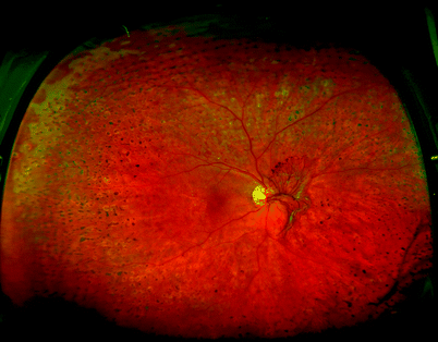
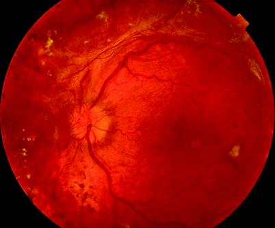
Fig. 12.1
NVD are present on this disc despite PRP
Ischaemic diabetic retinopathy characteristically affects the mid-peripheral retina outside the major temporal vascular arcades and nasal to the optic disc. Neovascularisation generally develops near the posterior limit of the ischemia, that is, at the optic disc and along the major vascular arcades. Vascular tissue, arising from intraretinal venules, grows out through the inner limiting membrane and proliferates within the most cortical part of the vitreous gel as a vascularised epiretinal membrane (flat new vessels). The vessels do not grow into the central gel except occasionally within Cloquet’s canal. The membranes incarcerate the gel on which they are proliferating, resulting in a vitreoretinal adhesion. The vitreous may stiffen and shrink secondarily to the retinopathy, increasing traction on blood vessels and membranes, or the vitreous may detach, rupturing blood vessels.
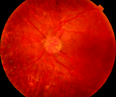
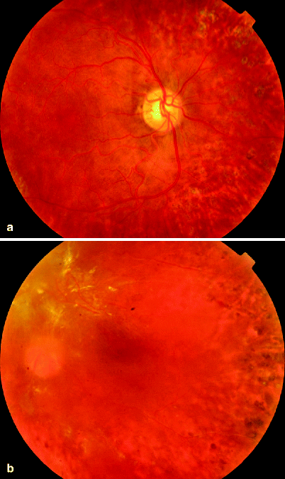
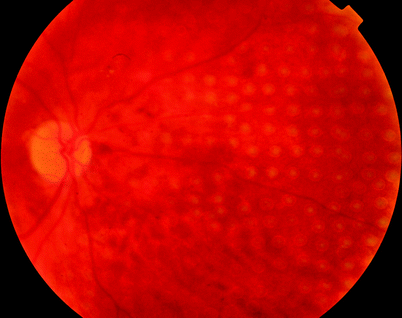
Fig. 12.2
Retinal neovascularisation requires a scaffold upon which to grow, that is, the vitreous. In the left eye in which the vitreous is present, extensive NVD are seen, whilst in the right eye which has had PPV, there is no structure (vitreous) upon which the vessels can grow (see Fig. 12.3)
Fig. 12.3
See previous figure
Fig. 12.4
Panretinal photocoagulation by pattern scan laser application
12.2.2 Diabetic Vitreous Haemorrhage
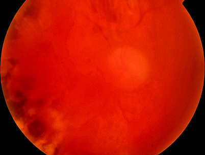
Fig. 12.5
Diffuse vitreous haemorrhage in diabetic retinopathy; visualisation with the indirect will reveal more detail in the retina than a 90D lens and the slit lamp
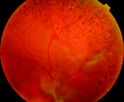
Fig. 12.6
Vitreous haemorrhage can be caused by neovascularisation or by avulsed blood vessels as in this patient
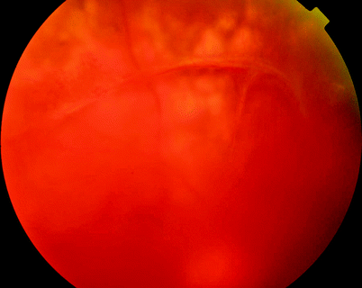
Fig. 12.7
A mature blood vessel is under traction by the vitreous causing it to haemorrhage. Laser will not regress this blood vessel, and only removal of the vitreous will prevent recurrent bleeds
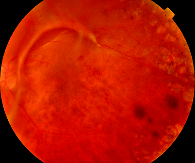
Fig. 12.8
The vitreous is applying traction to this blood vessel which may be a cause of recurrent haemorrhaging. Occasionally the blood vessel may be pulled out of the surface of the retina
12.2.3 Progression to Vitreous Haemorrhage and Tractional Retinal Detachment
In general, progression to vitreous haemorrhage or retinal detachment can be seen as a sign of failure in the systems of screening to detect diabetic retinopathy early, monitoring and treatment of the retinopathy but also in the care of the patients diabetes, blood pressure and other factors such as serum lipids by the diabetic physician and the patient themselves. The DRS and EDTRS showed reduction in sight loss by 50–70 % not elimination of sight loss (The Diabetic Retinopathy Study Research Group 1978; The Diabetic Retinopathy Study Research Group 1981; Early Treatment Diabetic Retinopathy Study Research Group 1991) because these patients develop maculopathy (the commonest cause of reduction in central vision), vitreous haemorrhage and tractional retinal detachment. Rates of progression to vitrectomy are reduced by good control of blood sugar, hypertension and lipids and timely and early PRP which reduced risk of PPV to 2.3 % from 4 % in the EDTRS (Early Treatment Diabetic Retinopathy Study Research Group 1991). Neovascularisation needs scaffold to grow on; therefore, PVD is protective, but most of these patients are too young to have PVD and have had laser therapy which may reduce the chance of PVD due to vitreoretinal adhesion at laser spots. Progression has been calculated as 5 %, requiring PPV over 5 years (Early Treatment Diabetic Retinopathy Study Research Group 1991). The prevalence of PPV in the diabetic population in South London has been estimated as 5 per thousand diabetic patients.
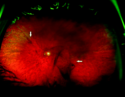
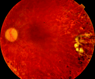
Fig. 12.9
A diabetic eye with NVE (vertical arrow) and central vitreous haemorrhage (horizontal arrow)
Demographics | |
Male | 54.1 % |
Type 1 | 36.8 % |
Type 2 | 63.2 % |
Insulin requiring | 71.4 % |
Ethnicity | |
Afro-Caribbean | 31.4 % |
Caucasian | 53.5 % |
Southeast Asian | 11.4 % |
Clinical Features
This is by far the commonest cause of haemorrhage into the vitreous cavity. The haemorrhage can occur into the gel or retrohyaloid space or rarely subretinal space (the last usually in association with tractional retinal detachment). Subretinal haemorrhage is associated with a poor visual outcome (Morse et al. 1997). Severe retrohyaloid haemorrhage may cause the posterior vitreous face to bulge forwards in bullae. Often diabetic haemorrhaging occurs spontaneously, that is, from action of the vitreous on the new blood vessels, but occasionally haemorrhage happens during vigorous isometric exertion. Approximately 50 % of the haemorrhages will clear spontaneously over 3 months. Untreated neovascularisation may progress over this time, risking further haemorrhage, TRD or neovascular glaucoma. Type one diabetics are particularly at risk of further complications if left too long before surgery (The Diabetic Retinopathy Vitrectomy Study Research Group 1985; Early vitrectomy for severe vitreous hemorrhage in diabetic retinopathy. Four-year results of a randomized trial: Diabetic Retinopathy Vitrectomy Study Report 5 1990), and early intervention is recommended. If waiting for clearance, it is important to monitor the intraocular pressure for erythroclastic glaucoma and to perform repeated ultrasound examinations for retinal detachment.
Causes of vitreous haemorrhage:
Active NV
Inactive NV
Vessel avulsion
Traction from vitreous
Pathological shrinkage
Mobility
PVD
Acutely raised BP
PRP | PPV |
|---|---|
Active NV and view rapidly clearing | Non-clearing VH 2-3/12 |
Inadequate PRP | |
INV | |
For visual rehabilitation |
Fig. 12.10
Tractional membranes often follow the vascular arcades
Indicators for early surgical intervention:
Iris neovascularisation | Urgent |
No previous PRP | Urgent |
Erythroclastic glaucoma | Soon |
Type 1 diabetes | Soon |
B-scan ultrasound examination is important to determine where the blood is situated, whether there is tractional retinal detachment, rhegmatogenous retinal detachment or posterior vitreous detachment, and is accurate in 90 % of cases (Genovesi-Ebert et al. 1998). Adhesion of the vitreous to the retina can be seen at the sites of neovascularisation often on the vascular arcades or at the optic disc. The extent of the vitreoretinal adhesion should be assessed to guide the surgeon on the complexity of the surgery. The role of vitrectomy in the management of the complications of diabetic retinopathy has been established for many years (Mandelcorn et al. 1976; Michels 1978; Aaberg 1979). The success rates of PPV are now high enough to offer surgery early in the clinical course of the haemorrhage without waiting for spontaneous clearance of the bleed.
In some circumstances, if the vitreous haemorrhage is mild, further laser can be applied to try to regress the neovascularisation without PPV. Some patients will bleed despite extensive PRP because of the presence of established neovascularisation which although gliosed has the capacity to bleed because of movement of attached gel or because the patient has an avulsed retinal blood vessel. These patients will not respond to further laser and will require PPV. The role of anti-VEGF injections in the treatment of vitreous haemorrhage has yet to be clarified although claims of more rapid regression of haemorrhage with injection alone have been made (Huang et al. 2009).
Surgery
Table 12.2
Difficulty rating for PPV for diabetic vitreous haemorrhage
Difficulty rating | Low |
Success rates | High |
Complication rates | Low |
When to use in training | Early |
Additional surgical steps
Remove the core vitreous and make a perforation in the posterior hyaloid.
Remove any retrohyaloid haemorrhage through the perforation.
Remove the remaining vitreous.
Apply panretinal photocoagulation.
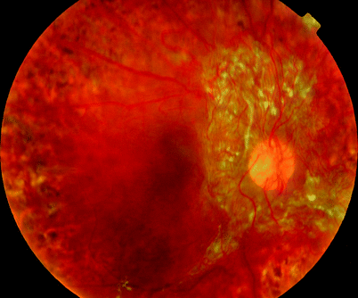
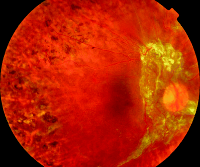
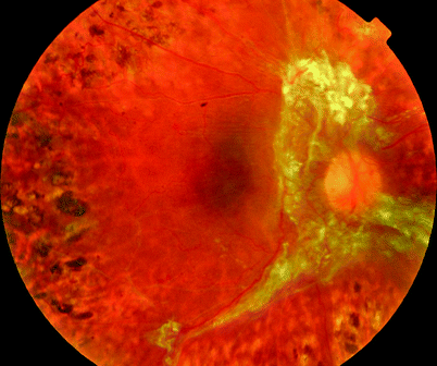
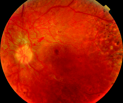
Fig. 12.14
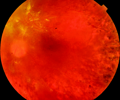
Fig. 12.15
See previous figure
Although vitreous haemorrhage from DMR may clear spontaneously after some months, pars plana vitrectomy is frequently required to rehabilitate the patient and can be associated with better visual prognosis (The Diabetic Retinopathy Vitrectomy Study Research Group 1988). The operation is more urgent in young diabetics to prevent irreversible loss of vision (The Diabetic Retinopathy Vitrectomy Study Research Group 1985). Preoperatively it is useful to determine with ultrasound whether the vitreous is fully detached or partially adherent to the retina or whether there is TRD or neovascularisation. A fully detached vitreous indicates an operation, which should be quick with a good prognosis for visual recovery, whereas extensive TRD indicates a longer operation with a poorer prognosis.
If the media are clear, subhyaloid blood can sometimes be treated with YAG laser (Celebi and Kukner 2001; Ulbig et al. 1998) without need for vitrectomy. More often, vitrectomy is required. During surgery, remove the central gel and then make a hole in the posterior hyaloid membrane. Remove the subhyaloid blood by placing a flute tip in the hole. This allows rapid removal of the subhyaloid blood without allowing it to enter the anterior vitreous cavity to spoil your view. Once the subhyaloid blood is removed, the retina can be identified, and the remaining gel removed with the reassurance that the retina is not detached and at risk of injury from cutter. Peripheral gel should be removed as much as possible as blood will leach out peroperatively and postoperatively, causing vitreous cavity haemorrhage and occasionally erythroclastic glaucoma (raised IOP from clogging of the trabecular meshwork with macrophages laden with red blood cell breakdown products).
Note: When removing blood from the vitreous base, usually, a very thin layer of clear gel at the vitreous base is encountered before hitting the retina.
Panretinal photocoagulation is applied to prevent iris neovascularisation (INV) because the removal of the gel allows easy access of the neovascular factors to the anterior segment, especially in aphakic eyes, increasing the risk of INV (Rice et al. 1983b; Blankenship 1980).
When using a blunt instrument near the retina, for example, removing preretinal blood, apply the instrument close to the retina in an anteroposterior fashion in a ‘daub’ like movement. Come away from the retina before moving to a new location. The reason for doing this is that it is difficult to cause an injury to the retina with a blunt instrument if it is moved directly onto the retina without moving laterally. If it is moved laterally whilst touching the retina resulting in a ‘scrape’, there is a high chance of causing a tear.
The spherical shape of the eye means that an anterior movement of the instrument tip must accompany any lateral movement of an instrument to avoid striking the retina.
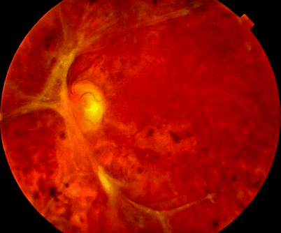
Fig. 12.17
This patients neovascular membranes are stable 6 years after receiving a pancreatic transplant
Be aware of these principles when:
Removing blood from the surface of the retina with a flute needle
Applying endolaser panretinal photocoagulation
Peeling epiretinal membranes
Detaching the posterior hyaloid membrane
Isolate and trim or dissect off any neovascularisation. Try not to leave any significant amount of tissue on the disc or elsewhere because these sites become small foci of contraction which may wrinkle the retina and fovea. Endodiathermy is usually unnecessary as the neovascularisation is small calibre and low flow, allowing rapid plugging with clot.
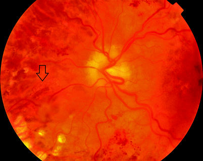
Fig. 12.18
Traction on the retina can create a retinal break as in this patient who has an avulsed blood vessel and an operculum on the nasal side of the disc overlying a retinal hole. Often as in this eye, progression to RRD does not occur; this may be due to the stiff, immobile vitreous in diabetes. The immobility of the vitreous prevents the creation of fluid currents thought to be necessary for SRF to accumulate
12.2.4 Diabetic Retinal Detachment
Clinical Features
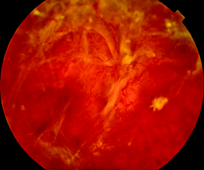
Fig. 12.19
Unfortunately, despite the advantages in screening and in treatment by panretinal photocoagulation, patients still attend with very severe tractional retinal detachments as seen in this picture. The appearance can be deceptive with dissections easier than at first apparent. As with all diabetic tractional retinal detachments, the key is determining the right layer and getting behind the posterior hyaloid, membrane and cortex. Elevation of cortex remnants in a vitreoschisis aids removal of the vitreous gel and reattachment of the retina
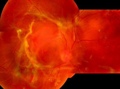
Fig. 12.20
The membranes in diabetic TRD usually form over the retinal vasculature, the optic disc and temporal to the macula encircling the macula. However, membranes can be seen anywhere in the retina
As in other epiretinal membranes, fibroblasts within the vascularised membranes contract, and the tangential traction so produced is stabilised and consolidated by collagen synthesis. The tangential traction results initially in folding of the inner retinal layers (internal limiting membrane and nerve fibre layer) and can then progress to traction retinal detachment.
Contraction of the neovascular membranes both anteroposteriorly and tangentially combined with shrinkage of the vitreous gel pulls the retina at its points of adhesion into the centre of the eye. Without a retinal hole to allow accumulation of subretinal fluid, the retina detaches with a concave configuration. Two forces are acting on the retina, one the action of the RPE to keep the retina flat and two the action of the shrinking vitreous and neovascular membranes pulling the retina centrally into the vitreous cavity.
During this process, the vitreous cortex often splits, leaving a thin layer on the retina that can be elevated during surgery to allow easier dissection of the fibrotic neovascular membranes (Schwatz et al. 1996; Chu et al. 1996). The vitreous detachment on the inner surface of the vitreoschisis is taut and stretches from the vitreous base to the neovascular membranes and between the membranes. The areas of detachment surround neovascularisation on the retinal arcades and are often multifocal. Eventually the macula detaches severely reducing the visual acuity, whilst the periphery remains flat. The extent of tractional retinal detachment (TRD) varies from a single focus to large areas of the retina. Similarly and more important to the surgeon, the areas of adhesion of the neovascular membrane to the retina vary, often being most pronounced on the vascular arcades and around the optic disc.
Traction on the disc can reduce vision by damaging the superficial nerve fibres (Kroll et al. 1999); indeed, axons are found in tissue removed from the disc surgically (Pendergast et al. 1995).
Traction on the retina may split the retina causing retinoschisis (Pendergast et al. 1995).
Occasionally a hole appears in the fragile ischaemic retina, allowing subretinal fluid accumulation. The retinal detachment then takes on a convex configuration and may extend further anteriorly in a bullous fashion. Application of panretinal photocoagulation to eyes with tractional retinal detachment can cause further contraction of the membranes and macular detachment (Ghoraba 2002). Therefore, in patients with established TRDs but with no PRP, it is often safer to proceed to PPV rather than try PRP alone.
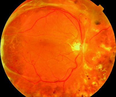
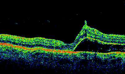
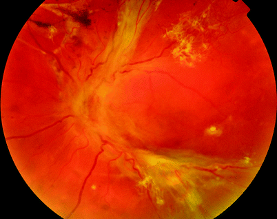
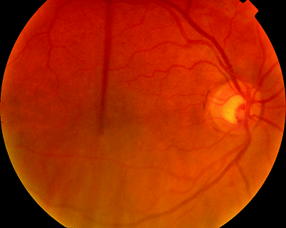
Fig. 12.21
The fovea is just being elevated by the TRD in this patient as shown by the OCT image (see Fig. 12.22)
Fig. 12.22
See previous figure
Fig. 12.23
If you see asymmetrical disease, fellow eye (Fig. 12.24) check the eye with less retinopathy for a PVD or check the carotids for stenosis (the worse stenosis is on the same side as the eye with less retinopathy)
Fig. 12.24
See previous figure
Surgery
Table 12.3
Difficulty rating for diabetic tractional retinal detachment
Difficulty rating | High |
Success rates | Moderate |
Complication rates | High |
When to use in training | Late |
Tractional Retinal Detachment
Additional surgical steps
Remove vitreous haemorrhage as above but retain any traction on the retina.
Create holes in the posterior hyaloid to allow easy access to the areas of retinal elevation and vitreoretinal adhesion.
Detect and elevate the posterior layer of any vitreoschisis.
Dissect the tractional membranes off the retina.
Complete the removal of the vitreous.
Apply panretinal photocoagulation.
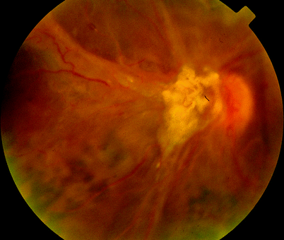
Fig. 12.25
The membranes can cause traction and damage on the optic nerve, reducing visual recovery postoperatively
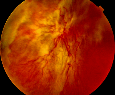
Fig. 12.26
A combination of rhegmatogenous and tractional retinal detachment may occur. Particular problems exist when the patient has a rhegmatogenous element in addition producing PVR. Other indicators of relative poor prognosis are subretinal haemorrhage and iris neovascularisation
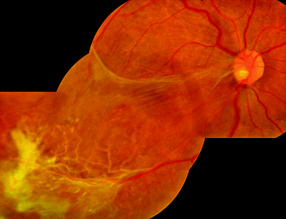
Fig. 12.27
Occasionally an eye in which neovascularisation has been stabilised by laser will have inactive membrane overlying the macula
Where significant neovascular membranes exist or retinal detachment is present dissection by delamination of any membranes is required. The membranes are best removed in total (‘en bloc’) to prevent future reproliferation and subsequent detachment (Kakehashi 2002; Williams et al. 1989; Abrams and Williams 1987). The core of the vitreous is removed whilst retaining the taut peripheral gel thereby maintaining traction upon the membrane. A hole is made in the cortical gel to allow access of instruments to the retinal surface. The outer portion of the vitreous cortex on the retinal surface is then elevated to find the plane of cleavage of the internal limiting membrane and posterior hyaloid membrane. The posterior hyaloid and the neovascular membranes are then dissected away from the retina to relieve traction and allow the retina to reattach. Modern cutters especially small gauge have orifices which are nearer the tip end of the cutter shaft.
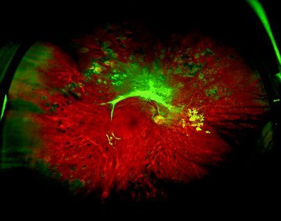
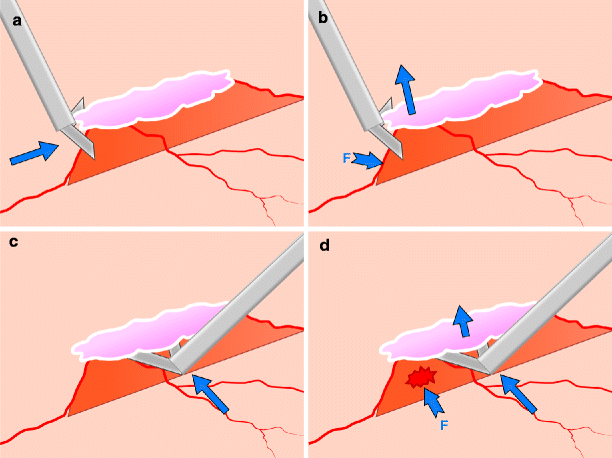
Fig. 12.28
Despite only minimal PRP, these membranes are stable perhaps indicating a ‘burnt out’ retinopathy
Fig. 12.29
Try to work along a ridge of adhesion (a, often along a large blood vessel) rather than across it because the scissors can straddle the ridge (b) and keep away from the retina, and any forces (arrows) are applied to the relatively strong blood vessel (F arrow). Going across the ridge risks the tips of the scissors incising the retina (c and d), and the forces are applied to the weak retina on the slope of the ridge
Note: In less complex TRD, the membranes can be trimmed down by these cutters effectively shaving down the membrane until the retina is cleared to membrane rather than using scissors to dissect off the membrane.
Elevating the cortical gel and posterior hyaloid which is still attached to the surface of the retina is the key manoeuvre of the operation. Having made the hole in the elevated vitreous cortex, use the edge of the scissors to run along the slope of an area of TRD. may catch the posterior hyaloid and see it lift both outwards towards the equator and towards the TRD. Elevate the PHM up to the edge of the TRD and then use the plane between this and the retina to elevate the membranes. Alternatively, the correct plane can sometimes be found by starting in the macular area and working outwards towards the arcades.
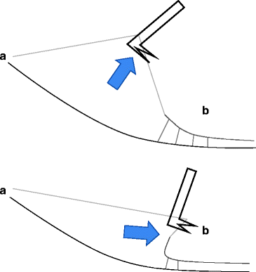
Fig. 12.30
When using the posterior hyaloid to lift a membrane (e.g. TRD membrane) to look for pegs at site (b), stay close to the edge of the membrane. Lifting further away from the membrane requires a vertical movement to avoid traction at point (a) to see the pegs. This will also put a force on pegs deeper under the membrane which you cannot see. If you stay close to the membrane edge, a tangential motion is possible without undue force at point (a) and only applying force to pegs at the edge of the membrane
Note: The PHM very often ends in the peripheral retina and does not often extend to the vitreous base in severe TRD.
In this plane, the membranes will separate much more easily. Cutting through cortical gel is much more difficult and will leave membrane on the retina, allowing later reproliferation. Some of the membrane will lift off, but sites of adhesion that will not lift must be cut. Keep your visualisation to a maximum by trimming any elevated membrane with the cutter whilst maintaining some anteroposterior traction on the TRD. As with any membrane, work around points of adhesion, lifting the membrane around a difficult site before tackling it. Usually I will elevate and PHM around the TRD 360°, if I can, before dissecting the area of TRD itself. When lifting the PHM, lift close to the edge of the TRD and then move tangentially out to elevate the PHM in the periphery. If using the PHM to lift the edge of the TRD, stay close to the TRD to achieve a large angle on the edge of the TRD without applying forces on the vitreous base.
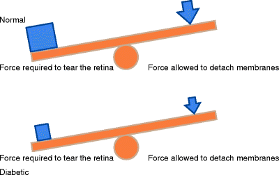
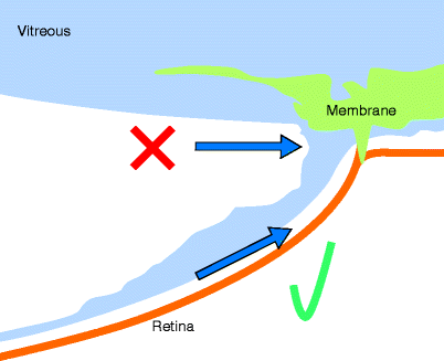
Fig. 12.31
The balancing act between pulling on an ERM and avoiding a retinal tear. In the normal eye, a large force is required before the retina tears, so that a relatively large force can be applied to pull off an ERM. In the ischaemic diabetic retina, a small force may tear the retina; therefore, only a small force can be applied to ERM to remove it
Fig. 12.32
It is important to dissect under the outer layer of a vitreoschisis which is easier than cutting through the vitreous to get to pegs of membranous attachment and avoids leaving vitreous on the retina
Most TRDs are made up of peg attachments except: at the optic disc where a larger area of adhesion approximately ½ a disc area is seen. This will usually peel off the disc with some cutting with scissors surprisingly easily though; be aware of the major blood vessels and vulnerability of the surface of the optic nerve head.
Where there is schitic retina, there can sometimes be a sheet of adhesion of the membrane to the retina.
Retinoschisis is an occasional feature in retina in TRD and is presumably due to splitting of the retina from traction. It may prevent flattening of the retina at the end of the operation and application of laser. Often partial-thickness inner retinal holes are seen. Dissection of membrane is often difficult from the surface of the schitic area.
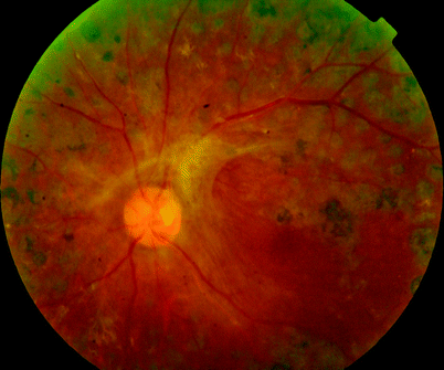
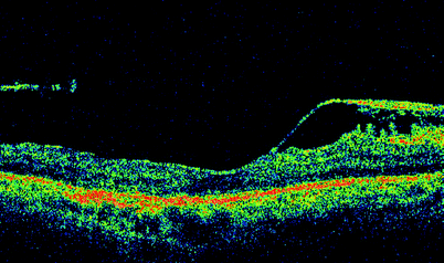
Fig. 12.33
The OCT of this patient shows the pegs of attachment between the membranes and the retina see 12.34.
Fig. 12.34
See previous figure