(1)
Department of Ophthalmology, St. Thomas’ Hospital, London, UK
15.1 Introduction
15.2 Dropped Nucleus
15.2.1 Clinical Features
15.2.2 Surgery
15.3.1 Clinical Presentation
15.3.2 Surgery
15.4.1 McCannell Sutured IOL
15.4.2 Iris-Clip IOL
15.4.3 Haptic Capture Method
15.4.4 Anterior Chamber IOL
15.4.6 The Aphakic and Aniridic Eye
15.5.1 Clinical Features
15.5.2 Surgery
15.5.3 Infective Organisms
15.5.4 Antibiotics
15.5.5 The Role of Vitrectomy
15.5.6 Success Rates
15.7 Needlestick Injury
15.7.1 Clinical Features
15.7.2 Surgery
15.9 Retinal Detachment
15.10 Chronic Uveitis
15.13.1 External Drainage
15.14 Summary
Abstract
Cataract surgery in the modern setting means phacoemulsification with a small incision and a sutureless wound. This is a rapid procedure with a high rate of success, approximately 95 %. There still remains a small complication rate of approximately 4 %.
Electronic supplementary material
The online version of this chapter (doi:10.1007/978-3-642-31872-6_15) contains supplementary material, which is available to authorized users.
15.1 Introduction
Cataract surgery in the modern setting means phacoemulsification with a small incision and a sutureless wound. This is a rapid procedure with a high rate of success, approximately 95 %. There still remains a small complication rate of approximately 4 %.
Some of these are serious complications and will often involve the vitreoretinal surgeon in remedying the situation.
15.2 Dropped Nucleus
15.2.1 Clinical Features
Dropped nucleus incidence 0.09–0.8 % (Aasuri et al. 2001; Kageyama et al. 2001; Mathai and Thomas 1999; Stilma et al. 1997)
The phrase ‘dropped nucleus’ has been used to describe dislocation of the nucleus (or part of the nucleus) of a cataract during phacoemulsification into the vitreous cavity. Dropped nucleus may happen to any case with an increased risk of capsular rupture or zonular dehiscence such as trauma, pseudoexfoliation or hard nuclei or because of the inexperience of the surgeon (Aasuri et al. 2001). The positive pressure applied by the infusion fluid during phacoemulsification means that a tear in the posterior capsule of the lens will result in the dislocation the contents of the anterior chamber (most often the lens) into the vitreous cavity. The higher density of the lens compared to the vitreous also encourages its dislocation into the posterior segment. The nucleus can be seen in the inferior vitreous accompanied by fluffy white soft cortical lens material.
Uveitis. The lens material stimulates uveitis which in the short term can be controlled, if the nucleus remains in the posterior segment for months a chronic uveitis is stimulated, which may persist after surgery.
Glaucoma, a severe rise in intraocular pressure can usually be controlled by topical medication in the short term till removal of the nucleus is performed. However, if the nucleus remains in the eye for a prolonged period, the glaucoma may not reverse after lens removal.
Retinal detachment. The disruption to the vitreous may produce retinal detachment by tearing the retina, 4–8 % of patients (Ross 1996; Oruc and Kaplan 2001). There is an increased risk of RRD approximately 4 % before PPV and 4 % after PPV with retinal tears also requiring treatment during PPV in a few patients (Hansson and Larsson 2002). RRD is reported earlier in these patients (mean 4 months) than after routine cataract extraction (mean 16 months) (Haddad et al. 2002).
The patient is understandably disappointed having expected a straightforward operation and improved vision. Instead, they often have reduced vision and discomfort from uveitis (56 %), raised IOP (52 %) and corneal oedema (46 %) (Gilliland et al. 1992). Pars plana vitrectomy and removal of the nucleus are required within 1 or 2 weeks to avoid glaucomatous damage or chronic uveitis or cystoid macular oedema (Rossetti and Doro 2002). Most patients if treated promptly will achieve good vision. However, occasionally, an eye will end up blind from associated choroidal haemorrhage or retinal detachment. Rarely dropped nucleus can present with endophthalmitis (Irvine et al. 1992). Also, the risk of endophthalmitis seems to be doubled over other surgery (Kim et al. 1996).
Note: Rapid referral for vitrectomy is required.
15.2.2 Surgery
Table 15.1
Difficulty rating for PPV for dropped nucleus
Difficulty rating | Moderate |
Success rates | Moderate |
Complication rates | Low |
When to use in training | Middle |
1.
Sew up the cataract wound.
2.
Clear the anterior segment and capsule of soft lens material.
3.
Use the vitreous cutter to remove the soft lens material in the vitreous cavity.
4.
Use the Fragmatome to remove the nucleus.
5.
Detach the posterior hyaloid if required.
6.
Insert an intraocular lens implant.
‘Fishing’ for the nucleus via the anterior segment by the phacoemulsification surgeon is not advisable as this can cause giant retinal tears from traction on the vitreous even after anterior vitrectomy (Aaberg et al. 1997). The timing of surgery by PPV has created controversy in the past (Hansson and Larsson 2002; Al-Khaier et al. 2001; Bessant et al. 1998; Borne et al. 1996; Hutton et al. 1978; Kim et al. 1994; Margherio et al. 1997; Stefaniotou et al. 2003), but it is now recommended that a patient who has suffered a dropped nucleus during phacoemulsification cataract extraction be operated on within 2 weeks from onset of the complication. This minimises the risk of secondary complications such as intractable glaucoma and cystoid macula oedema from the uveitis (Rossetti and Doro 2002; Yeo et al. 1999). Patients presenting with these complications in the short term usually have these reversed after PPV (Vilar et al. 1997).
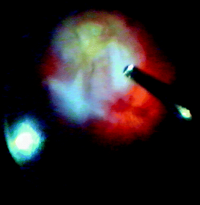
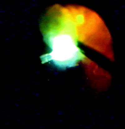
Fig. 15.1
Soft lens material can be removed easily by the vitreous cutter as can moderate amounts of lens nucleus
Fig. 15.2
The hard nuclear material is removed with a Fragmatome whilst supporting the lens with the light pipe. Whiter and fluffier softer lens material can be removed with the cutter
Primary Management
If the surgeon who has dropped nucleus has the capability to perform a vitrectomy and removal of the fragments, then this can be performed immediately. However, unless this facility is available, it is best to close the eye, tidy up the vitreous, leave lens implantation until the next surgery and ensure closure of the cataract wound. By delaying the surgery for 1 or 2 weeks, it allows the use of elective surgical time and provides time to discuss the problem with the patient and allow them a chance to gather their thoughts before proceeding to another operation. Uveitis during this period readily responds to topical steroid therapy and raised IOP responds to topical therapy with temporary use of acetazolamide orally if required.
Vitrectomy Surgery
At PPV surgery, the first thing to do is to secure the anterior chamber. Most wounds are now so small that they can usually be left without a suture but check the wound and rehydrate if necessary; the wound has not usually sealed by the time of the PPV. If the wound remains unstable, use a 10/0 absorbable suture to close the wound, any pressure on the posterior edge of the wound will open it during the vitrectomy. Create sclerotomies as usual, and excise any vitreous from the anterior chamber by passing through the posterior capsule into the AC (do not use old wounds if you want to go from the front, and create a new paracentesis for the cutter to enter). Remove any soft lens material from within the capsule using the cutter on aspirate only, in the same fashion for a normal SLM aspiration during cataract extraction but by accessing from the posterior segment the via the posterior capsular break. Preserve as much capsule, anterior and posterior, as is possible, as it is the intention to put in a posterior chamber lens implant at the end of the vitrectomy, and this capability will depend on the amount of supporting capsule. Then perform a PPV, taking note whether the vitreous has detached or not. Gain access to the nucleus, remove any SLM from around the nucleus and then assess the size of the nucleus to be removed.
If there is less than a quarter of the nucleus in the posterior segment, a chop and cut procedure will be adequate, whereby the nucleus is delaminated against the cutter, and small fragments of nucleus are chopped by the cutter. If there is more than a quarter of the nucleus in the back of the eye, then a Fragmatome is required to phacoemulsify the nucleus to speed its extraction. In this case, it is prudent to apply a small protective bubble of heavy liquid to the posterior pole, as it is likely that the nucleus will occasionally fall from the Fragmatome tip and may strike the macula or the optic disc (Wallace et al. 1993). Set the power of the Fragmatome to low, perhaps 20 % of normal, and use a pulse mode of 8 pulses/s. This makes it easier to hold onto the lens nucleus during phacoemulsification. The nucleus has a tendency to shoot away from the Fragmatome tip when using the ultrasound on continuous. It should be possible to manipulate the nucleus in a cartwheel fashion; using the light pipe and Fragmatome, remove as much of the nucleus as possible in one sweep.
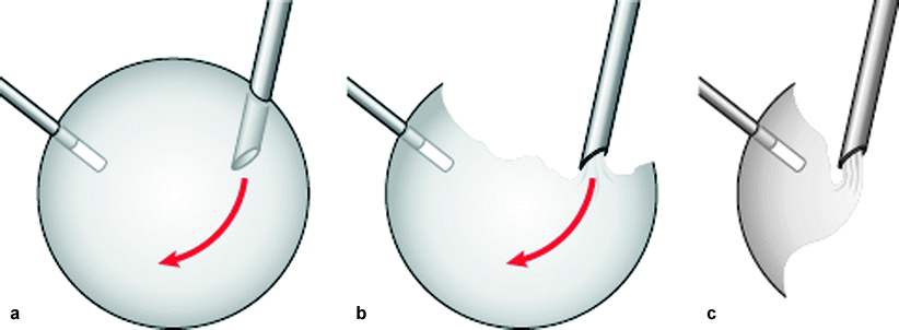
Fig. 15.3
Spike the lens or nucleus with the light pipe and, in a rotating manner, use the Fragmatome to emulsify and aspirate the nuclear fragments. Try to work towards the light pipe without dislodging the light pipe so that there is minimal need for re-engaging the nucleus, thereby reducing manipulation and time spent near the retina
Use the on-demand system for the Fragmatome to avoid ultrasound near the retina causing any damage to the retinal surface:
For linear aspiration, use foot pedal on direct depression.
When the nucleus has been engaged by aspiration only, lift the nucleus away from the retina into the mid-vitreous.
Once safely away from the retina, kick the foot pedal to the side to engage ultrasound on maximum to commence the phacoemulsification (Fragmatome).
If the nucleus disengages and drops, cease aspiration (lift your foot) and start again.
Note: The aspiration is high even at 150 mmHg (the bore of the Fragmatome port is larger than a cutter) for a fluid filled eye; therefore, rapid cessation of aspiration is required to prevent globe collapse, even if the infusion bottle height has been increased.
Eventually, the nucleus can be completely removed. If there is no posterior vitreous detachment, detach the posterior vitreous at this stage, difficult before as the nucleus is resting on the vitreous cortex. Perform an internal search to detect retinal tears or retinal detachment, and treat these as they arise, do not put gas in yet because insertion of intraocular gas may be problematic depending on the stability of the IOL.
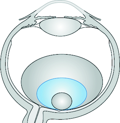
Fig. 15.4
Heavy liquids can be placed onto the posterior pole of the eye during removal of dropped nucleus. A small bubble shown in red has a very convex surface upon which the fragments will slip back down onto the retina. Increasing the size of the bubble flattens the surface of the heavy liquids, facilitating the maintenance of fragments in the mid-vitreous cavity. However, if the bubble becomes too large, then there is reduced space for working between the bubble and the anterior segment. An ideal size is indicated by the blue circle
Thereafter, perform secondary lens implantation, leaving the infusion on, and make a corneal wound with the keratome. Switch off the infusion and fill the anterior chamber with viscoelastic which acts as a plug, preventing the egress of fluid through the corneal wound. Assess the state of the capsule. If there is plenty of capsule available (i.e. enough to encapsulate securely the IOL), insert the lens implant into the bag. If not, place the lens implant into the ciliary sulcus (requiring at least two-thirds of the capsule remaining) and suture up the wound with 10/0 absorbable suture. If there is less than two-thirds of the capsule, insert an anterior chamber lens implant. A few surgeons recommend sulcus sutured lenses, but these have a high chance of dislocation in 6 years, approximately 25 % (Vote et al. 2006).
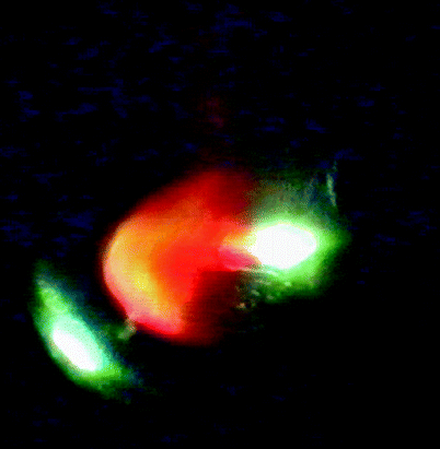
Fig. 15.5
This nucleus is blood stained because the eye has also suffered a choroidal haemorrhage
Difficult Situations
Concurrent retinal detachment and dropped nucleus: a mobile retina may cause problems whilst trying to perform Fragmatome extraction. In this case, heavy liquid will need to be inserted to stabilise the retina before removing the nucleus. Insert the IOL before the air exchange (with the posterior segment filled with heavy liquid) using viscoelastic in the anterior chamber as above. Remove the viscoelastic (it is difficult to do this after the air has been inserted), and then perform an air/heavy liquid exchange; you may need to wipe the posterior lens surface to avoid condensation (see Chap. 3). Treat any retinal breaks causing the retinal detachment either with the heavy liquid fill or under air.
Alternatively, if it is known that a retinal detachment is present, put the lens implant in early on, after clearing of the anterior chamber of SLM and vitreous.
Air may get into the anterior chamber in the presence of a large posterior capsule rent or zonule dehiscence and particularly if an anterior chamber lens implant is present; therefore, it is important to have most of your surgical manoeuvres completed before air insertion. If air enters the anterior chamber with further surgery still to be performed, fill the anterior chamber with viscoelastic to regain the view. However, when removing the viscoelastic, air will usually re-enter the anterior chamber. Although air in the anterior chamber is undesirable because of potential effects on the corneal endothelium, it is often necessary to leave this in and allow it to disperse postoperatively because you are unlikely to be able to refill the anterior chamber with fluid as this will fall into the air-filled posterior segment.
Note: If there is an unstable IOL in the presence of intraocular gas, it is often better to have the IOL sandwiched by gas, that is, gas in the anterior and posterior segments. If gas is in the posterior segment only, it tends to push the unstable IOL forwards and may cause capture of the pupil by the IOL edge.
If there is no support for the IOL and you anticipate needing an ACIOL or sutured or iris-clip IOL, do this at a second operation once the gas bubble has dispersed. These lenses could be problematic in the presence of gas with the possibility of:
Dislocation
Corneal touch
Displacement from visual axis
Very occasionally, the dropped nucleus is associated with a choroidal haemorrhage. In this circumstance, remove the nucleus and fill the eye with silicone oil, without lens implantation. Wait for resolution of the choroidal haemorrhage at a later date, and then determine whether implantation is possible. However, the prognosis for vision in an eye like this is often poor, and it may be that vision will be limited and lens implantation is not appropriate.
Success Rates
Visual outcome is approximately a 60 % chance of 20/40 vision or better, that is, at least 20 % less than after routine cataract extraction (Hansson and Larsson 2002; Kim et al. 1994; Smiddy et al. 2003).
Surgical Pearl of Wisdom
‘When you are performing combined vitrectomy and phako procedures, try to make the anterior rhexis a little smaller than usual so that the optic is entirely within the bag. This will prevent lens pupil capture and posterior synechiae forming in gas filled freshly pseudophakic eyes.
If some of the optic edge is not covered by anterior capsule, then all is not lost. Create a 4–5-mm posterior capsulotomy using the cutter and prolapse the lens optic through into the posterior segment; this seems to provide enough support to prevent anterior segment complications’.
D. Alistair H. Laidlaw, St Thomas’ Hospital, London, UK
15.3 Intraocular Lens Dislocations
15.3.1 Clinical Presentation
Prosthetic intraocular lens implants can also dislocate into the posterior segment, especially silicone plate IOLs after YAG capsulotomy (typically at 2 months after the laser procedure) (Agustin and Miller 2000; Schneiderman and Vine 1995). In some patients with weak zonular fibres (myopes, trauma, exfoliation), the whole lens and capsule may dislocate. This may occur after minor trauma to the eye.
15.3.2 Surgery
Usually, there is not enough capsular support to allow the implant to be inserted into the sulcus, and the IOL must either be removed or sutured into place.
Removal of the IOL
Insert a chandelier illumination.
Perform the vitrectomy (it may or may not be detached) and make sure the vitreous is cleared away from the IOL. It is possible to elevate the IOL off the retina with the negative pressure of the orifice of the end of a flute needle and placed perpendicular to the surface of the optic. The IOL can then be lifted into the vitreous cavity and transferred to a forceps. If the flute will not hold the IOL, use forceps to grasp the edge of the optic or one of the haptics. Always keep a watch on the haptics making sure they do not scrape the retina or pull on vitreous.
Dislocate the IOL into the anterior chamber but always be careful with the position of the IOL especially the haptics when under the iris and therefore unseen. There is the potential to engage the haptic into the vitreous base causing traction and retinal tears.
Once in the anterior chamber, if the optic is of a soft material, it is worth cutting 75 % of the way through the optic (lens cutting scissors are available) so that it can be removed through a smaller wound in the corneal periphery. Note: It is not necessary to cut the optic in two and risk posterior dislocation of one-half, with a cut through the optic the IOL can distort and therefore exit through a 3-mm wound.
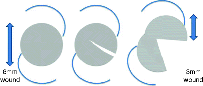
Fig. 15.6
To remove a flexible IOL from the AC through a small wound, first cut 75 % of the way across the optic; the IOL can then be removed through a smaller wound in one piece by pulling one-half through the wound and the other attached half follows behind
An alternative method is to fold the IOL in the AC. Make a paracentesis at 6 o’clock and insert a dialler needle under the optic centrally. Through the superior 3-mm wound, insert lens folding forceps and push these down onto the optic whilst the dialler needle remains stationery this folds the IOL over the needle. Remove the needle and then remove the folded IOL through the wound.
15.4 Surgical Options for the Aphakic Eye
15.4.1 McCannell Sutured IOL
There are a number of variations for this method. The key is to insert (or relocate) a PC IOL (e.g. three-piece foldable) with the optic in front of the pupil and the haptics behind. This holds the IOL in the pupillary plane and tents the iris over the haptics so that they can be seen. A long needle 10/0 non-absorbable suture is inserted through the peripheral cornea and is then passed through the mid-iris under the haptic and through the iris again and out through the peripheral cornea opposite. The suture is retrieved by inserting a hook through a paracentesis adjacent to the haptic and using the hook to retrieve the suture both near and distal to the haptic. The suture is now available to tie over the iris. Once done on both haptics, the optic can be reinserted into the posterior chamber.
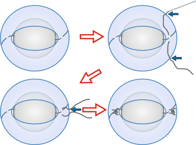
Fig. 15.7
For a McCannell suture (1) dislocate the optic through the pupil leaving the haptics posteriorly, (2) insert a suture using a long needle through the limbus (solid arrow) through the iris under the haptic and out through the limbus again (solid arrow) and cut off the needle, (3) create a paracentesis (solid arrow) and hook and draw out the loops of suture then tie over the haptic, and (4) repeat on other side and then push the optic through the pupil
Note: If repositioning a lens with capsule, the optic would need to be freed up from the capsule to allow anterior displacement through the pupil.
Problems:
Iritis
Secondary cystoid macular oedema
Suture lysis and dislocation
Haemorrhage from the iris
Pigment dispersion
15.4.2 Iris-Clip IOL
Iris-clip IOLs are available which can be inserted into the pupillary aperture. These have flanges into which the iris can be inserted to stabilise the IOL postoperatively. Possible complications:
Iritis
Secondary cystoid macular oedema
Dislocation
Haemorrhage from the iris
Pigment dispersion
15.4.3 Haptic Capture Method
Use a standard three-piece posterior chamber IOL:
Insert a 23-G infusion line.
Open the conjunctiva and create two scleral flaps (as for a trabeculectomy) at 180° to each other obliquely.
A sclerotomy is fashioned at 2 mm from the limbus under one of the flaps. Insert the folded IOL through a corneal wound.
Insert a 23-G forceps through the sclerotomy and grasp the tip of one of the haptics.
Draw the haptic out through the sclerotomy and push the tip into the sclera at the edge of the flap to stabilise the haptic. Sew up the flap tightly.
Repeat on the other side.
Insert the remaining 23-G sclerotomies to inspect the retina and vitreous base.
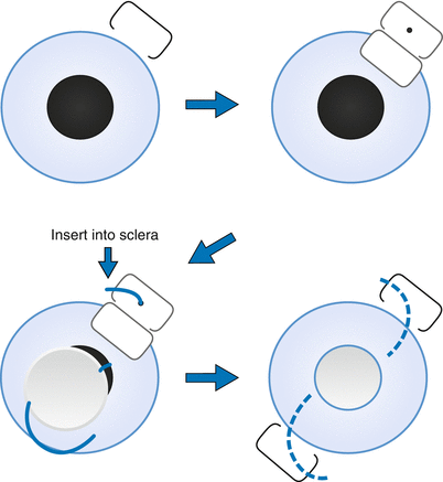
Fig. 15.8
The haptic capture technique can be used to place a standard three-piece foldable lens behind the iris in an aphakic eye. Create a scleral flap, then a sclerotomy in the bed of the flap, pull the end of the haptic through the sclerotomy and insert the end into the sclera at the edge of the flap incision, sew down the flap and repeat on the other side
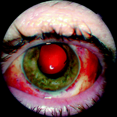
Fig. 15.9
An eye with a haptic capture IOL in situ 1 week after surgery
Complications:
Lens tilt
Lens iris touch or capture
Lens dislocation
Vitreous base damage and giant retinal tear
Hypotony and conjunctival bleb formation
15.4.4 Anterior Chamber IOL
The modern design of open loop AC IOL is less likely to cause corneal decompensation than older closed loop models. This may be because the open loop allows the haptics to flex on rubbing the eye rather than vaulting the optic forwards as with the closed loop systems. They require a large 6-mm wound to insert and will require 10/0 non-absorbable suture to the wound. The suture can be removed 3–4 months after surgery. Remember to do a peripheral iridectomy to avoid pupil block. Postoperative IOP rise is a possible complication.
15.4.5 Sutured Posterior Chamber IOLs
PC IOLs can be sutured into the ciliary sulcus using a variety of methods but employing:
Suture tied to the haptic of the IOL
Long needles to insert the Prolene suture
Trap doors of sclera under which the knot of the suture is buried
A simple method:
Tie the suture to the haptic, insert a 27-g needle through the sclera opposite and behind the iris and insert the long needle through orifice of the 27-g needle. Draw out the 27-g needle which pulls the long needle and suture with it.
The long needle is passed partially through the sclera circumferentially a few times and will then stay tight without suturing.
Repeat for the other side.
Problems:
Suture erosion
Lens tilt
Lens dislocation
Vitreous haemorrhage
15.4.6 The Aphakic and Aniridic Eye
Occasionally, you will be presented with an aphakic eye which has also lost its iris, for example, aniridia or post-traumatic. In these cases, the use of silicone oil tamponade is problematic because there is no iris diaphragm to hold the oil in the posterior chamber and the oil will fill the anterior chamber risking glaucoma and corneal touch and decompensation. A clever technique has been described whereby sutures are inserted to hold the oil away from the cornea (Gentile and Eliott 2010).
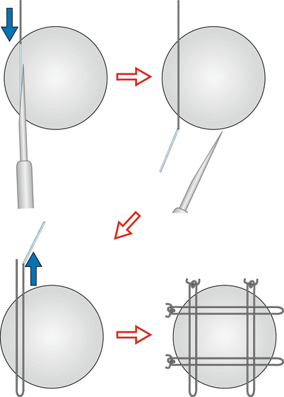
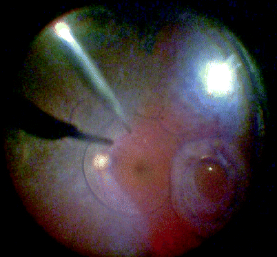
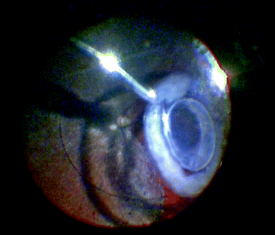
Fig. 15.10
In an aphakic–aniridic eye, a mesh of sutures can be used to prevent forward prolapse of silicone oil. First pass a 10/0 Prolene with a straight needle through the sclera 1-mm posterior to the limbus. Draw the needle out of the eye by engaging the suture needle into the lumen of a hypodermic needle, for example, 28 gauge. Pass the needle back in a similar fashion and then tie and bury the knot. Do this for two vertical and two horizontal sutures. The square mesh created will keep the oil in the posterior segment
Fig. 15.11
Removal of dislocated intraocular lens implants provides a number of challenges. In this patient, the capsule and lens have fallen backwards, but it remains hinged by the zonules at the 6 o’clock position. Many techniques exist to reposition such lenses including suturing of the lens to the sulcus, thereby avoiding complete removal of the lens. However, this runs the problems of suture erosion and breakage at a later date. Modern anterior chamber lenses are useful in the elderly if you wish to remove the lens in total, in which case this will need to be done through the anterior segment
Fig. 15.12
Silicone plate haptic lens implants may dislocate posteriorly after YAG capsulotomy. These can be removed through the anterior segment and cut inside the eye to allow a smaller exit wound for the implant
Fig. 15.13
If the zonular fibres give way, the whole lens and capsule can dislocate posteriorly. This can be seen years after PPV
Fig. 15.14
The IOL can be lifted into the AC for removal
15.5 Postoperative Endophthalmitis
15.5.1 Clinical Features
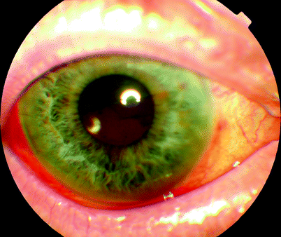
Fig. 15.15


A small hypopyon from postoperative endophthalmitis from Staphylococcus epidermidis
Stay updated, free articles. Join our Telegram channel
Full access? Get Clinical Tree
