CHAPTER 184 Characteristics of Normal and Abnormal Postnatal Craniofacial Growth and Development
An understanding of the abnormal first requires comprehension of the normal. Accordingly, this chapter is divided into two parts: A brief review of the main theories of postnatal craniofacial growth and the characteristics of normal postnatal facial growth and development is presented first. Subsequently, abnormal facial growth and development are examined through several clinical examples of relevance in pediatric otolaryngology. Embryology of the head and neck is covered in Chapter 181.
Theories of Craniofacial Growth
Postnatal facial growth appears to be a complex, multifactorial phenomenon. It is clear from the resemblance of children to their parents in both facial structure and soft tissue development that genetic factors play a significant role in basic facial pattern. However, “environmental” factors most certainly influence facial growth as well. These factors can range from intercellular influences to biomechanical forces (i.e., physical forces acting on bone). This essentially is Wolff’s law (1870), which states that “changes in the function of a bone are attended by alterations in its structure.”1 Bone is a tissue that responds to the stresses placed on it. Bone remodels.
Genetic Preprogrammed Theory (Growth Sites)
Early theorists believed that the sizes and shapes of bones were genetically determined within their cells and that sutural patterns were therefore predetermined. Because bone size and shape are defined by sutures in the head, these pioneers theorized that the position of sutures limits and controls the expansion of bones. Baume2 described “growth centres” as places of endochondral ossification with tissue-separating force, resulting in an increase in skeletal mass, and “growth sites” as regions of periosteal or sutural bone formation and modeling resorption that are adaptive to environmental influences.3 The cells of the sutures grew into the void left as the bone segments pulled apart from the tissue-separating force. In other words, growth sites are where growth literally takes place, and growth centers are areas in which genetically predetermined growth takes place. This theory was tested by experiments involving suture removal and transplantation, and the conclusion was that sutures are not primarily responsible for growth.4 This theory is no longer accepted as correct.
Cartilage-Mediated Growth (Scott)
In 1953, Scott theorized that the cartilage of the nasal septum “is an extension of the cartilage of the cranial base.”5 In its growth, the cartilage of the nasal septum separates the facial bones from one another and from the cranial portion of the skull and allows growth to take place at the sutures by the ordinary mechanism of surface deposition. He held that the nasal septum and also the mandibular condyles and synchondroses (otherwise known as cephalic cartilages) are growth centers. As the nasal septum grows, it puts tension on the sutures of the nasomaxillary complex, thereby displacing it forward and downward, with resultant growth occurring at pressure-related growth sites. Tension occurring in the maxillary sutures causes new bone growth when the sutures are pulled apart. Therefore, suture growth is passive and secondary.
Latham6 theorized that a septomaxillary ligament (connection between the septum and maxilla) in the fetus causes the maxilla to be pulled forward by the septum as the maxilla grows. It is unknown whether this ligament plays a role in postnatal growth, and experimentation related to the role of the nasal cartilage in this concept is unknown. Animal experimentation involving removal of septal cartilage shortly after birth7 or secondary to trauma seems to result in midface hypoplasia, but such experiments cannot clearly prove that the cartilage removal causes the growth deficiency. Cartilage seems to play some role that has not yet been precisely determined.
The Functional Matrix Hypothesis (Moss)
The most fundamental dictum of the functional matrix hypothesis was stated by Moss,8,9 as follows:
The origin of all skeletal units, all of their subsequent changes in size, shape and location and their subsequent maintenance and being, are always, and without exception, secondary, compensatory and mechanical obligatory responses to the morphogenetically and temporally prior and primary alterations of the operational (functional) demands of their specifically related functional matrices. Put more succinctly, “Bones do not grow, they are grown.”10
Facial growth involves intimate morphogenic and biomechanical relationships among all of its component tissue parts, each influenced by and in turn influencing the other. A fundamental principle is that no part is developmentally independent or self-contained.11
Bone Growth Concepts
Osteogenesis
Endochondral Bone Formation
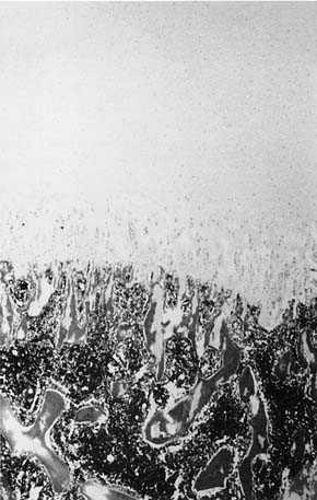
In endochondral bone formation, simultaneous removal of cartilage and subsequent formation of bone occur. Endochondral bone formation is more complex than intramembranous bone formation. Various zones exist in active endochondral bone formation, including the zones of reserve cartilage, proliferating cartilage, hypertrophic cartilage, calcified matrix, and resorption. The hypertrophic cartilage cells enlarge, and the surrounding matrix becomes calcified. A separate collar of bone forms around the circumference of the center of this cartilage bar. This is periosteal or, more precisely, perichondral bone. A vascular bud migrates into the cartilage and a primary marrow cavity forms. Meanwhile, activity at the various zones continues. Erosion of the cartilage takes place at the two ends of the expanding primitive marrow cavity, and bone forms on the spicules of the cartilage at the erosion front. The epiphyseal plate of long bones allows simpler visualization of this process (Fig. 184-1).
This descriptive review of bone formation can be supplemented by a molecular view of bone formation.12
Remodeling and Displacement
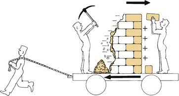
Enlow and Hans11 described the two basic types of growth movement: remodeling and displacement. This concept pertains to both bone and the surrounding soft tissues in the craniofacial complex. Remodeling involves changes in the size and shape of bone, the joining of separate bones and how they fit together, the adaptation intrinsically and extrinsically of these newly joined bones, and finally the relocation of these bones—each influenced by their overall enlargement. Displacement is the process whereby a space is created between bones as they enlarge. Resorption and deposition are ongoing in bones throughout the craniofacial complex during growth as part of the remodeling and displacement processes.
According to Rodan, “bone formation and bone resorption are finely regulated processes, controlled by local and systemic factors, carried out by a family of different interacting cells which communicate via cytokines, cell contacts, and elaboration of matrix which feeds back on cellular behavior.”13
Facial Changes during Normal Growth and Development
General
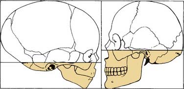
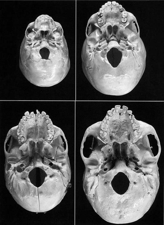
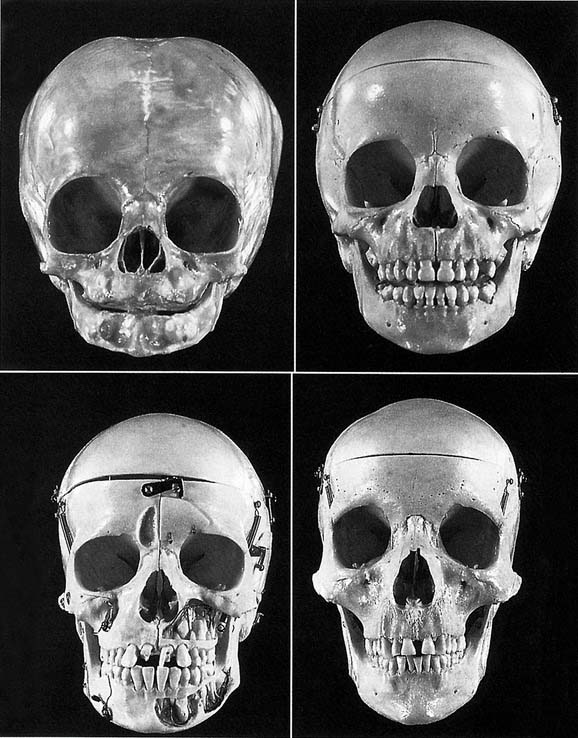
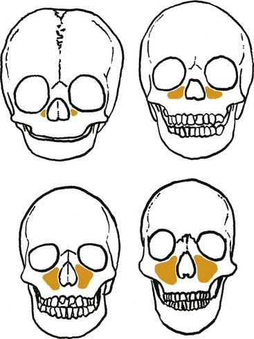
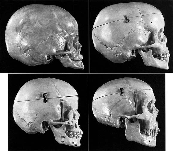
Infants have a large head relative to their body size. Their facial complex, which includes the nasomaxillary complex and mandible, is much smaller than the calvaria and basicranium (Figs. 184-2 through 184-4). With growth and development of the facial complex come changes in the size ratio of the cranium to the face. In infants the cranium-to-face ratio is approximately 3 : 1; whereas in adults it is approximately 2 : 1 (see Fig. 184-4).

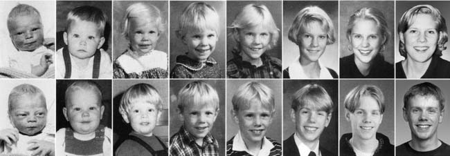
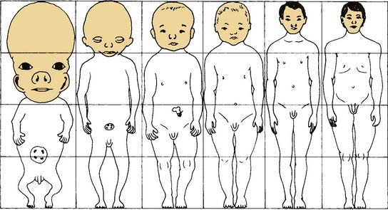
With growth, increasing demands are made on the respiratory and masticatory systems. As part of the growth of the facial complex, the nose, mandible, and maxilla increase in size—resulting in the larger, coarser features of the adult face in comparison with the child’s (see Fig. 184-3). The facial complex increases in size relative to the cranium. Similarly, body size increases relative to head size, to result in normal adult body and head proportions (Fig. 184-5).
Craniofacial Regions of Growth
The craniofacial complex can be divided into three regions: (1) the neurocranium, which is further divided into the calvaria and basicranium; (2) the nasomaxillary complex; and (3) the mandible. Figures 184-2 and 184-3 show the changes that occur in the craniofacial complex from birth to adulthood. The embryonic human skull has 110 ossification centers, many of which fuse, to result in 45 bones in the neonatal skull and ultimately 22 bones in the young adult skull.1 Table 184-1 summarizes the changes in size of various anatomic sites that take place during growth and development from infancy to adulthood.1,14–25
Table 184-1 Normal Craniofacial Growth and Development
| Growth Aspect | Age/Development |
|---|---|
| Neurocranium growth | Birth/25% of ultimate growth |
| 6 months/50% of ultimate growth | |
| 2 years/75% of ultimate growth | |
| 10 years/95% of ultimate growth | |
| Facial skeletal growth | 10 years/65% of ultimate growth |
| Neurocranium/face size ratio | Birth/8 : 1 |
| 2 years/6 : 1 | |
| 5 years/4 : 1 | |
| Adult/2.1 : 1 to 2.5 : 1 | |
| Brain size | Birth to adulthood/3.5∞ increase |
| Fontanelle closure | 3 months after birth/anterolateral |
| Second year of life/posterolateral | |
| 2 months after birth/posterior | |
| Second year of life/anterior | |
| Orbits | 2 years/50% of ultimate growth |
| 7 years/100% of ultimate growth | |
| Inner ear | 6 months of fetal life/full adult size |
| Brain weight | Full-term infant/25% of eventual weight |
| 6 months/50% of eventual weight | |
| 2 years/80% of eventual weight | |
| 10 years/almost eventual weight | |
| Postnatal brain growth | 1 year/60% of ultimate growth |
| 2 years/80% of ultimate growth | |
| 8 years/95% of ultimate growth | |
| Minimal growth continues to at least age 25 years | |
| Facial height (males) | Birth/40% to 61% of ultimate growth |
| Upper facial height | 1 year/63 to 86% of ultimate growth |
| 3 years/72 to 90% of ultimate growth | |
| 5 years/82 to 100% of ultimate growth (mean, 89%) | |
| Total facial height | Birth 38% to 45% of ultimate growth |
| 1 year/50 to 66% of ultimate growth | |
| 3 years/66 to 78% of ultimate growth | |
| 5 years/68 to 82% of ultimate growth (mean, 77%) | |
| Facial dimension | 3 months/equivalent to 40% of adult size |
| 2 years/equivalent of 70% of adult size | |
| 5 years/equivalent of 80% of adult size | |
| Nasal height and nasal bridge length | Fully mature in boys at age 15, girls at age 12 |
| Nasal tip protrusion | Boys age 15, females age 13 |
| Upper and lower nasal dorsum | Males age 14, females age 12 |
| Anterior and posterior nasal depth | Males age 14, females age 12 |
| Foramen magnum | 1 year/80% of transverse adult size |
| 1 year/74% of sagittal adult size | |
| Skull base (basion = nasion) | Birth/56% of adult length |
| 2 years/70% of adult length | |
| 10 years/90% of adult length | |
| Prechordal part contributes 75% of the postnatal length and parachordal (basioccipital) contributes to 25% | |
| Dentition eruption | 8 months-2.5 years/primary teeth |
| 6 years-20 years/permanent teeth | |
| Maxillary antrum height | Birth/20% |
| 7 years/50% | |
| 10 years/55% | |
| Bizygomatic width | Birth/60% |
| 3 years/80% | |
| 5 years/82% | |
| 10 years/85% | |
| Cranial width | Birth/65% |
| 3 years/85% | |
| 5 years/92% | |
| 10 years/95% | |
| Cranial length | Birth/60% |
| 3 years/85% | |
| 5 years/90% | |
| 10 years/95% |
Data from references 1, 14–25.
Neurocranium (Calvaria = Roof and Basicranium = Floor)
Calvaria
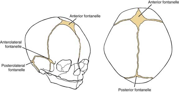
The calvaria comprises the frontal, parietal, and parts of the temporal, occipital, and sphenoid bones. These bones are formed by intramembranous ossification, with the exception of noncalvarial parts of the occipital, temporal, and sphenoid bones, which are formed through endochondral ossification. These bones initially are separated by fontanelles, which develop into sutures early and then finally fuse in adult life (Fig. 184-6).
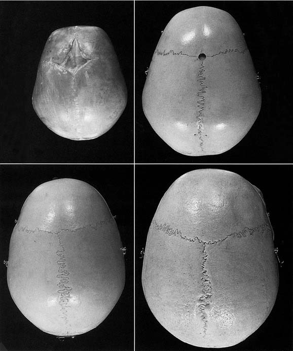
As the child’s brain grows, the calvarial bones are displaced. This displacement theoretically results in the deposition of new bone on the sutural edges. Growth of bone is seen on both the internal and external tables of the calvaria. This also results in an expanded medullary space. Significant thickening occurs from infancy to adulthood. The ascendancy (dominance) of the neurocranium over the facial complex during the early stages of life is clearly related to the inordinately early development of the brain.21 The major portion of calvarial growth is completed by the age of 4 years (Fig. 184-7).
Basicranium
Growth of the cranial base creates space for the enlarging brain and oronasal structures with their associated airway. The basicranium grows more slowly than the neurocranium. At birth and at 10 years of age, the cranial base exhibits 63% and 95%, respectively, of its ultimate size.21 The basicranium comprises parts of the occipital, temporal, sphenoid, and ethmoid bones. The occipital, temporal, and sphenoid portions form through endochondral ossification. The ethmoid and inferior nasal concha form solely by intramembranous bone formation.
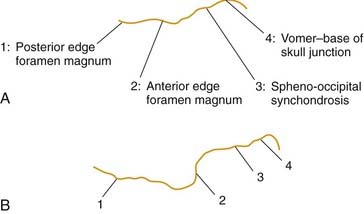
In the midline, synchondroses are present (Fig. 184-8). These synchondroses remain after the chondrocranium (cartilage precursor of the basicranium) has ossified. During childhood, the spheno-occipital synchondrosis is the principal growth cartilage of the basicranium, and its growth persists into early adolescence.3,11 Other synchondroses of the basicranium are the sphenoethmoidal, intersphenoidal, sphenofrontal, and frontoethmoidal synchondroses, all of which play a minimal role in postnatal growth. The synchondroses have zones similar to those in the epiphyseal cartilage of long bones. However, growth occurs in two directions, resulting in elongation of the basicranium until its ultimate ossification is completed (Fig. 184-9).
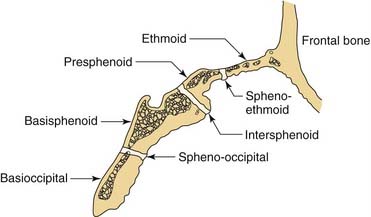
Figure 184-8. Diagrammatic representation of the synchondroses of the cranial base, showing the location of these important growth sites.
(From Proffit WR. Contemporary Orthodontics. 2nd ed. St Louis: Mosby; 1993.)
The development of ossification center sutures and synchondroses in the chondrocranium throughout childhood hase been chronicled by means of computed tomography (CT). These data can provide standards for skull base maturity. In addition, postnatal normal CT variants and magnetic resonance imaging (MRI) assessment of the cranial base have been characterized.26,27
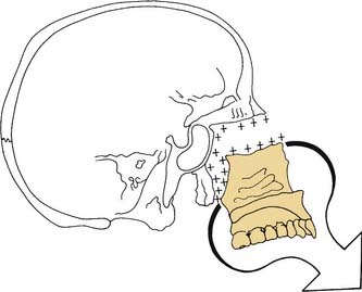
In newborns and infants, the cranial base is quite flat. With growth into childhood, a more convex superior or flexed appearance emerges, with further enlargement during adolescence and adulthood. The spheno-occipital synchondrosis becomes more flexed as the maxillary portion of the nasomaxillary complex moves toward the foramen magnum and moves under the anterior cranial fossa (Fig. 184-10). Components of the cranial base can be seen more clearly in older pediatric skulls than in infant skulls. The foramen magnum approaches adult size and the primary dentition has erupted by 3 years of age. By 8 years of age, the occipital bone has fused around the foramen magnum, and the secondary dentition is erupting. The major difference between the adolescent and adult skull is fusion of the spheno-occipital synchondrosis. Fusion of this synchondrosis begins at approximately 15 years of age for boys and at 12 or 13 years of age for girls on the cerebral surface. Ossification on the external aspect is complete by age 201 (Fig. 184-11). The chondrocranium-basicranium is a junction between both the neurocranial and facial complex bones. Its alteration affects not only neurocranial and skull growth but also facial morphology.3
Nasomaxillary Complex
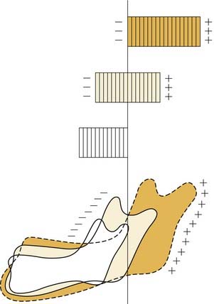
The nasomaxillary complex is displaced forward and downward from the cranial floor (basicranium) as a result of growth of the associated soft tissues. New bone growth occurs at sutural contact surfaces on both sides of the suture, with a resultant increase in the size of both bones involved (Fig. 184-12). Bone is added at the frontal maxillary, zygomaticotemporal, zygomaticosphenoidal, zygomaticomaxillary, ethmoidomaxillary, frontoethmoidal, nasomaxillary, nasofrontal, frontolacrimal, palatine, and vomerine sutures. Interaction between the basicranium and the nasomaxillary complex constantly occurs. Rotatory displacement is caused by the downward and forward growth of the middle cranial fossa, resulting in greater or lesser amounts of displacement and remodeling growth in the posterior and anterior parts of the maxilla, depending on the biomechanical forces involved.11
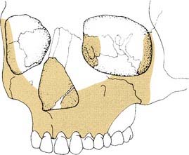
The anterior surface of the maxilla is an area of significant resorption (Fig. 184-13). Even though bone is being resorbed here, the maxilla is still growing forward. This is a good example of remodeling and displacement (Fig. 184-14). Growth and development of the nasomaxillary complex are shown in Figure 184-15. Vertical nasomaxillary growth occurs at the frontomaxillary, frontozygomatic, frontonasal, ethmoidomaxillary, and frontoethmoidal sutures as a result of orbital and nasal growth.
The orbits, which are linked to cranial growth, are quite large at birth because of their relationship with the globe. Orbital remodeling is quite complex—50% of postnatal orbital growth is achieved by the age of 2 years, and it is almost complete by age 7 years.21 The superior aspect of the orbit in toddlers is similar to that in adults, although the inferior aspect differs. The floor of the nose is almost at the level of the inferior orbital rim in infants; then the distance progressively increases so that the floor is significantly lower in adults. The eyes in the adult face are not much farther apart than those in a child’s. According to Enlow and Hans,11 however, “because of the larger nose, higher nasal bridge, increase in the vertical facial dimension and the widening of the cheekbones, the eyes of the adult appear much closer together.” The main contribution to the widening face is from the lateral expansion of the zygomaticomaxillary sutures and the intermaxillary suture growth. Eruption of the primary and secondary teeth results in an increase in the size of the maxilla. As the maxilla moves downward and forward and secondary teeth erupt, the maxillary sinuses increase in size as well.
Sinuses
At birth, the maxillary sinus is large enough to be seen radiologically. Relatively rapid growth occurs between ages of 1 and 4 years; by age 5, the sinus has started to expand laterally beyond the infraorbital foramen, although its floor does not extend below the nasal cavity. As the permanent dentition erupts, there is growth inferiorly of the floor of the sinus. By the age of 16 years, the maxillary sinus reaches full size (Fig. 184-16). The ethmoid sinuses are present at birth and have attained adult size by the early teens. The frontal sinus distinguishes itself from the ethmoids around age 5 and demonstrates a variable growth rate until reaching adult size in late puberty. The sphenoid sinus is the last to develop and is present radiologically at approximately age 6. Growth of the sphenoid sinus continues well past puberty into the early adult years.28
Shah and coworkers reviewed 91 CT scans in 66 patients (16 obtained serially over time) between the ages of 0 and 12 and demonstrated periods of significant growth in the paranasal sinuses in children. The ethmoid sinuses were the first to fully develop, followed by the maxillary, sphenoid and frontal sinuses.29
Mandible
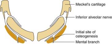
The mandible forms in relation to the first branchial cartilage, otherwise known as Meckel’s cartilage. Meckel’s cartilage is interesting in that it is not replaced by bone. Instead, membranous bone forms lateral to Meckel’s cartilage, and Meckel’s cartilage then disappears30 (Fig. 184-17). At birth, the mandible comprises two bones with the symphysis menti between them. This suture closes by the age of 1 year, after which the major portion of mandibular growth occurs posteriorly.
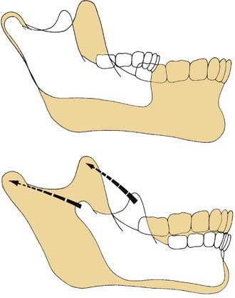
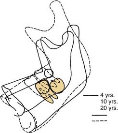
Resorption occurring anteriorly and deposition occurring posteriorly result in the overall growth of the mandible (Fig. 184-18). Although the condyles are a major site of growth, they are not a master growth center, as was previously thought.
Variation of endochondral bone growth occurs at the articular contact portion of the condyle. The main contribution of condylar cartilage is to provide regional adaptive growth by maintaining the condylar area in a proper anatomic relationship with the temporal bone as the whole mandible is simultaneously being carried downward and forward.11 The mandible grows by enlarging and widening in response to the increase in size of the muscles of mastication and an enlarging oropharynx, which accommodates the concomitant growth of the nasomaxillary complex. The ramus grows posteriorly and at the same time increases the inter-ramal dimension along with the laterally expanding cranial floor. The coronoid processes grow superiorly and buccally, with the anterior edges of the rami being continually resorbed in the process. The condyles grow posteriorly, superiorly, and laterally (Fig. 184-19). Even though it is a single bone, Sperber1 described the mandible functionally as six skeletal subunits. These subunits are the alveolar, coronoid, and angular bones, the condylar process, and the chin, all of which are attached to the “body” of the mandible. Moss’s concept of functional matrices then influences the growth of each subunit (teeth: alveolar unit; action of the temporalis muscle: coronoid process; masseter and medial pterygoids: angle and ramus; lateral pterygoid–condylar process, perioral muscles, tongue, and expanding oropharyngeal cavity: entire mandible).
The infant mandibular arch is U-shaped; whereas the adult arch is more V-shaped (Fig. 184-20). The gonial angle (the angle between the ramus and the body) is more obtuse in infants than in adults (Fig. 184-21). Mandibular growth takes place in two specific growth spurts, the first between the ages of 5 and 10 years and the second between ages 10 and 15 years.31 Growth in mandibular width is completed during early adolescence. Growth in mandibular length is complete 2 to 3 years after menstruation in females and approximately 4 years after attainment of sexual maturity in males. Growth in mandibular height is complete in the late teens in females and early twenties in males (see Fig. 184-21).
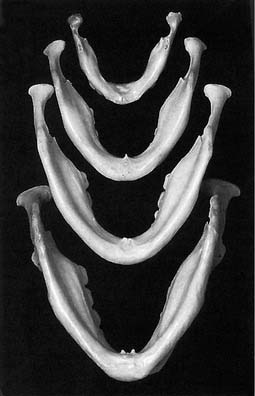
Figure 184-20. With mandibular growth, the U shape of the infant mandible (top) changes to more of a V shape, as seen in the adult (bottom).
Internal and external rotational changes of the mandible occur during growth and, of course, in conjunction with rotational changes in the maxilla (Fig. 184-22). Bjork and Skieller32 described this in detail using metallic implants. Miller and Kerr31 supported their findings but used a digitizer program for longitudinal follow-up with cephalograms in a number of patients. Overall mandibular growth is not uniform and steady, because corpus length and ramus height grow in an irregular pattern.25,34 Jaw rotation influences the degree of tooth eruption.
Craniofacial cephalometric and anthropometric studies are abundant in the literature, and normative data are available. A review of these topics is beyond the scope of this chapter; however, these measurements may be used to assist in the interpretation of optimal growth and development in the clinical situation.35–42
Abnormal Facial Growth and Development
Asymmetry of the human face is well recognized as a normal finding and in fact is an intrinsic facial characteristic. According to Ferrario and colleagues, “the skeletal structures show a greater degree of asymmetry than their soft tissue drape.”43 Age is certainly one factor that influences asymmetry. Melnik44 found that the left side of the face was larger at the age of 6 years, whereas the right side of the face was larger at age 16. Disagreement exists in the literature regarding the degree, side, and localization of normal facial asymmetry, although some researchers believe that this is because of methodologic differences.43 Normal facial asymmetry involves many subtle differences that are not readily apparent to the viewer. Conversely, abnormal facial asymmetry typically is quite obvious.
Our culture is obsessed with physical attractiveness, which to a large extent is based on facial symmetry and what is perceived to be “normal.” Langlois45 found that the preference for attractive persons is evident early in life and involves all age groups and races. As a result, people whose appearance is divergent from the “norm,” with or without intellectual impairment, are viewed differently in our society.
Radiotherapy
Ionizing radiation is widely recognized as detrimental to the growth of developing tissue. In a majority of cases, this consequence is a result of the application of full doses to radiosensitive tumors.46 Rhabdomyosarcoma and undifferentiated sarcomas constitute the most common type of malignant tumor of soft tissue of childhood. Of these sarcomas, 35% are found in the head and neck region. Retinoblastoma is the most frequent malignant ocular tumor in childhood and is a frequent indication for radiotherapy in the pediatric population.47,48 Multimodal therapy (radiotherapy and chemotherapy) has been effective in the treatment of these tumors with a 3-year relapse-free survival rate of between 70% and 100%.49–52 The value of radiotherapy has not been questioned given its success rate,53 but as more children have been cured of cancer and the survival rates have increased, concerns about the long-term side effects of these therapeutic regimens (alone or in combination) have arisen.50,51
The effects of radiation therapy on facial growth, as well as radiation’s collateral effects, are detailed in Table 184-2. Virtually 100% of children who have received radiotherapy suffer from alterations in the development of facial soft tissues, and in more than 60% of these children, the facial bones are affected.47,54 The younger the patient at the time of irradiation, the greater the effects on craniofacial development.51,55,56 These side effects are more frequent and more severe within the first 10 years after irradiation.54
Table 184-2 Side Effects of Radiotherapy
| Category | Manifestations |
|---|---|
| Skin | Dryness, abnormal pigmentation, telangiectasis, malignant degeneration, atrophy, alopecia, sebaceous cysts |
| Mucosa | Xerostomia, atrophy, fibrosis |
| Soft tissues | Atrophy, hypoplasia, sensitivity to infections |
| Facial bones | Growth retardation, hypoplasia, dysplasia, appositional growth restriction, weak resistance to infections, loss of cortical bone density, atrophy of alveolar process, micrognathia, facial asymmetry, condylar hypoplasia, “bird face,” abnormal occlusion |
| Teeth | Failure to erupt, disturbed eruption, damage to tooth buds, anodontia, hypodontia, pseudoanodontia, mineralization, caries, root foreshortening, enamel dysplasia, atypical root morphology |
| Orbit | Bone and soft tissue hypoplasia, persistent infections. |
| Eyes | Cataracts, xerophthalmia, retinopathy, corneal lesions, loss of vision, keratoconjunctivitis, dryness of globe, enophthalmos, stenosis of lacrimal duct |
| Head and neck muscles | Atrophy, trismus, fibrosis |
| Risk of second malignancy | Osteogenic sarcoma, soft tissue sarcomas usually within radiation fields, thyroid, breast, acute myeloid leukemia, histiocytoma |
| Brain | Cerebral function damage |
| Middle ear | Serous otitis media, chronic otorrhea |
| Paranasal sinuses | Bone hypoplasia, chronic sinusitis |
| Audiology | Sensorineural or conductive hearing loss |
| Speech | Velopharyngeal incompetency |
| Hormonal | Growth retardation, growth hormone deficiency |
| Psychologic | Psychologic disorders |
| Cosmetic | Bone or soft tissue hypoplasia, facial asymmetry, severe neck fibrosis, nuchal asymmetry |
| Various | Nutritional and vitamin deficiency |
Data from references 46–48, 50, 51, 53; and from Berkowitz RJ, Neuman P, Spalding P, Novak L, Strandjord S, Coccia PF. Developmental orofacial deficits associated with multimodal cancer therapy: case report. Pediatr Dent. 1989;11:227; Larson D, Kroll S, Jaffe N, Serure A, Goepfert H. Long-term effects of radiotherapy in childhood and adolescence. Am J Surg. 1990;160:349; Sklar CA. Overview of the effects of cancer therapies: the nature, scale and breadth of the problem. Acta Paediatr Suppl. 1999;88:1; and Sonis AL, Tarbell N, Valachovic RW, Gelber R, Schwenn M, Sallan S. Dentofacial development in long-term survivors of acute lymphoblastic leukemia. A comparison of three treatment modalities. Cancer. 1990;66:2645, 1990.
It has been difficult to determine the safest dose of irradiation that keeps side effects on growing tissue to a minimum but at the same time is effective in eradicating the cancer.46,53,54 Techniques for radiotherapy administration have changed during the past 20 years. The principal aim is to achieve a balance between cure and late toxicity of the treatment.54 Changes to minimize undesirable effects include radiotherapy with megavoltage, hyperfractionation, three-dimensional therapy, intensity-modulation, and conformal radiation with multiple fields.47,55 In all children undergoing radiotherapy of the head and neck, it is mandatory that multidisciplinary follow-up be part of the ongoing management.
Functional Endoscopic Sinus Surgery
FESS has become the standard of care in the surgical management of chronic sinusitis and other sinus disorders in the pediatric population. Its effectiveness has been accepted when clear indications for surgery are established.57,58 However, concerns have arisen regarding potential negative effects on the growth and development of paranasal sinuses and the nasomaxillary complex in the pediatric patient.59
A knowledge of sinus anatomy during different stages of development, as well as of pathologic circumstances such as chronic sinusitis, is imperative in decisions regarding FESS in the pediatric population.28,60–62
The absolute and relative indications for FESS in the pediatric population varied during the 1990s and into the millennium. These changes were related to the evolving definitions of sinus disorders and short- and long-term outcome measures.60,63–68 This topic is covered in Chapter 195.
Results of animal studies have varied depending on the species and the intervention. Kraut and Kronman examined 36-day-old New Zealand White rabbits and unilaterally filled their maxillary sinuses with polyhydroxyethylmethacrylate without adverse effect on craniofacial growth.69 Mair and coworkers studied eight newly weaned piglets subjected to unilateral FESS. The researchers examined the piglets 6 months after surgery, noting significant reduction of the facial growth and maxillary-ethmoidal sinus development on the operated side.70 Carpenter and associates found significant restrictive effects on piglet facial skeletal development after FESS.71 Verwoerd-Verhoef and Verwoerd demonstrated that controlled resection of the bony sinonasal wall and partial resection of anterior ethmoid did not affect later development of nasal bones and maxilla on 20 young New Zealand White rabbits.72
Several studies in children have been undertaken. Mair and coworkers analyzed the uncinate processes and ethmoids of 84 children (mean age of 6 years; age range, 1 to 18 years) who previously had undergone FESS. In children younger than 9 years of age, woven and lamellar bone were present, in contrast with the presence of exclusively lamellar-mature bone in the children older than 9 years of age. These results suggest that the safest age at which a pediatric patient should be operated on is 9 or older.70 Kosko and colleagues in 1996 presented five cases of children (mean age of 30 months) who had undergone bilateral FESS for refractory chronic sinusitis. At mean postoperative month 42, four children showed unilateral maxillary hypoplasia and one child showed bilateral hypoplasia without apparent changes in facial symmetry.59 Sinus hypoplasia can be from primary causes related to individual development or secondary causes associated with a response to facial trauma, chronic sinusitis, or FESS, which has affected the activation of bone formation factors or the removal of growth centers.59,61,73
Stay updated, free articles. Join our Telegram channel

Full access? Get Clinical Tree


