Care of the Eye in Facial Paralysis

Chapter 61 Care of the Eye in Facial Paralysis


Rehabilitation of a patient with facial paralysis depends on restoration of optimal lid position and function.13 This chapter summarizes techniques that I have found to be most helpful in achieving that goal, based on more than 5000 patients with facial paralysis on whom I have operated during the past 3 decades.


The chapter is divided into sections for lid reanimation procedures, lower lid reapposition procedures, and ancillary procedures. In practice, a combination of these techniques may be performed during the same operation. When the procedures are combined, the reanimation procedure is performed first because it is most influenced by the lid swelling that occurs during the course of the surgery. The lower lid reapposition procedure is performed next. Upper lid entropion correction is performed just before the upper lid incision is closed, and brow elevation is performed last. The final section of the chapter explains two useful temporizing procedures.


CRITERIA FOR SURGERY


The following three groups of patients with facial paralysis require lid surgery for functional reasons:


Patients with short-term problems (≤3 months to anticipated recovery of orbicularis oculi function) can usually be treated by conservative means. Patients with significant paralytic deficits who require 6 months or more to recover, or who are not expected to recover, are generally best served by early surgical intervention.


The group of patients whose prognosis is unclear, such as patients who might improve in 3 months, but could conceivably require 6 months or more to recover, poses the greatest challenges in surgical selection. In such patients, criteria such as the reliability of follow-up; the accessibility of medical care; the ability of the patient or family to care for the eye; and the patient’s own needs, desires, and lifestyle all play a role in decision making. When it is safe and feasible, a prolonged trial of conservative management may allow the patient and the physician to decide if they are on the correct course.


ANESTHESIA AND SURGICAL PREPARATION


In lid reanimation procedures, in which the patient’s cooperation is required, any medication that might make the patient drowsy and unable to cooperate fully throughout the operation is not used. Short-acting intravenous medication, such as propofol (Diprivan), methohexital sodium (Brevital Sodium), or a similar agent is given at the beginning of the surgery in amounts just adequate to cover the discomfort of the local injection. Lidocaine 2% (Xylocaine) with epinephrine (unless contraindicated by hypertension or cardiac problems), to which sodium bicarbonate 7.5% (Neutra-Caine) has been added (1 part sodium bicarbonate to 9 parts lidocaine with epinephrine) is used at the beginning of the surgery. Bupivacaine 0.5% (Marcaine) is used at the end of the surgery to reduce pain during the immediate postoperative period. It may also be included in the initial injection, using equal parts of bupivacaine and lidocaine with epinephrine.


Local infiltration is placed in the areas to be operated, such as along the upper lid fold and along the lateral orbital rim for spring implantations, and at the canthi and brow areas, if surgery is to be performed there. Excessive infiltration should be avoided because it paralyzes the levator, impairs extraocular motility (making it harder to judge lid position), and distorts lid anatomy. The eyelids, both sides of the face above the mouth, and the forehead are prepared with green soap and then with povidone-iodine (Betadine), which is washed off.


DRAPING


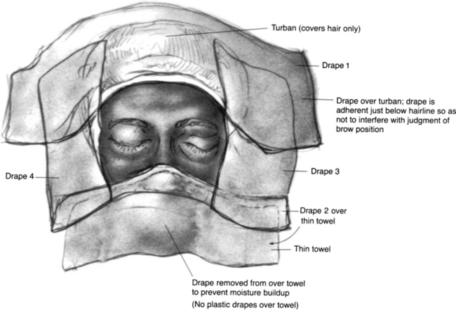
The hair is covered with a small drape formed into a turban and secured with a clamp. A second small sheet is incorporated with that drape to cover the superior end of the table. A body sheet is also placed. The eyelids and brow are isolated by means of two No. 1000 Steri-Drapes cut in half (Fig. 61-1). Each of the four drapes forms a border of the surgical field, which includes both eyes and the forehead area. Before the Steri-Drape is placed over the nose, a thin cloth towel is placed over the nose to avoid the suffocating feeling resulting from plastic over the nose. In addition, the plastic drape over the towel inferior to the adhesive area is excised to prevent moisture accumulation. Care is taken to avoid distorting the lower lid anatomy or brow areas by undue traction from the drapes.


The body drape is fastened to the head drape on both sides with a towel clip so that it does not slip down when the patient is brought to the seated position. The patient is secured on the table with a safety belt, and the belt is positioned to provide access for loosening it as needed when the patient is brought to the seated position.


The Mayo stand is brought over the drapes for easy access to the instruments. Although the tent effect obtainable by draping over the Mayo stand would be desirable, it does not lend itself readily to moving the stand away when the patient needs to be brought to the seated position.


GENERAL CONSIDERATIONS


Procedures are generally performed on an outpatient basis, unless the patient is already hospitalized because of the neurotologic or head and neck surgery. Bipolar or unipolar cautery may be used for hemostasis. Unipolar cautery should not be used if the patient has an auditory brainsterm implant (ABI) or cochlear implant. Cutting cautery with a fine needle tip is useful for performing the lid dissection. The eye is constantly protected with a scleral shell. At the end of the procedure, antibiotic ophthalmic ointment is applied to the wounds. Ophthalmic ointment is used because it is not irritating if any gets into the eye. The lids are not bandaged. An ice pack is applied to the closed lids and kept in place for 48 hours, after which time warm tap water compresses are used for at least 20 minutes four times a day until the swelling subsides. The antibiotic ointment is applied to the wounds twice a day until they are healed, and appropriate lubricating drops are prescribed for the eye.


UPPER LID REANIMATION PROCEDURES


The four procedures that I believe are the most useful in reanimating paralyzed lids are the following, in order of preference:


In all of these procedures, the principle is to create an external force that opposes the levator palpebrae superioris, the opening muscle of the lid. The respective forces are spring tension, gravity acting on the gold weight, and elasticity of the silicone rod.


The relative merits and limitations of the various procedures are related to how they develop external closing forces and the consequences of increasing those forces. The greater the force required by any device, the greater the pseudoptosis (lid droop in the primary position of gaze that results from the implant). If the palpebral spring, the gold weight, and the silicone rod prosthesis all are adjusted to provide the same closing force in a given patient, the pseudoptosis should be the same with each device. In the enhanced palpebral spring implantation procedure, the levator muscle is strengthened to balance the spring force, and less pseudoptosis is possible with the same closing force compared with any of the other three procedures. Similarly, by tightening the levator and using a stronger spring force, the surgeon makes increased blink speed possible.


Because the gold weight is gravity dependent, very large and unsightly gold weights may be required in lids that need a strong closing force. Also because the gold weight is gravity dependent, lid closure may not be guaranteed when the patient is supine, as in sleep. Blink speed is limited with the gold weight. Patients whose work or lifestyle requires their being in a very cold or hot environment may experience pain from the cold or hot gold weight in their eyelid. A surgeon who does these procedures only infrequently can more easily master the techniques of the gold weight implant and silicone rod prosthesis, however, than the technique of the palpebral spring implant or the enhanced palpebral spring implant.


The silicone rod prosthesis has the advantage of providing support for the lower lid as well. Its major disadvantage is its inevitable loss of elasticity over months or years. If facial nerve function does not recover before the prosthesis runs out of elasticity, it will need to be replaced. By contrast, a gold weight or palpebral spring can function over many years. A small percentage of springs may fail over time because of fatigue and breakage, necessitating replacement.


An additional advantage of the palpebral spring is that the tension on the wire can be adjusted postoperatively, either externally or through a small incision. This technique allows loosening of the spring when the patient recovers partial function, but is not yet well enough to have the spring removed.


All prosthetic devices are subject to the potential hazards of extrusion or infection over the long-term. With the techniques currently in use, these complications have been sufficiently infrequent, however, as not to limit the usefulness of the devices.


In recent years, since I devised the enhanced palpebral spring procedure, I have used it with increasing frequency instead of nonenhanced spring implantation. The extra surgical effort is usually well rewarded by the diminished pseudoptosis and increased blink speed obtained by this procedure. I prefer the enhanced palpebral spring implantation in most cases of significant upper lid closure deficits. When the patient has a strong levator or when the spring is being used only as a short-term remedy, the nonenhanced palpebral spring procedure may be used. Surgeons who are just beginning to undertake palpebral spring implantation should start off with the nonenhanced procedure and then move on to the enhanced procedure.


I find the gold weight beneficial to patients whose closure problem is minimal, but nevertheless just exceeds the limits of conservative management. Patients who require definitive, reliable closure, such as patients with coexistent poor Bell’s phenomenon or CN V involvement, are better protected with springs than with weights. Patients whose ocular management failed with weights in place have been successfully treated by removing the weights and replacing them with springs. The silicone elastomer (Silastic) elastic prosthesis is most useful in patients with an excellent prognosis for recovery in about 6 months in whom significant lower lid lagophthalmos coexists because stretching of the prosthesis over time may decrease its function after 6 months.


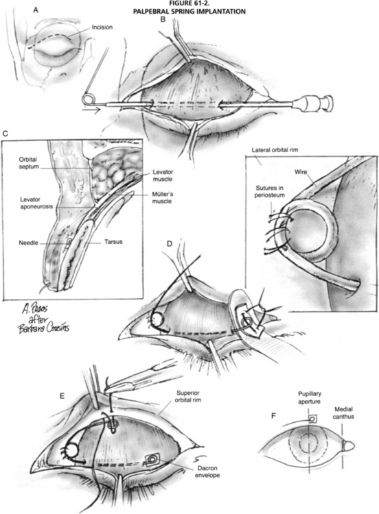
Palpebral Spring Implantation


The palpebral spring414 is built preoperatively either in the office or at the bedside. Building the spring is time-consuming, and the surgeon and the patient should be comfortable during the procedure. Good light must be available, and if possible, the patient should be seated so that lid movement can be best evaluated.


Each spring is constructed from a plain piece of wire that is shaped to conform to the individual lid anatomy of the patient. Generally, a 0.011 inch wire provides suitable tension for most patients. Patients with very strong levators may require the use of 0.011 inch or 0.012 inch wire, and patients with weak levators (especially when the levator is not going to be tightened) may benefit from the use of 0.009 inch or 0.008 inch wire. A new wire used in pacemaker leads, the alloy 35NLT (Fort Wayne Metals), has been made with sufficient tensile strength to replace the previous alloy. With this wire, the 0.011 inch wire diameter often does need to be varied. Because of its track record in biomechanical applications, it is anticipated to have excellent longevity.


The construction is begun by forming an 8 mm loop at what is to become the fulcrum of the spring. The posterior aspect of that loop should be the superior arm of the spring. Because loosening the spring intraoperatively is easier than tightening it, the two arms should form an angle of about 120 degrees as they leave the fulcrum.


The fulcrum is placed over the lateral orbital rim and held in position by the surgeon’s fingers. Curves are created in the lower arm to match the patient’s lid anatomy. Curvature is also provided to accommodate the fact that the upper eyelid opens up and back, not straight up and down. Slight variations in spring position and curvatures may enhance its effect; these factors should be varied in the evaluation of the spring preoperatively.


Sometimes, making more than one spring with slightly different curvatures is useful in determining which model would work best. Usually, the fulcrum should be placed as far laterally as possible without lengthening the spring so much that its design and placement are difficult. The completed spring is stored until the day of surgery, when it is placed on a gauze pad to prevent loss and sterilized in a low-temperature chemical unit and not a steam autoclave, to subject the wire to less heat stress. Alternatively, the wire may be sterilized in gas sterilizer 24 hours or more before surgery, and retained in a sterile envelope.


Surgical Technique


The eye is protected with a scleral shell. An incision is made along the lid fold at the junction of the medial one third and lateral two thirds of the palpebral aperture and is carried across the orbital rim (Fig. 61-2). Dissection is carried superolaterally, in the plane between the septum and orbicularis, to expose the orbital rim. Dissection is carried downward at the medial aspect of the incision to expose the tarsus.


image

FIGURE 61-2 Palpebral spring implantation. A, With protective scleral shell in place, incision is made along the lateral two thirds of the lid crease and is carried across the orbital rim laterally. Dissection is carried downward at the medial end of the incision to expose tarsal plate. Dissection is also carried upward and laterally to expose orbital rim. B, A 22 gauge blunted spinal needle with the stilette in place is passed from the medial end of the dissection to emerge laterally in the plane between orbicularis and the tarsus. Passage should be carried out overlying the midtarsus, and needle is angulated slightly downward at its lateral extent. Exit of the needle tract should be close to lateral orbital rim periosteum. The lid is everted to confirm that needle has not inadvertently perforated the tarsus. The previously prepared wire spring, sterilized in low-temperature chemical sterilizer or gas sterilizer, is passed through the needle, and needle is withdrawn. C, Cross section of lid illustrates placement of needle over the midtarsus in the plane between tarsus and orbicularis. Wire spring should be resting on the epitarsal surface. D, Scleral shell is removed, and fulcrum of the spring is brought into the desired position along the orbital rim. The spring should be placed in a position in which its curves conform perfectly to the eyelid contour. (Inset: Fulcrum of spring is secured to lateral orbital rim periosteum with three 4-0 Mersilene sutures, and an extra bite of the periosteum is taken with each stitch.) Loops are fashioned at each end, and the spring is cut to size. Loops should be flat and tightly closed to leave no sharp edges. The medial loop is enveloped in 0.2 mm thick polyester (Dacron) patch material, to which it is secured by means of three 8-0 nylon sutures tied internally. Polyester patch is creased in Gelfoam press before surgery and is autoclaved with the other instruments. The folded polyester envelope is cut to size at surgery. The crease in the patch material should be directed downward so that the spring and patch together provide a smooth inferior surface. The loop at the end of the inferior arm is directed upward for the same reason. Suturing of the loop to the polyester is facilitated by resting the polyester on a retractor. E, The end of the spring with its polyester envelope is replaced into the lid between tarsus and orbicularis. In time, the end of the spring becomes fixed to tarsus by granulation tissue integrating into the polyester patch. Securing the patch to the tarsus directly with an additional running 8-0 nylon suture helps to provide fixation until connective tissue grows into the polyester. Tension on the spring is checked, with the patient in the upright and supine positions. The tension can be adjusted by grasping the upper end of the spring with forceps and changing its position. When the correct tension has been determined, the upper loop of the spring is secured to the orbital rim periosteum with a 4-0 Mersilene suture. An extra bite of the periosteum may be taken in the stitch before it is tied. When sutures are placed to secure either the fulcrum or the upper loop of the spring to the orbital rim periosteum, it is safer to sew in the direction away from the globe. Spring tension is checked again with the patient seated and supine. Additional adjustments can be made by bending the wire or repositioning the loop. When the adjustments are completed, two additional 4-0 Mersilene sutures are placed through the upper loop in a manner similar to that of the initial suture. Deeper tissues overlying the spring are closed with 5-0 plain gut suture to ensure that the spring and Mersilene sutures are well covered. Skin and muscle are closed with running 6-0 plain gut fast-absorbing suture. F, The end of the spring should be at the pupillary axis, with eyes in the primary position of gaze.


(From Tse D, Wright KW [eds]: Oculoplastic Surgery. Philadelphia, Lippincott, 1992.)


A blunted 22 gauge spinal needle is passed, beginning in the area of the exposed tarsus, 5 mm superior to the lid margin. The needle passes in the plane between the orbicularis and the tarsus to a point 2 mm above the lid margin at the lateral aspect of the lid. It continues until it emerges at the anterior aspect of the lateral orbital rim. The stylette is then removed. The undersurface of the lid is inspected to ensure that the needle has not inadvertently perforated the tarsus.


The end of the lower arm of the previously prepared palpebral spring is passed into the needle, and the needle and spring are withdrawn medially, bringing the spring into the lid. The scleral shell is removed, and the spring is positioned so that its previously determined curvature conforms to the lid anatomy. The upper arm of the spring is placed in position, and its length is determined. It usually needs to be about ¾ inch long. A loop is fashioned at the point that is to become the end of the upper arm, and the wire is cut to size. The loop is closed to leave no sharp ends. The upper loop is made at a 90 degree angle to the fulcrum loop so that the upper loop can be tucked under the superior orbital rim. The loop is primarily held in place by pushing against the bone, decreasing the role of the sutures and helping to prevent late slippage. The loop at the fulcrum is held in place with forceps, and the patient is asked to open and close the eye. The position at which the spring curvature best conforms to the lid anatomy is then found, with the eye opened and closed, and the loop at the fulcrum is sutured in place. The suturing is accomplished with 4-0 Mersilene suture, and an extra bite of periosteum is taken with each stitch. Three such sutures are generally placed for the nonenhanced procedure, and five are placed for the enhanced procedure because of the greater tensions involved.


The lower arm of the spring is cut to size, and a loop (which is also meticulously closed) is formed on its end. The loop should be formed upward to maintain a smooth inferior surface to the spring. The end of the spring should be at the pupillary axis, with the eyes in the primary position of gaze. Before the end of the spring is covered with polyester (Dacron) patch material, the angulation of the loop should be checked with the patient’s eyes open and closed to ensure that the spring tracks well with lid movement, and that the loop stays relatively parallel to the tarsus during opening and closing.


A piece of 0.2 mm polyester patch material, which has been creased by its placement in a press that is used for compressing absorbable gelatin sponge (Gelfoam) before it is autoclaved, is cut to size to fit over the inferior loop. This piece is converted into a pouch by closure of the sides with 8-0 nylon sutures tied internally. The creased side is directed downward. The open lateral side is slipped over the spring, to which it is secured with an 8-0 nylon suture beginning within the pouch. The suture is passed through the spring loop and the posterior end of the pouch and is terminated by passing through the anterior side of the pouch. The knot is tied internally to prevent erosion. The polyester envelope is secured to the tarsus with one or more 8-0 nylon sutures, as needed, to prevent slippage of the polyester until granulation to the tarsus occurs.


Spring tension is adjusted to just close the eye, by moving the upper arm of the spring closer or farther from the orbital rim. At the desired tension, the upper arm is bent so that the loop can be tucked under the orbital rim; it is secured with 4-0 Mersilene sutures to periosteum, taking an extra bite of periosteum with each stitch. Tension is checked with the patient seated and supine. Additional adjustments can be made by bending the wire of the upper arm to loosen or tighten it.


Bending the wire of the lower arm near the fulcrum should be avoided at this time because such adjustments may be required during the postoperative period, and excess bending of the wire may increase its chance of breakage. When the final position of the upper loop has been determined in the nonenhanced procedure, two additional 4-0 Mersilene sutures are placed, and an extra bite of periosteum is taken with each stitch. Four additional sutures are used in the enhanced procedure.


The deeper aspect of the wound overlying the orbital rim is closed with 5-0 plain gut suture to cover the spring and Mersilene sutures at the upper loop and fulcrum. The lid fold incision is closed with running 6-0 plain gut suture. The eye is dressed with antibiotic ointment and an ice pack.


Jun 14, 2016 | Posted by in OTOLARYNGOLOGY | Comments Off on Care of the Eye in Facial Paralysis

Full access? Get Clinical Tree

Get Clinical Tree app for offline access