 Bitot’s spots are considered a sign of past or present vitamin A deficiency; however, some patients do not respond to vitamin therapy. Patients with Bitot’s spots appear with disfigured conjunctiva, and some may have night blindness and punctate keratopathy. Histologically, keratinization with granular cells appears with irregular maturation, inflammatory infiltration of the conjunctival substantia propria, and loss of goblet cells. Prominent Bitot’s spots may represent massive accumulations of gram-positive bacilli and keratin debris.
For patients who are nonresponsive to vitamin A therapy, the available treatment for Bitot’s spots is excision of the defective conjunctiva. We have used the Fugo blade to remove these unsightly conjunctival spots.
CASE STUDIES
Case 1
The patient was a 19-year-old army recruit who was rejected because of the bilateral presence of Bitot’s spots.
TECHNIQUE
Anesthesia
Administer surface anesthesia. Then puncture the conjunctiva with a 100-µm Fugo blade tip close to the edge of the incision, and inject 2% lignocaine through it using a thin cannula to avoid possible subconjunctival hemorrhage from a needle prick. Balloon the conjunctiva with anesthetic fluid.
Surgical Technique
Sweep a 600-µm Fugo blade tip at the lowest energy setting over the surface of the lesion from one edge to the other until the conjunctiva appeared to be reasonably clear of the spot. Perform this in both eyes at the same setting (Fig. 14.1).
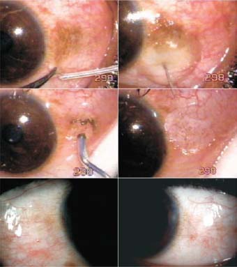
Figure 14.1. The conjunctiva is punctured close to the edge of lesion with a 100-µm tip. Lignocaine is injected through this hole to raise the conjunctiva. A 600-µm Fugo blade tip is swept over the lesion. Bottom: The two eyes 10 days after treatment.
Do not apply a dressing. Prescribe antibiotic–steroid ointment to be used twice a day for 10 days. In our case, the eyes were clear enough for the recruit to join the military service.
Case 2
The patient in this case was a well-nourished 5-year- old boy in whom Bitot’s spots had been seen since the age of 6 months, soon after he suffered a bout of severe diarrhea. His classmates at school often commented on the odd appearance of his eyes.
The patient was given general anesthesia for the procedure. The Bitot’s spots were easily removed with the sweeping motion of the 600-µm Fugo blade tip over the conjunctiva. It took a couple of seconds to treat each eye. Steroid–antibiotic ointment was prescribed. Ten days after treatment, the conjunctiva looked normal (Fig. 14.2).
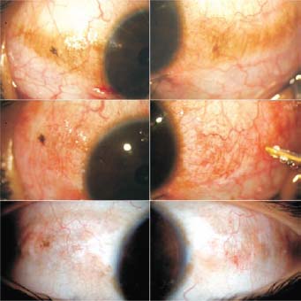
Figure 14.2. The right and left eyes at the start of surgery, at the end of surgery, and 10 days after surgery.
Summary
The Fugo blade is particularly effective in removing Bitot’s spots and similar surface lesions with great ease.
Suggested Reading
Ahed MA, et al. Bitot’s spots following hemicolectomy. Eye 2003;17:671–673.
Baker BM, Bender DA, eds. Vitamins in medicine, vol. 2, 4th ed. London: Heinemann, 1982; p. 222.
Emran N, Tjakrasudjatma S. Clinical characteristics of vitamin A responsive and nonresponsive Bitot’s spots. Am J Ophthalmol. 1980;90:160–171.
Stay updated, free articles. Join our Telegram channel

Full access? Get Clinical Tree


