Benign Diseases of the Salivary Glands: Introduction
The salivary glands consist of two parotid glands, two submandibular glands, two principal sublingual glands, and a large number of minor salivary glands. Combined, the salivary glands produce serous secretions, mucous secretions, or both. The serous saliva of the parotid gland and the predominantly mucous secretions of the submandibular, sublingual, and minor salivary glands provide digestive enzymes, bacteriostatic functions, lubrication, and hygienic activities. The secretions of the parotid and submandibular glands are primarily stimulated by the autonomic nervous system.
The salivary glands consist of multiple secretory units that include an acinus at the proximal end and a distal ductal unit. The ductal unit combines a sequential array of ductal elements extending away from the acinus: the intercalated duct, the striated duct, and the excretory duct. Myoepithelial cells surround the acinus and extend to the intercalated duct. These myoepithelial cells contract, enabling the glandular cells to expel their secretions. Benign disorders of the salivary glands involve abnormalities of saliva production and secretion.
Saliva is produced by the clustered acinar cells and contains electrolytes, enzymes (eg, ptyalin and maltase), carbohydrates, proteins, inorganic salts, and even some antimicrobial factors. Approximately 500–1500 mL of saliva is produced by the acinar cells daily and transported through the ductal elements at an average rate of 1 mL per minute. Human saliva is generally alkaline.
Benign diseases of the major and minor salivary glands can often be classified as nonneoplastic and neoplastic. Most clinically significant benign diseases involve primarily the parotid and submandibular glands and less frequently the paired principal sublingual and widely distributed minor salivary glands.
The parotid gland is the largest of the paired major salivary glands, with an average weight of 25 grams. Each gland is located lateral to the masseter muscle anteriorly and extends posteriorly over the sternocleidomastoid muscle behind the angle of the mandible. The dermis lies laterally to the gland, and the lateral parapharyngeal space lies medially. Each encapsulated gland is artificially divided into a superficial lobe and a deep lobe by the branches of the seventh cranial nerve. The parotid duct, or Stensen duct, courses anteriorly from the parotid gland over the masseter muscle and pierces the buccinator muscle to enter through the buccal mucosa, usually opposite the second maxillary molar. The Stensen duct can be found approximately 1.5 cm below the zygoma.
The parotid gland has two layers of draining lymph nodes. The superficial layer lies beneath the capsule, and the deeper layer lies within the parotid parenchyma.
The paired submandibular glands are the second largest salivary glands in the body, each weighing approximately 10–15 grams. Each submandibular gland is divided into superficial and deep lobes by the posterior edge of the mylohyoid muscle and occupies the submandibular triangle. The submandibular duct, also known as the Wharton duct, courses anteriorly above the mylohyoid muscle and ends in the anterior floor of the mouth. The submandibular duct is inelastic and therefore, when obstructed, causes pain.
The principal sublingual glands are paired and located in the submucosa, superficial to the mylohyoid muscle. Each gland is bounded laterally by the inner cortex of the mandible and medially by the styloglossus muscle; the paired glands meet in the midline. The sublingual glands have multiple small or “minor” sublingual ducts, referred to as the ducts of Rivinus, which open directly into the oral cavity. Some of these ducts unite to form the major ducts of Bartholin. These major ducts can also join the submandibular ducts.
The lingual nerve descends laterally to the anterior end of the sublingual gland and runs along its inferior border. Anteriorly, the lingual nerve and submandibular duct run parallel until the lingual nerve ascends into the tongue.
The hard and soft palates contain the greatest concentration of minor salivary glands; however, these glands are also located in the oral cavity, lips, tongue, and oropharynx. Minor salivary glands may be identified in groups, such as the anterior lingual glands of Blandin–Nuhn.
Nonneoplastic Diseases
Infectious Disease
|
Noninfectious, Inflammatory Disease
|
Noninflammatory Disease
|
Infections can occur in an otherwise normal salivary gland or result from prolonged abnormalities of salivary function. Infections can be acute, subacute, or chronic. The primary etiologic agents include viruses and bacteria. However, infections may result secondarily from trauma, radiation, or duct obstruction, as is the case with acute sialadenitis.
- Acute, bilateral swelling of the parotid glands accompanied by pain, erythema, tenderness, malaise, fever, and occasionally trismus.
- Peak incidence in young children aged 4–6 years.
- Incubation period is 14–21 days.
- Disease is contagious.
- Diagnosis can be confirmed with serologic testing.
Mumps (paramyxovirus) is the most common viral disorder causing parotitis (ie, inflammation of the parotid gland). The peak incidence occurs in children aged 4–6 years. The incubation period is 14–21 days, and the disease is contagious during this time.
In an acute viral inflammation of the parotid gland, bilateral swelling may be accompanied by pain, erythema, tenderness, malaise, fever, and occasionally trismus if an extensive inflammation of the adjacent pterygoid musculature exists. After a thorough history and physical exam, checking the antibodies for the mumps S, mumps V, and hemagglutination antigens can confirm the diagnosis.
The differential diagnoses of viral parotitis include the coxsackie A virus, cytomegalovirus, influenza A virus, and echoviruses. A serologic screen to test for these viruses may verify the diagnosis.
Complications of acute viral parotitis may involve other organs. Rare sequelae include meningitis, encephalitis, hearing loss, orchitis, pancreatitis, and nephritis.
The disease course of viral parotitis is self-limiting and treatment is primarily symptomatic. The administration of the mumps vaccine has likely decreased the incidence of mumps. Viral infections in immunocompetent individuals often resolve with excellent prognosis.
- Acute painful swelling of the salivary glands with fever.
- Can occur in postoperative patients and in elderly patients with chronic medical conditions.
- Risk factors include dehydration, trauma, immunosuppression, and debilitation.
- Skin overlying the parotid may be warm, tender, and edematous.
- Untreated acute suppurative sialadenitis may lead to an abscess.
- Saliva from the affected gland should be cultured.
In addition to viruses, bacteria can cause symptoms of acute painful swelling of the salivary glands, especially the parotid gland. Acute suppurative sialadenitis accounts for 0.03% of hospital admissions and can occur in up to 30–40% of postoperative patients.
An underlying pathogenesis begins with the stasis of salivary flow in patients; stricture, or obstruction of the ducts then follows. The stasis decreases the ability of saliva to contribute to oral hygiene and promote antimicrobial activity.
Predisposing factors for acute suppurative sialadenitis include dehydration, immunosuppression, trauma, and debilitation. Therefore, a higher incidence of this infection is found in postoperative and elderly patients, as well as in patients who have undergone chemotherapy or radiation.
In addition to acute parotid swelling in parotitis, there may be overlying skin erythema, pain, tenderness, trismus, purulent ductal discharge, induration, accompanying fevers, or any combination of these symptoms and signs. The common bacteria cultured from purulent saliva include Staphylococcus aureus, Streptococcus pneumoniae, Escherichia coli, and Haemophilus influenzae. Other organisms obtained from chronically ill, hospitalized patients are Klebsiella, Enterobacter, Pseudomonas, and Candida.
If left untreated, acute suppurative sialadenitis can progress to an abscess, a potentially fatal complication in severely debilitated patients. Clinical palpation of the parotid gland may reveal significant induration and a doughlike consistency of the gland. An ultrasound or a computed tomography (CT) scan of the parotid gland may aid in locating an area of loculation.
The principal treatment of acute suppurative sialadenitis includes rehydration, intravenous antibiotics with penicillinase-resistant gram-positive coverage, warm compresses, massage, sialogogues, improved oral hygiene, or a combination of these therapies. If there is no clinical improvement within 48 hours of nonsurgical therapy, then an abscess may be presumed. Incision and drainage using a parotidectomy incision may be performed. Care must always be used to avoid injury to the facial nerve. An alternative method may use CT- or ultrasound-guided imaging to perform a fine-needle aspiration of an abscess.
Most patients with acute suppurative sialadenitis respond to medical therapy. However, mortality rates may be higher in patients with severely debilitating or complicated medical conditions. In the case of submandibular sialadenitis, failure of improvement warrants consideration of other pathology: duct obstruction, abscess, salivary stones, or tumors. Submandibular abscesses can mimic Ludwig angina, a severe infection involving the floor of mouth and the submental and submandibular spaces. If not treated, Ludwig angina can lead to airway obstruction.
- Painless, bilateral enlarged parotid glands.
- Xerostomia.
- Known risk factors for HIV.
- Associated cervical lymphadenopathy may be associated.
- Presence of amylase in the cyst fluid helps confirm the diagnosis.
Lymphoepithelial cysts associated with human immunodeficiency virus (HIV) occur almost exclusively in the parotid gland; however, anecdotal reports cite some occurrences of these cysts in the submandibular glands as an unusual finding. One possible explanation for the predominant presence of these cysts within the parotid gland is that this gland, unlike the submandibular gland, has intraglandular lymph nodes.
HIV infection should be considered in a young individual with bilateral symmetric parotid swelling, especially if the parotid swelling appears multicystic; this finding may be the initial presenting symptom of HIV infection for some patients.
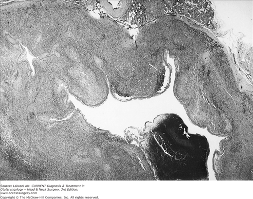
A CT scan or ultrasound may reveal bilateral multiple cystic masses in the parotid gland. Serologic testing for HIV antibodies confirms the diagnosis. Fine-needle aspiration of these cysts can reveal amylase in the fluid, which also leads to the diagnosis (see Figure 18–1).
Observation or serial drainage of symptomatic cysts is the recommended treatment. A recent treatment modality includes sclerotherapy of the cysts. Rarely is parotidectomy indicated; however, when it is performed, the histopathology often shows multiple lymphoepithelial lesions and florid follicular hyperplasia with follicle lysis. Similarly, cysts involving the submandibular gland may require gland excision.
The parotid cysts found in HIV-infected patients are often associated with the histologic finding of benign lymphoepithelial lesions. There is little malignant transformation.
- Chronic unilateral or bilateral salivary gland swelling.
- Minimal pain.
- Fine-needle aspiration biopsy of the gland can aid in diagnosis.
- Risk factors such as exposure to tuberculosis, animal exposure, trauma, and multiorgan system involvement should be considered.
- Uveitis, facial palsy, and parotid enlargement are suggestive of sarcoidosis.
Granulomatous disorders may present with acute salivary gland swelling or chronic unilateral glandular swelling. The glandular mass is not usually accompanied by significant pain. Primary tuberculosis should be considered if there are risk factors for exposure.
The diagnosis of tuberculous sialadenitis may be made with acid-fast staining for organisms, a culture of the saliva, and placement of a purified protein derivative skin test. A fine-needle aspirate of the gland helps to obtain material for diagnosis. Treatment of primary tuberculous sialadenitis includes multidrug antituberculous medications.
The differential diagnoses of granulomatous sialadenitis include animal cat-scratch disease, sarcoidosis, actinomycosis, Wegener granulomatosis, and syphilis.
Cat-scratch disease does not directly involve the parotid gland; instead, it affects the periparotid and intraparotid lymph nodes. In the submandibular gland, it can present as an acute submandibular mass without causing ductal obstruction, which suggests the involvement of the adjacent lymph nodes. The offending organism is a gram-negative rod, Bartonella henselae, and diagnosis may be made using indirect fluorescent antibody tests with rising IgG titers, or with the Warthin–Starry silver stain looking for gram-negative bacilli. Cat-scratch disease is usually self-limiting and treatment is supportive while the mass lesions slowly resolve.
Sarcoidosis is noninfectious and involves the parotid gland in less than 10% of cases. It is a diagnosis of exclusion and is confirmed by histologic findings of noncaseating granulomas. Sarcoidosis may occur as part of a syndrome known as uveoparotid fever or Heerfordt syndrome. This syndrome is characterized by parotid enlargement, facial palsy, and uveitis. The involvement of the parotid and lacrimal glands leads to xerostomia and xerophthalmia. The disease often affects adults in their twenties and thirties with spontaneous resolution occurring in the ensuing months to years.
Actinomycosis is easily diagnosed with special histologic stains that demonstrate sulfur granules. Actinomycosis should be suspected if a patient has painless parotid swelling with a history of recent dental infection or trauma. Trismus may develop with progression of the infection. Penicillin is the drug of choice for treatment of actinomycosis.
Wegener granulomatosis can present as an acute unilateral mass in the gland, often with pain. This diagnosis, characterized histologically by necrotizing inflammation and vasculitis, is confirmed with serologic testing for the cytoplasmic antineutrophil cytoplasmic antibody (C-ANCA) and histopathologic examination.
The treatment of Wegener disease depends on the involvement of other organs; Wegener granulomatosis can be a rapidly fatal disease if it is untreated and involves other major organs. The initial treatment consists of several weeks of steroids with the addition of cyclophosphamide or other immunosuppressive agents. A more indolent subtype of Wegener, as often seen in the head and neck region, can be controlled with immunosuppressive therapy. The prognosis is excellent for many of the granulomatous diseases.
- Acute, painful swelling of the major salivary gland, especially the submandibular gland, which may be recurrent.
- Aggravation of symptoms with eating; swelling may subside after approximately 1 hour.
- History of gout or xerostomia.
- A stone in the floor of the mouth may be palpated; treatment depends on the location of the calculus.
- Calculus may be extracted intraorally, or if distal, then the submandibular gland may be indicated.
- Complications include acute suppurative sialadenitis, ductal ectasia, and stricture.
Approximately 80–90% of salivary calculi occur in the submandibular gland, whereas only 10–20% is reported in the parotid gland; a very small percentage of salivary calculi are found in the sublingual and minor salivary glands. Sialolithiasis is a common cause of salivary gland disease and can occur at any age with a predilection in men. Risk factors for salivary stone obstruction include long illnesses with dehydration. There are also associations with gout, diabetes, and hypertension.
Normal saliva contains abundant hydroxyapatite, the primary compound in salivary stones. Aggregates of mineralized debris in the duct can form a nidus, promoting calculi formation, salivary stasis, and eventually obstruction. The submandibular gland is more susceptible to calculi formation than the parotid gland because of the longer course of its duct, higher salivary mucin and alkaline content, and higher concentrations of calcium and phosphate.
Submandibular calculi consist primarily of calcium phosphate and hydroxyapatite; because of the high calcium content of these calculi, the majority is radiopaque and visualized on X-rays. Parotid calculi are less likely to be radiopaque. Approximately 75% of the time, a single stone is found in the gland. If the obstruction is not relieved, local inflammation, fibrosis, and acinar atrophy ensue.
Recurrent swelling and pain in the submandibular gland exacerbated with eating is the common presentation of salivary calculi. Prolonged obstruction can lead to acute infection with increasing pain and erythema of the gland. Patients may also report a history of xerostomia and occasionally gritty, sand-like foreign bodies in their oral cavity. A physical exam is essential as stones often are palpated in the anterior two thirds of the submandibular duct. In addition, an induration of the mouth floor is sometimes observed. Stones located within the body of the gland are not easily palpated.
Stay updated, free articles. Join our Telegram channel

Full access? Get Clinical Tree


