Hemangiomas of Infancy & Vascular Malformations: Introduction
Hemangioma of Infancy
Hemangiomas are true tumors with pathologic endothelial cell proliferation; vascular malformations are distinguished by this distinct absence.
- Absent at birth or history of small premonitory mark at birth.
- Rapid neonatal growth of the lesion.
- Cutaneous lesions develop either a typical “strawberry” appearance or a bluish hue (“deep bruise” appearance).
- Magnetic resonance imaging (MRI) is diagnostic when the diagnosis is uncertain or when serial exam is not possible.
- Visceral involvement is suspected if there are more than three cutaneous lesions.
- Progressive stridor in the appropriate age group (2–9 months) is suspicious for airway hemangioma.
Hemangiomas are the most common tumors of infancy. They are more common in females than in males (3:1), in white populations, and in premature infants. Most of these neoplasms are located in the head and neck. Additionally, most are single lesions; however, about 20% of patients have multiple lesions. Hemangiomas exhibit a period of rapid postnatal growth. The duration of the proliferative period is variable, but is usually confined to the first year of life. The proliferative period rarely extends to 18 months. The involutional phase is also quite variable, occurring over a period of 2–9 years. After complete involution, normal skin is restored in about 50% of patients. In other patients, the skin may show evidence of telangiectasia, yellowish hypoelastic patches, sagging or fibrofatty patches, and scarring if the lesion has ulcerated.
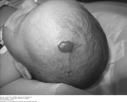
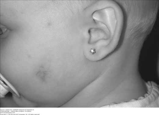
Hemangiomas can be classified as superficial (Figure 7–1), deep (Figure 7–2), or combined. The term superficial hemangioma replaces the older terms capillary hemangioma and “strawberry” hemangioma and refers to hemangiomas located in the papillary dermis. The deep hemangioma, often slightly blue in color, originates from the reticular dermis or the subcutaneous space and, in the past, was referred to as a cavernous hemangioma. The combined hemangioma has elements of both the superficial and the deep hemangioma.
Proliferative hemangiomas have been shown to express high levels of indolamine 2,3-dioxygenase (IDO), basic fibroblast growth factors (β-fgf), proliferating cell nuclear antigen, type IV collagenase, urokinase, and, most recently, insulin-like growth factor 2. Involuting hemangiomas have been characterized by exhibition of tissue inhibitor of metalloproteinase 1 (TIMP1), thrombospondin, interferon-α, and decreased levels of other factors seen in the proliferative hemangioma.
In addition, it has recently been shown that endothelial cells are of clonal origin and the defect that leads to tumor growth and the altered expression of growth factors is intrinsic to the endothelial cell. These clonal endothelial cells have also been shown to have characteristics similar to placental endothelial cells, which may suggest that hemangiomas are of placental origin. There is a higher rate of hemangioma in children whose mother underwent chorionic villis sampling, giving additional weight to placental origin theories.
Most commonly, the diagnosis is determined by history and physical examination. The history will typically reveal that more than 50% of hemangiomas are seen at birth as a prominent cutaneous mark. This mark may manifest as a whitish patch, an anemic nevus, a faint telangiectasia, or a blue spot. The rapid proliferation of this initial lesion is highly suggestive of a hemangioma. A superficial hemangioma will assume the typical “strawberry” appearance, making the diagnosis obvious. In the case of a subcutaneous, intramuscular, or visceral tumor, the diagnosis may be uncertain. In these instances, various radiologic modalities can be very helpful. MRI is the most informative of the available modalities.
When an infant aged 2–9 months presents with progressive stridor or persistent croup-like symptoms, consideration should be given to the possibility of a subglottic hemangioma. This neoplasm is said to be more common in children with a cutaneous hemangioma in a facial or “beard” distribution. The diagnosis of a subglottic hemangioma should be made with a direct laryngoscopy and a bronchoscopy.
Special consideration should be given to children with three or more hemangiomas. In these children, abdominal ultrasounds should be obtained to evaluate for visceral hemangiomas and, most especially, hepatic hemangiomas. If the screening ultrasound is positive, MRI of the entire body is indicated to detect other internal hemangiomas.
Another special diagnostic situation arises when a child presents with extensive facial hemangiomas, sometimes referred to as segmental hemangiomas. The term segmental hemangioma relates to the approximate distribution that may correspond to sensory innervation patterns. The acronym PHACE can help the clinician recall the findings seen in these children, which include the following:
- Posterior fossa malformations
- Hemangiomas
- Arterial anomalies
- Coarctation of the aorta and cardiac defects
- Eye abnormalities
Congenital hemangiomas are rare vascular tumors that are fully developed at birth and in that way are distinguished from the more typical hemangioma of infancy. There are two types of congenital hemangioma. One does not involute, the non-involuting congenital hemangioma (NICH), and the other does involute quickly, rapidly involuting congenital hemangioma (RICH). These tumors are also pathologically distinguishable from the hemangioma of infancy, in that they are glucose transporter 1 protein (glut-1) negative.
A vascular malformation is another typical diagnostic alternative to consider when attempting to diagnose a potential hemangioma. The natural history of the hemangioma (not present at birth with rapid growth in the first months of life) is usually adequate evidence to support a confident diagnosis.
A pyogenic granuloma, which is neither a vascular malformation nor a hemangioma, is often confused with a hemangioma. A pyogenic granuloma is often the result of a minor trauma. The lesion is usually sessile, and as it grows it becomes pedicled, often bleeding impressively. The treatment is surgical excision.
Kaposiform hemangioendothelioma (KHE) is a rare vascular tumor with close association with Kasselbach–Merritt syndrome. Differentiation from hemangioma of infancy is typically based on recognition of aggressive behavior such as compression and invasion of surrounding tissue. These are large abnormal vascular tumors, and early recognition and treatment can be life saving.
Tufted angiomas (angioblastoma of Nakagawa) are benign erythematous plaques that grow slowly over several years. They will often stabilize after the slow-growth period. A pathologic specimen is usually diagnostic.
Magnetic resonance imaging with contrast is the most useful of all radiologic evaluations of hemangiomas. MRI can differentiate a hemangioma from a vascular malformation. A discussion of clinical suspicions with the radiologist may help determine the need for concomitant magnetic resonance angiography, which is especially helpful in locating feeder vessels of high-flow arteriovenous malformations.
The ultimate method of differentiating all diagnostic possibilities is with a histologic study of the tissue. A biopsy should be done whenever there is a possibility that the lesion in question is a malignant tumor; however, a biopsy is rarely necessary as there is usually ample epidemiologic, clinical, and radiologic information that can facilitate a reliable diagnosis.
Although rare, the complications of hemangiomas dictate a need for treatment. These complications include:
Ulceration (most common in the perineum and lip/perioral area).
Airway obstruction.
Visual loss. Obstruction of the visual axis for one week in the first year of life can cause permanent amblyopia.
External auditory canal obstruction.
Bleeding. Bleeding is usually low flow and therefore can be managed simply with pressure.
Heart failure. This complication is managed with medical therapy (usually by a cardiologist) and with attempts to control the growth of the hemangioma. Steroids should be the initial medical therapy, with vincristine and other chemotherapies used for steroid failures. Surgical therapy combined with embolization would be a second tier of therapy if medical treatment was not effective and the problem became life threatening.
The decision to intervene and attempt to treat the patient without an active or inevitable complication must be weighed against the fact that most hemangiomas will resolve completely or with minimal long-term sequelae. For hemangiomas with active or inevitable complications, multiple treatment options exist. The most appropriate treatment will depend on the location and the nature of the impending complication as well as the child’s specific medical and social situation. For example, if follow-up is not possible, early definitive surgical management may be more strongly considered.
Steroids are the usual first line of treatment. Typical initial doses are 2–5 mg/kg/d of prednisolone or prednisone. Steroids are best administered in a single dose in the morning. This initial therapy is usually used for 4–12 weeks. This dose is then tapered over the next several months, according to what the patient can tolerate. Rebound growth may necessitate a second course of therapy. Alternate-day dosing or rest periods of several weeks may lessen troublesome side effects such as cushingoid appearance, growth retardation, decreased appetite, and susceptibility to infection. Monitoring of blood glucose and blood pressure are recommended. Adrenal suppression can be a result of therapy. Concomitant use of a proton pump inhibitor is also suggested.
Intralesional steroid injections may be used as an initial therapy, especially for orbital or periorbital lesions, tumors of the nasal tip, and globular tumors of the lips, ears, and cheeks and parotid hemangiomas. A 1:1 ratio of long-acting steroids (eg, triamcinolone 40 mg/mL) and short-acting steroids (eg, betamethasone 6 mg/mL) yields the best results. Three injections of triamcinolone, at doses of 3–5 mg/kg per procedure spaced 4–6 weeks apart, are the suggested course. Injections of long-acting corticosteroids in a suspension in the periorbital tissues can result in blindness. Great caution is needed in this area, especially in the upper lid. A low-pressure injection technique is thought to decrease risk of embolization. When effective, injection therapy usually leads to a dramatic reduction in the size of the lesion within one week. In general, steroid therapy (systemic or intralesional) can be extremely effective in one-third of patients, partially effective in another third, and ineffective for the final third of patients.
A new treatment gaining rapid acceptance is treatment of hemangiomas with propranolol. This treatment seems both effective and safe. Typically, infants are treated at a dose of 1 mg/kg/dose. The medication is given twice daily. Prior to initiating therapy the children are screened for cardiac defects with an echocardiogram. The medication is typically prescribed for 1 year. This therapy, although new, is now widely used and under study at many institutions. The mechanism of action is unknown.
Interferon alfa-2a is a comparatively new agent for the treatment of hemangiomas. Although it is effective in most cases, it is generally considered a second-line drug because of cost, the route of administration, and the potential side effects. The treatment is generally reserved for pulmonary hemangioma, life-threatening hemangioma, and diffuse neonatal hemangioma. Transient side effects include fever, elevated liver enzymes, and neutropenia. Spastic diplegia and other permanent neurologic complications associated with the use of interferon alfa-2a have resulted in the cautious application of this therapy. The typical dose is 3 million units/m2 injected subcutaneously daily. The therapy is generally administered for 6–12 months.
Vincristine is gaining popularity as another efficacious treatment for complicated or refractory hemangiomas. There are relatively few side effects as compared to interferon. The therapy should be coordinated by someone experienced in using the medication. One drawback of the therapy is the need for central venous access for up to 12 weeks.
Laser therapy for hemangiomas is becoming widely practiced to combat mucosal lesions and cutaneous lesions with or without ulceration. In the United States, laser debulking of mucosal lesions is the typical treatment of obstructing lesions such as subglottic hemangiomas. The goal is to reduce the lesion size to allow for an adequate airway. Recurrence is anticipated, and the treatment is repeated until the hemangioma stops proliferating and involutes. Various laser therapies are employed, but all share the drawback of causing a mucosal ulceration in the airway.
Ulceration is a controversial indication for cutaneous laser therapy. The yellow light emitted by pulsed dye lasers is selectively absorbed by hemoglobin and melanin. In an ulcerated hemangioma, the laser light does not need to pass through the skin and the melanin within the skin to reach the hemangioma; therefore, the risks of scarring due to absorption by melanin are considered lessened. Recent advances in the flashlamp pulsed dye laser include longer wavelengths, longer pulse durations, and the very important dynamic cooling of the surface tissues. These advances have allowed for higher energy treatments, deeper penetration, fewer complications, and better overall responses. These advances have led to increased confidence in using flashlamp pulsed dye laser for the treatment of select non-ulcerated cutaneous lesions. The KTP and Nd:YAG lasers have been employed for intralesional therapy by using bare fibers to deliver high energies to the deep components of the lesions. The use of these laser technologies, although gaining in acceptance and recognition of their utility, is not standardized and is limited by the experience of the practitioner.
It is commonplace to consider excision in a completely involuted lesion, when the residuum causes a functional or aesthetic problem. Baggy fibrofatty tissue is recontoured for improved cosmesis.
The early surgical excision of an actively proliferating lesion is appropriate in an area (eg, the glabella, eyelid, airway, the nasal wall) that will certainly lead to complications or impaired function. This may also avoid the need for protracted systemic therapy and spare the child and family the anticipated psychosocial difficulty.
Some surgeons also advocate the surgical intervention of lesions that have stopped proliferating instead of waiting for a protracted involution phase. Physicians who advocate this earlier removal do so with the hope of diminishing psychosocial stress. This technique also takes advantage of the natural tissue expansion of surrounding skin and soft tissue, which occurs in the proliferative phase.
Regardless of the timing, the procedures are typically accomplished using routine techniques. Special preoperative planning consideration should be taken when operating on actively proliferating or recently quiescent lesions to minimize blood loss such as embolization. In addition to standard techniques, circular excision with purse-string closure, with subsequent lenticular removal of scar, has been advocated. This technique may lead to smaller eventual scarring.
Stay updated, free articles. Join our Telegram channel

Full access? Get Clinical Tree


