CHAPTER 210 Aspiration and Swallowing Disorders
Swallowing involves multiple, highly integrated, and partially overlapping actions; however, for discussion purposes, it is frequently separated into three phases—oral, pharyngeal, and esophageal. During the oral phase, food is processed into a “swallow-ready ball” (bolus), and then transported to the back of the mouth. This phase is limited to sucking fluid from a nipple during infancy. After approximately 6 months of age, this phase is sometimes subdivided to include the oral preparatory phase, which conditions solid foods requiring chewing. The pharyngeal phase comprises a series of complex and interrelated events that direct and propel boluses through the pharynx into the esophagus while the airway is protected. During this phase of swallowing, the velum rises and approximates the pharyngeal walls, breathing stops, the larynx rises, the vocal folds adduct, and the base of tongue and pharyngeal muscles propel the bolus through a relaxed upper esophageal sphincter. The act of swallowing results in mechanical closure of the airway and cessation of breathing. The esophageal phase begins when the bolus enters the esophagus and ends when it passes into the stomach.
Typical swallowing maturation involves transformation from the primitive sucking and swallowing reflexes used during infancy into the mature and volitional functions of biting, chewing, and bolus formation necessary for the safe and adequate delivery of nutrients in older children and adults. Appropriate adjustments to growth and developmental changes of the aerodigestive system and changes in airway protective responses are essential to competent postnatal maturation processes. Changes in the anatomic relationships of the oral cavity, pharynx, and larynx occur throughout the first few years of life and are well described. In addition, neurodevelopmental, cognitive, and sensory inputs modulate deglutitive function, and consequently may influence the maturation process.
Competent deglutition (the act of swallowing) is crucial to survival. Two primary functions of swallowing are to direct oral secretions, liquids, and food from the mouth to the stomach while protecting the airway, and to provide sufficient amounts of nutrients and fluids for children to grow and develop optimally. Appropriate adjustments to growth and increases in nutritional needs are crucial to successful postnatal deglutition. Because breathing and swallowing share common conduits, such as the oral cavity and pharynx, their functions are intertwined.
Dysphagia (swallowing dysfunction) may be caused by any condition that interferes with structural integrity of the structures comprising the aerodigestive tract or their coordination. Congenital or acquired structural or anatomic anomalies may cause airway and swallowing defects. This chapter focuses on the evaluation of infants and children with suspected dysphagia and the associated primary aerodigestive tract anomalies. Management approaches to swallowing and airway-related problems are reviewed.
Evaluation of Infants and Young Children with Suspected Dysphagia
Clinical or Bedside Evaluation
Evaluation of all children with feeding and swallowing difficulties begins with taking a thorough history and performing a physical examination. Children with anatomic anomalies are at increased risk for dysphagia when the underlying conditions interfere with the structural integrity of the oropharynx or the complex coordination of neuromuscular and airway processes involved in deglutition; consequently, the focus of the clinical evaluation is the identification of diagnostic tests that determine the nature and extent of the swallowing impairments, and assist with management decisions. Children who present with structural anomalies as one component of a complex medical history or syndrome (e.g., bronchopulmonary dysplasia or Smith-Lemli-Opitz syndrome) may require other specialized evaluations (e.g., direct laryngoscopy and bronchoscopy) to determine the impact of the associated comorbidities on swallowing.
Instrumental Evaluation
When the clinical evaluation identifies problems that may be caused by or reflect problems that are invisible, an instrumental evaluation is typically recommended. Common examinations that allow for direct visualization of the structures involved in swallowing include radiologic (e.g., upper gastrointestinal series [UGI] and videofluoroscopic swallow studies [VFSS]) and endoscopic (e.g., flexible endoscopic evaluation of swallowing [FEES]) procedures. Clinicians need to be cautious about the interpretation of negative findings of aspiration during UGI, VFSS, and FEES. Each of these evaluations may establish a diagnosis of dysphagia by identifying specific aspects of the pathophysiology of the dysfunction; however, they may fail to detect aspiration even in children who aspirate, particularly when aspiration is episodic.
Upper Gastrointestinal Series
UGI may provide important information for children with anatomic anomalies, particularly when the anomaly is distal to the oropharynx. UGI provides information that allows for assessment of the anatomy and physiology of the esophagus, stomach, and duodenum; defines gastrointestinal obstructions; and identifies malrotation. In addition, it serves as a screen of the oropharyngeal structures and their functions. Decisions about whether liquid barium should be administered orally or by nasogastric tube are determined by the specific diagnostic questions, and whether children are at risk for aspiration secondary to swallowing dysfunction.
Videofluoroscopic Swallow Study
VFSS, sometimes referred to as a modified barium swallow study, may be useful in children suspected to have oropharyngeal dysphagia because it images the structures of the oral cavity, pharynx, and cervical esophagus during deglutition.1 VFSS may screen gastrointestinal structures distal to the cervical esophagus. The goals of VFSS are to provide information that helps determine whether anatomic or structural abnormalities are present; ascertain if coordination of the structures and functions of the upper aerodigestive tract supports safe and efficient bolus passage; and identify strategies that enhance the safety and efficiency of feeding, while minimizing the dysphagic problems. During VFSS, children ingest liquids or foods or both impregnated with barium contrast material to simulate functional feeding as closely as possible. Consequently, children must be ready, willing, and able to cooperate with VFSS.2,3
Flexible Endoscopic Evaluation of Swallowing
FEES is an extension of the routine flexible fiberoptic nasopharyngolaryngoscopy examination, and may be used to evaluate the structures and functions of the nasopharynx, oropharynx, and larynx during phonation, spontaneous swallows, and swallows of liquids and foods.4,5 FEES may be particularly useful in children who are nonoral feeders, who are unable to cooperate with VFSS, who have vocal fold dysfunction, or in whom their ability to handle their secretions is questionable.
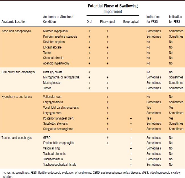
FEES may provide sensory information when the endoscope touches adjacent mucosa or structures. To standardize evaluations of responses to sensory input, calibrated air pulses have been administered endoscopically.6 The utility of FEES plus sensory air pulse testing (FEES-ST) has been shown during the evaluation and treatment of children with specific diagnostic conditions (e.g., type I laryngeal clefts) and during preoperative evaluations for pediatric airway reconstruction.7,8 Elevated laryngopharyngeal sensory thresholds have been documented in children with clinical diagnoses of recurrent pneumonia, neurologic disorders, and gastroesophageal reflux disease (GERD).6 Table 210-1 outlines the utility of using VFSS and FEES based on the anatomic or structural condition.
Other Diagnostic Evaluations
Other diagnostic tests may be needed to determine the cause of the swallowing dysfunction. Imaging of the brainstem may be needed to diagnose abnormalities of the skull base or spine as potential underlying conditions. Specialized evaluations may be needed for children with refractory respiratory conditions. Chest radiographs, pulmonary function tests, high-resolution computed tomography (CT), and bronchoscopy provide information regarding the extent of lung injury. Endoscopy of the gastrointestinal tract may be beneficial for patients with histories or presentations suggestive of gastrointestinal conditions.
Four Anatomic Sites of Dysphagia
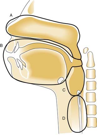
The four primary anatomic sites with anomalies of the aerodigestive tract that may adversely affect swallowing and contribute to dysphagia are the nose and nasopharynx, oral cavity and oral pharynx, hypopharynx and larynx, and trachea and esophagus (Fig. 210-1).

Figure 210-1. Four anatomic sites of dysphagia. A, Nose and nasopharynx. B, Oral cavity and oral pharynx. C, Hypopharynx and larynx. D, Trachea and esophagus.
Nose and Nasopharynx
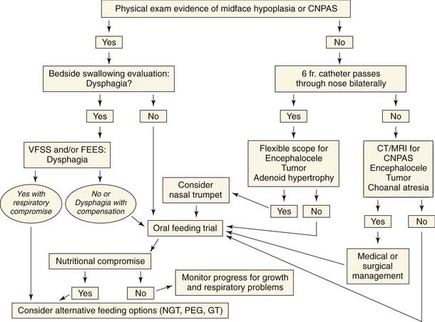
Anomalies of the nose and nasopharynx affect the oral and pharyngeal phases of swallowing. Dysphagia from anomalies of the nose and nasopharynx is secondary to upper airway obstruction and the caliber of the upper airway and the child’s ability to compensate while swallowing (Fig. 210-2). Bilateral disease has a more significant impact on airway patency and swallowing than unilateral disease.

Figure 210-2. Evaluation and management of dysphagia with suspected nose and nasopharynx anomaly. CNPAS, congenital nasal pyriform aperture stenosis; FEES, flexible endoscopic evaluation of swallowing; GT, gastrostomy tube; NGT, nasogastric tube; PEG, percutaneous endoscopic gastrostomy; VFSS, videofluoroscopic swallow studies.
Any cause of nasal airway obstruction can lead to feeding difficulties, particularly during infancy when nasal airflow is an important component of breathing and feeding. Nasal obstruction can result from myriad conditions, including midface hypoplasia, congenital nasal pyriform aperture stenosis, septal deviation or hematoma, chronic or acute rhinitis, congenital midline nasal masses (including dermoids, encephaloceles, and gliomas), nasal or nasopharyngeal tumors, choanal atresia, and adenoidal hypertrophy. These children frequently have difficulty coordinating feeding and breathing, and may have failure to thrive or recurrent aspiration.9
Unilateral nasal disease may not be detected for years and is often associated with unilateral nasal congestion or rhinorrhea. Bilateral disease has a more significant impact on airway patency and swallowing than unilateral disease and is usually recognized soon after birth because the child commonly has severe respiratory difficulties with cyclical cyanosis relieved by crying.10 These children are also unable to feed effectively, and may have coughing and choking with cyanotic spells when attempting to eat. Diagnostic evaluation often starts with attempted passage of a 6F feeding tube through the nares. A thorough evaluation with flexible fiberoptic endoscopy and imaging should be carried out if nasal obstruction is suspected. Figure 210-2 outlines the evaluation for children with dysphagia that is suspected to originate from the nose and nasopharynx.
Oral Cavity and Oral Pharynx
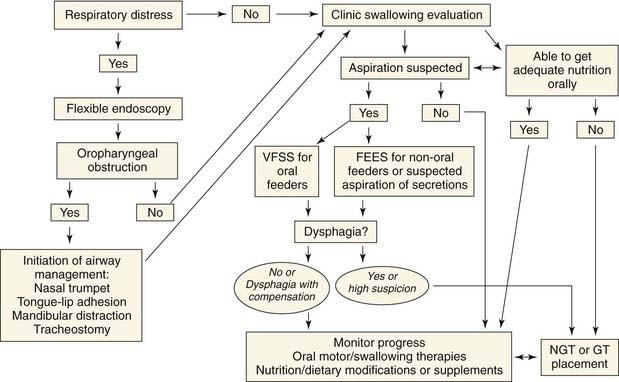
Anomalies of the oral cavity and oral pharynx may affect the oral and pharyngeal phases of swallowing, and many have the potential for upper airway obstruction. The respiratory issues should be evaluated and stabilized before initiating the feeding efforts or evaluations for suspected dysphagia (Fig. 210-3).

Figure 210-3. Evaluation and management of dysphagia with suspected oral cavity and oropharynx anomaly. FEES, flexible endoscopic evaluation of swallowing; GT, gastrostomy tube; NGT, nasogastric tube; VFSS, videofluoroscopic swallow studies.

Stay updated, free articles. Join our Telegram channel

Full access? Get Clinical Tree

