Anterior and Subtemporal Approaches to the Infratemporal Fossa

Chapter 54 Anterior and Subtemporal Approaches to the Infratemporal Fossa


The infratemporal fossa (ITF) is a potential space bounded superiorly by the greater wing of the sphenoid and the temporal bone. Neurovascular foramina, including the carotid canal, jugular foramen, foramen spinosum, foramen ovale, and foramen lacerum, connect the ITF with the middle cranial fossa. Medially, the ITF is contained by the superior constrictor muscle, the pharyngobasilar fascia, and the pterygoid plates. Medially, the ITF communicates with the pterygopalatine fossa via the pterygomaxillary fissure, which is continuous with the inferior orbital fissure and the orbit. Laterally, the ITF is bounded by the zygoma, mandible, parotid gland, and masseter muscle. The pterygoid muscles constitute the anterior boundary; posteriorly, the ITF is confined by the articular tubercle of the temporal bone, glenoid fossa, and styloid process. Using this definition, the ITF contains the parapharyngeal space (i.e., internal carotid artery [ICA], internal jugular vein, CN IV to XI) and the masticator space (i.e., internal maxillary artery, pterygoid venous plexus, and pterygoid muscles).


The presence of neurovascular structures within the ITF (e.g., ICA) or adjacent to it (e.g., CN VII) is the limiting step for designing a surgical approach to the ITF. Surgical approaches often center on the preservation and identification of these neurovascular entities. The first report in the English literature of a surgical approach to the ITF is attributed to Barbosa,1 who in 1961 described his approach for advanced tumors of the maxillary sinus. Transtemporal approaches described by Fisch and preauricular approaches by Schramm and Sekhar are the basis for other modifications.28 Subsequent approaches follow the surgical and anatomic principles shown by these authors.


PREOPERATIVE EVALUATION


Diagnostic and Staging Work-up


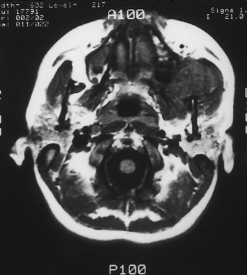
Owing to the inaccessibility of the ITF to physical examination, radiographic imaging is a vital component of the evaluation. Computed tomography (CT) and magnetic resonance imaging (MRI) provide valuable information and are obtained using standard skull base protocols. CT is superior to MRI, showing the remodeling or erosion of neurovascular foramina or other bones of the skull base. MRI better delineates the soft tissue planes, the tumor–soft tissue interface, and the presence of tumor along neural and vascular structures (Fig. 54-1). CT and MRI are often complementary.


Another crucial question is the relationship of the tumor to the ICA. Magnetic resonance angiography (MRA) and computerized tomography angiography (CTA) provide a noninvasive assessment of the vasculature of the ITF and brain. If preoperative embolization of the tumor is indicated (e.g., juvenile nasopharyngeal angiofibromas, paragangliomas), angiography is preferred over MRA. Angiography provides important information regarding the vascularity of the tumor, its relationship to the ICA, and the cerebral circulation and its collateral blood supply. Neither angiography nor MRA is adequate, however, to predict reliably the adequacy of the collateral intracranial circulation if sacrifice of the ICA is necessary.


If the risk for injury or sacrifice of the ICA is high, the collateral cerebral blood flow may be evaluated using angiography-balloon occlusion with xenon CT (ABOX-CT). A nondetachable balloon is inserted in the ICA via the femoral artery. The balloon is inflated for 15 minutes while the awake patient is monitored for any neurologic deficit. If the patient does not develop any deficit, the balloon is deflated, and the patient is transferred to a CT suite. A mixture of 32% xenon/68% oxygen is administered via facial mask for 4 minutes. CT shows the distribution of xenon, which reflects the blood flow within the cerebral tissue, providing a quantitative assessment of milliliters of blood flow per minute per 100 g of brain tissue. The process is repeated after reinflation of the arterial balloon. A computer calculates the differential of the xenon diffusion in the brain before and after the balloon inflation, identifying patients at risk for an ischemic stroke secondary to reduced blood flow after occlusion of the ipsilateral ICA (Table 54-1).9


TABLE 54-1 Xenon Computed Tomography


Cerebral Blood Flow (mL/min/100 g Tissue) Risk Implication
>35 Low Carotid may be sacrificed
21-35 Moderate Patient would tolerate occlusion under controlled circumstances; reconstruction is recommended
≤20 High Patient would not tolerate occlusion of internal carotid artery

Despite a negative finding from ABOX-CT testing, patients can sustain ischemic brain injury because of the loss of collateral vessels that are not assessed by balloon occlusion testing (“watershed area”), or because of embolic phenomena. In addition, this test is performed under ideal and controlled circumstances, and does not account for the possibility of episodes of hypoxia, hypotension, or electrolyte and acid-base disturbances that may alter the brain’s hemodynamics. Every effort should be made to preserve or reconstruct the ICA and to diminish the possibility of embolus formation during the surgery. Other techniques that provide information regarding collateral cerebral blood flow include single photon emission CT (SPECT) with balloon occlusion and transcranial Doppler.


Histologic diagnosis should be obtained before the extirpative surgery whenever possible. Tumors amenable to a punch or open biopsy are approached in this manner. Tumors that are in deeper planes may be sampled by fine-needle aspiration biopsy. Rarely, a histologic diagnosis cannot be obtained before the approach because of the intrinsic limitations of fine-needle aspiration biopsy. Under these circumstances, a frozen section analysis, obtained via a skull base approach, may be sufficient to justify the resection of the tumor. Vital neurovascular structures, such as the ICA, the eye, and cranial nerves, should not be sacrificed, however, based on a frozen section analysis.


The extent of the evaluation to rule out regional or distant metastasis or to determine that the ITF tumor is a metastasis is dictated by the histologic type and stage of the tumor. CT scan of the neck is more sensitive than physical examination for the detection of regional lymphadenopathy. Patients presenting with tumors that metastasize hematogenously (sarcoma, melanoma) should undergo CT scan of the chest and abdomen and a bone scan. Cerebrospinal fluid (CSF) cytology is advised for patients with tumors that have invaded the dura. These patients are also at risk for “drop metastasis,” which should be ruled out by spinal MRI.


Rehabilitation Considerations


Functional or neurologic deficits that are identified preoperatively should be taken into consideration during the surgical planning and during postoperative care. These deficits often have a significant impact on the recovery and functional rehabilitation of the patient.


Dysfunction of the trigeminal nerve or the masticator muscles or both is commonly underdiagnosed. Cutaneous and corneal sensation should be assessed preoperatively. Corneal anesthesia associated with concomitant facial nerve palsy requires aggressive measures to prevent corneal injury.


Lateral deviation of the jaw on opening may reflect weakness or paralysis of the ipsilateral pterygoid muscles, invasion of the muscles, or dysfunction of the temporomandibular joint (TMJ). Likewise, trismus may be due to mechanical restriction caused by the bulk of the tumor, ankylosis of the TMJ, scarring, tumor tethering, or pain. The nature of the trismus is an important consideration in the perioperative management of the airway. Trismus secondary to pain resolves with the induction of general anesthesia, allowing safe oral endotracheal intubation. In patients with mechanical trismus, an awake nasotracheal intubation may be performed if it is anticipated that surgery would correct the trismus. Otherwise, a tracheostomy, performed under local anesthesia, is the safest perioperative airway.


Neoplastic invasion of the facial nerve may manifest with facial weakness or paralysis, facial spasms, epiphora, facial spasms, and dysgeusia. Significant destruction of the facial nerve by tumor may occur before the patient develops these clinical signs. A gold weight, implanted in the upper eyelid, or surgical tightening of the lower lid may be necessary to protect the cornea.


Hearing loss caused by a tumor of the ITF may be either conductive, resulting from eustachian tube dysfunction, or sensorineural, resulting from tumor involvement of the temporal bone or posterior cranial fossa. A myringotomy or amplification or both facilitate communication with the patient.


Deficits of the lower cranial nerves (CN IX, X, XI, and XII) are associated with tumors that originate in the parapharyngeal space or tumors that extend to the jugular foramen, or both. Patients with deficits of CN IX, X, and XII present with varying degrees of swallowing or speech problems, such as hypernasal or slurred speech, nasal regurgitation, dysphagia, aspiration, and dysphonia. Findings on physical examination reflect the involvement of specific cranial nerves, and include decreased elevation of the palate, decreased mobility and strength of the tongue with deviation to the involved side on protrusion, decreased supraglottic sensation, pooling of secretions in the hypopharynx, ipsilateral vocal cord paralysis, and decreased bulk and strength of the sternocleidomastoid and trapezius muscles. Patients with partial deficits of the lower cranial nerves (paresis) often experience a complete deficit (paralysis) after surgery, resulting in increased dysphagia and aspiration. Consequently, a tracheostomy for tracheal toilet and a gastrostomy tube for nutrition and hydration are often necessary during the perioperative period.


Laryngeal framework surgery (thyroplasty) performed during the extirpative surgery or the early postoperative period improves the glottic closure and decreases the risk for aspiration, often obviating the need for a tracheotomy for the sole purpose of tracheopulmonary toilet.1012 Laryngeal framework surgery allows the patient to compensate for the deficits using the remaining function (contralateral side) more effectively. Laryngeal framework surgery does not restore the motor or the sensory function. These patients remain at a higher risk for aspiration and nutritional deficiencies. Collaboration with an experienced speech-language pathologist, who can assist with the monitoring of the patient and the diet modifications and provide intensive swallowing therapy, is crucial to prevent the pulmonary and nutritional complications of aspiration. In patients with severe deficits or in patients with cognitive problems, strong consideration should also be given to placement of a gastrostomy tube to facilitate postoperative feeding and decrease the risk of prandial aspiration.


Velopharyngeal insufficiency may be ameliorated by a palatal lift prosthesis that pushes the soft palate against the posterior pharyngeal wall. Alternatively, a pharyngeal flap or a palatopexy may be performed in patients who do not tolerate the prosthesis.


Reconstructive Considerations


Most commonly, a temporalis muscle transposition flap is adequate to separate the cranial cavity from the upper aerodigestive tract and obliterate the dead space. Microvascular free flaps, such as rectus abdominis flap (for soft tissue defects), latissimus dorsi flap (for myocutaneous or massive defects), and iliac composite flap (for defects requiring bone reconstruction), are indicated when the temporalis muscle or its blood supply is sacrificed as part of the oncologic resection, when the patient requires a complex resection involving composite tissue flaps with skin or bone or both, or when the extirpative surgery leads to a massive soft tissue defect and dead space. These needs are usually anticipated during the surgical planning, and the patient and consultants (e.g., the microvascular surgeon) are informed accordingly.


Ideally, functional and cosmetic deficits created by the tumor or the surgery should be addressed in a single stage, concomitant with the oncologic resection. When a temporary facial palsy is anticipated, corneal protection using lubricants or a temporary lateral tarsorrhaphy or both is usually adequate. Grafting of the facial nerve involves a longer recovery period, however. Insertion of a gold weight implant into the upper eyelid is advisable. When an immediate reconstruction of the facial nerve is impossible, static fascial slings or muscle transpositions are indicated. Lower cranial nerve deficits may be ameliorated by laryngeal framework surgery, tracheotomy, or laryngotracheal separation, as previously discussed.


Other Perioperative Considerations


Preoperatively, the patient’s blood is typed and crossmatched for 2 to 6 U of packed red blood cells, according to the extent and nature of the tumor and surgery. Autologous blood banking is used when feasible, although it is frequently impractical. A Cell Saver autologous transfusion device may be used during the resection of benign vascular tumors.


Perioperative antibiotic prophylaxis with a wide spectrum against the flora of the skin and upper aerodigestive tract and that exhibits good penetration of the blood-brain barrier is administered before the surgery and is continued for 48 hours after the surgery. The use of a broad-spectrum cephalosporin with good CSF penetration (e.g., ceftriaxone) seems to be as effective as multiple antibiotic regimens.


Somatosensory evoked potential monitoring using the median nerve is indicated whenever surgical manipulation of the ICA is anticipated. Lower cranial nerve monitoring is not routinely employed. It may be useful for the identification and preservation of nerve function when the tumor is in close proximity to these nerves. Conversely, facial nerve monitoring is routinely used for transparotid or transtemporal approaches. Monitoring electrodes and lines for vascular access should be secured with sutures, staples, or adhesive dressings.


The choice of anesthetic agent is influenced by the extent of intracranial dissection, potential for brain injury, systemic hemodynamics, the need for monitoring of cortical and brainstem functions (e.g., brainstem-evoked response, somatosensory evoked potentials, electroencephalography), and the need for cranial nerve monitoring (CN VII, and X to XII). All these factors should be thoroughly discussed with the anesthesiologist.


When changes of head position during surgery are anticipated, the endotracheal tube should be secured with a circumdental or circum-mandibular wire ligature (No. 26 stainless steel wire). The operating table is positioned perpendicular to the anesthesia staff, and if intradural dissection is anticipated, a spinal drain is inserted and secured with sutures and adhesive dressing (e.g., Tegaderm, Op-Site). Other measures to diminish the intracranial pressure, such as hyperventilation, osmotic diuresis, and corticosteroids, are used as needed throughout the surgery.


A nasogastric tube and Foley catheter are passed and secured after adequate placement is corroborated. Antiembolic sequential compression stockings are recommended to prevent deep venous thrombosis.


SURGICAL APPROACHES


The head of the patient is positioned on a horseshoe headrest or, if necessary for intracranial neurovascular or neurosurgical work, on a three-pin head fixation system. When the horseshoe headrest is used, it is important to use additional “egg-crate” foam padding because the scalp may develop a decubitus ulcer during prolonged surgery. If the ICA is at risk, the head should be positioned in slight extension to provide access to the neck for proximal control of the ICA. Tarsorrhaphy sutures are placed for protection of the eyes. The scalp is shaved following the planned incision line (e.g., bicoronal), and the incision line is infiltrated with a solution of lidocaine and epinephrine (1:100,000 to 1:400,000).


Preauricular (Subtemporal) Approach


The preauricular approach is suited for tumors that originate in the ITF and intracranial tumors that originate at the anterior aspect of the temporal bone, or greater wing of the sphenoid bone, and that extend into the ITF.5,13,14 It may also be combined with other approaches, such as a subfrontal approach to expose massive tumors that extend to the anterior and middle skull base. The preauricular approach does not provide an adequate exposure for the resection of tumors that invade the tympanic bone, however, and does not provide control of the intratemporal facial nerve or jugular bulb.


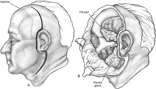
An incision, following a hemicoronal or bicoronal line, is carried through the subcutaneous tissue, galea, and pericranium (Fig. 54-2). Over the temporal area, the incision extends down to the deep layer of the temporal fascia. The anterior branches of the superficial temporal artery are preserved, ensuring adequate blood supply to the scalp flap. Ipsilateral to the tumor, the incision is extended following into the preauricular crease down to the level of the tragus. When proximal control of the ICA is warranted, the incision is extended into the neck using a lazy S pattern, or, alternatively, a separate cervical incision is performed. The scalp is dissected following a subpericranial plane, separating the attachments of the pericranium to the deep layer of the temporal fascia. The scalp flap is elevated from the deep temporal fascia using a broad periosteal elevator.


Above the zygoma, the deep temporal fascia splits into superficial and deep layers, which attach to the lateral and medial surfaces of the zygomatic arch. To continue the surgical exposure, the superficial layer of the deep temporal fascia is incised following an imaginary line that joins the superior orbital rim to the zygomatic root. The dissection continues deep to this plane, elevating the superficial layer of the deep temporal fascia off the zygomatic arch (see Fig. 54-2). Fascia and periosteum are reflected anteriorly with the scalp flap. This maneuver protects the frontal branches of the facial nerve that are just lateral to the superficial layer of the deep temporal fascia. Elevation of the periosteum from the lateral surface of the zygomatic arch and malar eminence completes exposure of the orbitozygomatic complex. The periorbita is elevated from the lateral orbit using a Penfield No. 1 dissector, exposing the roof and lateral wall of the orbit down to the inferior orbital fissure.


The fascial attachments of the temporalis and masseter muscles to the zygomatic arch are transected using electrocautery. The attachments of the temporalis muscle to the cranium are transected with the electrocautery, and the muscle is elevated off the temporal fossa. If the temporalis muscle is to be returned to its original position at the completion of the surgery, a curved titanium plate (1.5 to 1.7 mm) is screwed at the temporal line, leaving some screw holes empty to facilitate suturing from the plate to the muscle (Fig. 54-3). Then the masseteric fascia is dissected from the masseter muscle, elevating the overlying parotid gland with a broad periosteal elevator (see Fig. 54-2

Only gold members can continue reading. Log In or Register to continue

Stay updated, free articles. Join our Telegram channel

Jun 14, 2016 | Posted by in OTOLARYNGOLOGY | Comments Off on Anterior and Subtemporal Approaches to the Infratemporal Fossa

Full access? Get Clinical Tree

Get Clinical Tree app for offline access