12
Orbital Inflmmation
Evana Valenzuela Scheker and C. Robert Bernardino
INTRODUCTION
Orbital inflammatory disease (OID) is a general term encompassing all inflammatory diseases that affect some or all of the structures contained within the orbit external to the ocular globe. In rare cases, the areas involved by the inflammatory process may extend beyond the orbit, such as into the cavernous sinus through the orbital apex or to the eyelids through the orbital septum. OID needs to be differentiated from neoplastic and infectious processes that may have similar presentation.
Pain is a prominent complaint in OID. Involvement of the extraocular muscles can result in diplopia. Lacrimal gland involvement often results in painful superolateral orbital swelling, while generalized orbital tissue involvement may present with proptosis, chemosis, periorbital or lid swelling, and, in severe cases, loss of vision due to optic nerve compression.
PATIENT HISTORY
Patients typically complain of periorbital pain, diplopia, a “bug-eyed” appearance, ocular injection, grittiness, and tearing. In advanced cases in which optic nerve compression is present, the patient may have altered color perception, enlarged blind spot, transient complete visual loss, or a visual field defect (Fig. 12.1A and B).
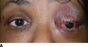
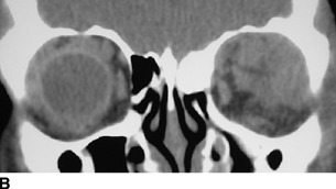
Figure 12.1 A: An example of OID, in which this patient presented with acute onset of painful proptosis. Note the proptosis, hypoglobus, and chemosis on the left side consistent with OID. B: A coronal cut of an MRI of the patient in image A—infiltrative lesion in the superior lateral orbit on the left side. A biopsy confirmed the diagnosis of orbital inflammatory pseudotumor.
The examination of patients with orbital pain includes attention to the following:
- A full ophthalmic assessment of the anterior and posterior segments
- Orbital assessment including exophthalmometer measurements
- Determination of lid retraction
- Assessment for lid lag/lagophthalmos
- Evaluation for limitation of extraocular muscles
- Examination for signs of corneal exposure
- An assessment of optic nerve function, testing for an afferent papillary defect visual field aberration and dyschromatopsia
In general, diagnostic tools useful in the evaluation of OID include
- CBC—an elevated white blood cell count can suggest infection
- Inflammatory markers
- Erythrocyte sedimentation rate (ESR)
- C-reactive protein (CRP)
- Erythrocyte sedimentation rate (ESR)
- Thyroid function panel
- Antithyroid antibodies
- Antineutrophil cytoplasmic antibodies (ANCA)
- Urinalysis—proteinuria suggests a systemic vasculitis with renal involvement
- Imaging (to help guide therapy)
Overall, imaging is helpful in differentiating between thyroid-associated ophthalmopathy and OID. In addition, the appearance of OID may overlap with that of malignant processes, in which case orbital biopsy is recommended.
Computed tomography (CT) scanning offers better visualization of bone, calcification, and acute hemorrhage.
Ultrasound scanning is a real-time dynamic imaging technique and may be superior to CT in the assessment of soft-tissue disease. It is highly operator dependent, and for most cases is not superior to MRI, with the exception of its role as an adjunctive means of assessing orbital blood flow.
AUTOIMMUNE CAUSES OF OID
Idiopathic Orbital Inflammation (IOI, Orbital Pseudotumor)
This is a diagnosis of exclusion based on clinical, radiological, and histopathological findings. It is the third most common cause of orbital inflammation, accounting for roughly 5% of cases. It is a nongranulomatous, usually acute to subacute or chronic inflammatory disease with no systemic manifestations that may affect teenagers to the elderly with peak in fourth to fifth decade and no gender predilection. The most common location is the anterior to mid orbit, frequently involving the lacrimal gland (50%). It is usually unilateral. There is a great deal of variability in the presentation, clinical course, and outcome. Association with infections and systemic disease has been reported.
Clinical Manifestations
- Inflammation/mass effect with or without pain
- Abrupt unilateral pain (89%)
- Rapid proptosis
- Conjunctival injection
- Chemosis
- Periorbital edema (75% of patients)
- Erythema
- Motility restriction
- Palpable mass in 50% of cases
- Rarely, loss of visual acuity
Diagnosis
- Imaging—inflammation and infiltration of involved orbital structures including the extraocular muscles, lacrimal gland, retrobulbar fat, and sometimes the sclera as well. There may be intracranial extension via the orbital apex or superior orbital fissure and involvement of the cavernous sinus. If extraocular muscles are involved, it involves the entire muscle including the tendinous insertions, resulting in a more tubular appearance on imaging. Diffuse IOI may result in inflammatory “streaking” of the orbital fat; it could be reminiscent of lymphoma. Atypical bony involvement should alert to the possibility of an infectious or malignant process.
- CT—enlargement of the involved structures, streakiness in the adjacent fat, and enhancement post administration of contrast. There may be separate soft-tissue nodules.
- MRI—enhancement in areas of involvement. T2-weighted images are iso- or hyperintense to that of extraocular muscles but are often less hyperintense than other orbital diseases due to the cellular nature of the infiltrate. Involved areas show moderate enhancement with gadolinium. Enhancement often correlates with disease activity and therefore a lower signal corresponds to disease that is less responsive to therapy. There may be thickening and enhancement of the posterior episclera. The lacrimal gland can have diffuse enlargement without alteration of its structure.
- Echography—low to medium reflectivity, regular acoustic structure, and weak sound attenuation. Edema in Tenon capsule may appear as an area of lucency behind the globe.
Pathology
Gray rubbery mass composed of varying degree of nongranulomatous inflammation, polymorphic infiltrate of lymphocytes, eosinophils, plasma cells, PMN leukocytes, and sclerosis.
A sclerosing form exists, which histopathologically shows scarification and collagen deposition in orbital fat within extraocular muscles around the optic nerve and within the lacrimal gland. This form carries a worse prognosis, involving slow, progressive course that damages orbital structures and responds poorly to therapy, sometimes with permanent visual loss.
Treatment
Systemic corticosteroids, usually prednisone 1 mg/kg/day with a slow taper over the course of months are recommended to avoid breakthrough inflammation. In general, the prognosis is excellent with cure rates estimated at 37% and relapse rates at 52%.
Cytotoxic agents including methotrexate, azathioprine, cyclophosphamide, chlorambucil, and mycophenolate mofetil are sometimes used to maintain inflammatory control. Tumor necrosis factor-alpha antagonists, intravenous (IV) immunoglobulin, plasmapheresis, and radiation have also been employed. Rarely, surgical removal of chronically inflamed tissue is indicated.
Prognosis
Excellent, with complete resolution; fellow orbit involved in 25% of cases
Inflammation in Associated Structures
- Dacryoadenitis: localized inflammation of the lacrimal gland
- Presentation: painful, firm, upper temporal orbital mass often associated with ptosis or S-shaped deformity (Fig. 12.2A,B–C)
- Differential diagnosis: sarcoidosis, Sjögren syndrome, lymphoma, or primary glandular tumor
- Myositis: acute or subacute inflammation of extra-ocular muscles
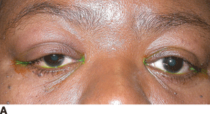
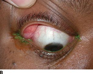
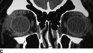
Figure 12.2 A: Dacryoadenitis presenting with upper eyelid swelling, pain localized around the lacrimal gland, and S-shaped deformity. B: Eversion of the right upper eyelid reveals an inflamed lacrimal gland. C: Coronal cut of a CT scan demonstrates inflammation in the right lacrimal gland and surrounding orbital soft tissue.
Clinical Presentation
- Any age group. Women more than men
- Unilateral, one muscle (typically superior or lateral rectus)
- Recent upper respiratory tract infection
- Acute to subacute diplopia and unilateral pain that worsens with movement of the eye
- Motility restriction
- Exophthalmos
- Globe displacement
Diagnosis
- CT or MRI shows enlargement of an entire extraocular muscle, from origin to insertion.
- Echography shows a thickened muscle with low internal reflectivity and a regular acoustic structure.
Episcleritis
This entity is described in detail in Chapter 11. It is, in short, hyperemia of the fibrous coat that surrounds the sclera and is frequently seen associated with OID.
Scleritis
Also discussed in Chapter 11, this is inflammation of the sclera proper. Pain is generally the hallmark of the presentation. B-scan ultrasonography will show fluid in the episcleral space adjacent to the scleral inflammation.
Tolosa-Hunt Syndrome (THS)
This is idiopathic granulomatous inflammation of the cavernous sinus and superior orbital fissure. The condition affects structures traversing the cavernous sinus and may also affect the optic nerve. Some cases of IOI may extend to the cavernous sinus resulting in some overlap with THS.
Clinical Presentation
- Acute, unilateral orbital pain.
- Deficits of the third, fourth, fifth, or sixth cranial nerves may coincide with the onset of pain or develop subsequently up to 2 weeks later.
Diagnostic Criteria
- Steady, gnawing pain behind the eye with ophthalmoplegia.
- Involvement of nerves running the cavernous sinus.
- Duration of symptoms for days to weeks.
- Spontaneous remission with residual deficit.
- Recurrent attacks at intervals of months or years.
- Exclusion of all other disease processes.
Hunt and colleagues suggested responsiveness to corticosteroid treatment as a diagnostic indicator, although recent reports have cautioned against using this clinical pattern as a diagnostic criterion. Life-threatening diseases, such as lymphoma, aneurysm, and cavernous sinus metastasis, may be missed if corticosteroid responsiveness is considered pathognomonic for this syndrome.
Imaging
CT only excludes bony erosion but may not differentiate THS from other causes of inflammation MRI may be more sensitive for cavernous sinus pathology. It shows an enhancing cavernous sinus soft tissue mass that tends to resolve after corticosteroid therapy.
Treatment
THS responds favorably and rapidly to corticosteroids, but at least half of patients will experience recurrences, sometimes in the contralateral side. For this reason, patients typically require at least 2 years of follow-up and therapy before the disease process subsides.
Differential Diagnosis, Causes of Granulomatous Orbital Inflammation
- Tuberculosis (TB)
- Syphilis
- Sarcoidosis
- Wegener granulomatosis (WG)
- Malignant tumor like lymphoma and carcinoma
- Ruptured dermoid cyst
- Foreign body
- Blunt orbital trauma
- Idiopathic
Thyroid Eye Disease, Thyroid-Associated Orbitopathy, or Grave Ophthalmopathy
This is the most common cause of OID in adults, accounting for nearly 60% of cases in the 21- to 60-year-old age group and 40% of all orbital disease in those older than 60. It is an immunological disorder that affects the orbital muscles and fat. Thyroid eye disease (TED) and Grave thyrotoxicosis are commonly synonymous. Enlarged extraocular muscles are evident in most patients with Graves disease, but only 25% to 50% of patients will develop clinical signs of TED.
Clinical Presentation
- Middle-aged adults (30 to 50 years) affected more frequently.
- More common in females (3–4:1 F:M ratio).
- More severe disease in men and onset after age 50.
- Almost always bilateral but often asymmetrical; onset usually insidious and painless.
- Multiple muscles involved simultaneously, most commonly inferior and medial rectus. The increase in the extraocular muscle volume correlates with the severity of optic neuropathy.
Clinical Manifestation (Fig. 12.3A)
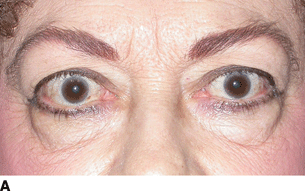
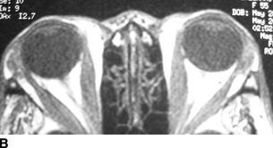
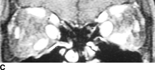
Figure 12.3 A: TED in which this patient has proptosis and eyelid retraction of all four eyelids. B: Axial cut of an MRI demonstrating fusiform enlargement of the medial and lateral rectus muscles is classic for TED. C: Coronal cut of an MRI demonstrates enlargement of multiple rectus in TED. Notice that the medial rectus and the inferior rectus are enlarged on both sides, while the lateral rectus is less involved.
- Lid retraction (hallmark of the disease)
- Dry eyes
- Foreign body sensation
- Photosensitivity
- Exophthalmos
- Lid lag
- Restricted extraocular movement
Acute Phase
- Conjunctival injection
- Chemosis
- Lid edema
- Corneal exposure
- Optic nerve compression (rare)
- Less than 5% suffer from intense pain, diplopia, or vision loss
Hyperthyroidism
Hyperthyroidism is frequent
- Nervousness
- Tremor
- Fatigue
- Heat intolerance
- Diaphoresis
- Palpitations
- Weight loss
- Hair thinning
- Roughly 10% of patients will be euthyroid in presentation
Disease Course
The disease course is self-limited, with an active or acute inflammatory phase lasting 6 months to 2 years. Patients require close observation for the progression to advanced disease in which vision may be acutely threatened, followed by the quiescent or “burnt-out” phase. This phase involves residual fibrosis and scarring that leads to permanent but non-progressive deficits.
Diagnosis
- TED is a clinical diagnosis. Imaging studies and blood abnormalities can support a clinical diagnosis.
- Increased fat lucency, increased amount of orbital fat.
- Extraocular muscle enlargement sparing the insertions and origins (Fig. 12.3B and C).
- Muscle involvement—first inferior rectus, followed by medial, superior, and lateral recti muscles. Oblique muscles are rarely involved. Therefore isolated rectus muscle involvement should suggest other diagnoses such as myositis, or infiltrative diseases like lymphoma.
Imaging
- CT: Helps evaluate bony orbital anatomy for planning surgical interventions. Also helps evaluate the orbital apex in relationship to enlarged rectus muscles.
- MRI: T1 isointense and T2 isointense to slightly hyperintense to muscle. It is useful at assessing optic nerve compression due to thyroid-associated orbitopathy. It can predict response to therapy.
- Low T2 signal corresponds to a more fibrotic form, which is less responsive to therapy.
- Echography: Thickened muscle to medium to high internal reflectivity and an irregular acoustic structure.
Pathology
Early histopathology will show inflammatory cell infiltration, glycosaminoglycan deposition, and increased water content, resulting in variable amounts of extraocular muscle enlargement and orbital adipose tissue expansion. Variable amounts of edema and infiltration with inflammatory round cells.
Tumor necrosis factor-alpha and other proinflammatory cytokines are prominent in orbital tissue and may be involved in the stimulation of glycosaminoglycan synthesis.
Treatment
Thyroid dysfunction should be stabilized. However, normalization of thyroid function is not associated with reversal of eye disease. If active hyperthyroidism is present, the use of antithyroid medications (propylthiouracil and methimazole) is indicated. Radioactive iodine therapy is associated with worsening orbital inflammation and is generally avoided in patients with moderate to severe eye disease. Concomitant use of corticosteroids may prevent exacerbations. Surgical thyroidectomy is not associated with worsening TED, and therefore indicated for hyperthyroidism poorly controlled with medications and in the presence of TED.
Ocular disease is managed with symptomatic therapy until the disease stabilizes. Systematic corticosteroids and/or radiotherapy for acute orbital inflammation and congestion may be of benefit.
Mild Disease
- Intensive ocular lubricants/artificial tears
- Topical corticosteroids for chemosis
Moderate to Severe Disease
- The use of corticosteroids or radiotherapy is controversial.
- Both appear equally effective.
- Use of corticosteroid-sparing agents such as methotrexate after good response with corticosteroids but unable to taper off corticosteroids or having severe side effects from corticosteroid use.
- Eyelid recession, strabismus surgery, or orbital decompression after stabilization to improve function and cosmesis.
- Orbital decompression: urgent indication if TED that continues to progress despite corticosteroid use or irradiation, especially when there is of vision loss (either by corneal exposure or compressive optic neuropathy).
- Correction of residual proptosis, followed by lid or strabismus surgery during the inactive phase, after the disease remains unchanged for greater than 6 months. The order of surgeries should be decompression followed by strabismus surgery and finally eyelid surgery.
Vasculitis
WG is the most common systemic vasculitis affecting the orbit. Other diseases, including Churg-Strauss syndrome, polyarteritis nodosa, and giant cell arteritis, have occasionally been implicated as well. WG is discussed in greater detail in Chapter 15.
Diagnosis
The diagnosis of WG is based on
- Biopsy for granulomatosis and necrotizing inflammation in the respiratory system
- Biopsy verified necrotizing vasculitis in small- to medium-sized vessel or glomerulonephritis or positive ANCA testing (see Chapters 4 and 15)
Imaging in WG-associated orbital disease shows diffuse orbital mass or masses that may be bilateral and may involve the adjacent nasopharynx.
MRI can depict granulomas and better delineate mucosal inflammation and ulceration in the sinuses, nasal cavity, and orbits. Most commonly, extension of sinonasal involvement is associated with osseous destruction.
Differential Diagnosis
- Bacterial and fungal orbital inflammatory infiltration
- Lymphoma of the orbit
- Sarcoidosis
- TED
- Orbital pseudotumor
Treatment
- Medical therapy as discussed in Chapter 19.
- Orbital decompression is indicated in cases of severe orbital inflammation with proptosis, especially in case of optic nerve impairment. Occasionally, the orbital inflammation is refractory to therapy despite successful management of disease elsewhere.
Temporal Arteritis (TA) or Giant Cell Arteritis
TA classically appears in patients of at least 50 years of age with new-onset headache, jaw claudication, scalp tenderness, anterior ischemic optic neuropathy, myalgias, fatigue, and weight loss. Temporal artery tenderness and elevated sedimentation rate are characteristic.
Ocular Manifestations
Neuro-ophthalmic manifestations include posterior ischemic optic neuropathy, central retinal or cilioretinal artery occlusion, and cranial neuropathies. Orbital manifestations include orbital ischemic syndrome and, very rarely, orbital inflammation. Orbital inflammation causes include periocular pain, decreased vision, and proptosis. Reported sites of orbital involvement include the extraocular muscles, optic nerve sheath, and intraconal fat. It is important to highlight the utility of temporal biopsy in the face of an inaccessible orbital mass.
Criteria for the Diagnosis
- Age at disease onset more than 50 years.
- New onset or type of localized head pain.
- Temporal artery tenderness or decreased pulsation.
- ESR >50 mm/hour (Westergren). Elevated CRP and platelet count is also seen. A rule of thumb is a normal ESR is half a patient’s age in males or half the age plus 10 in females.
Biopsy
Temporal artery biopsy should be obtained, with a biopsy specimen at least 20 mm in length. It is conventional, though not strictly necessary, to biopsy the temporal artery on the more clinically affected side of the head. Note that patients are generally started on empiric corticosteroid therapy as soon as TA is suspected, and we feel that 7 days is the longest one may wait before obtaining a biopsy in this situation. Histopathologic findings of TA include vasculitis characterized by any of the following:
- Mononuclear cell infiltration
- Granulomatous inflammation
- Multinucleated giant cells
- Defects or absence of the elastic lamina
- “Skip lesions”—inflammation may be interspersed between noninvolved tissue
We often repeat the biopsy on the contralateral side if the first biopsy was negative and our clinical suspicion is high.
Treatment
If we feel a patient might have TA, we start prednisone or prednisolone 1 mg/kg/day immediately and obtain a biopsy within 7 days. While a positive biopsy establishes the diagnosis incontrovertibly, a marked clinical improvement from prednisone is highly suggestive, and merits either repeat biopsy on the other side or empiric therapy. If the diagnosis of TA is made, definitely or presumptively, we seek rheumatologic consultation for long-term management. Immunosuppression should be continued until normalization of inflammation indices is seen.
Churg-Strauss Syndrome
Churg-Strauss syndrome is characterized by a history of allergies and crescendo asthma, an eosinophilic pulmonary vascular infiltrate, peripheral eosinophilia (>10%), and high serum levels of IgE. It classically affects the lung and paranasal sinuses, but extrapulmonary manifestations, namely, skin, cardiac, and gastrointestinal. Ocular features are unusual.
Orbital involvement of Churg-Strauss syndrome presents with proptosis. In the case of myositis, patients have a red, swollen, tender, muscle insertion. Imaging often demonstrates lacrimal gland and extraocular muscle swelling. The biopsied tissue reveals extravascular infiltration of eosinophils.
Orbital Sarcoidosis
This disease is discussed in detail in Chapter 15. Most cases of orbital sarcoid are discrete orbital masses, usually in the inferior quadrants of the orbit. These are more common in patients with active systemic sarcoidosis. Within the orbit, sarcoidosis can involve the lacrimal gland, soft tissue of the orbit, and the optic nerve (Fig. 12.4A and B). Orbital soft tissue involvement is distinctly uncommon. In one large series, orbital involvement was seen in 1% of patients with ophthalmic manifestations. In two other series, none were reported. Inferior orbit is the most commonly reported in cases with orbital involvement. Extraocular muscle involvement may be seen in association with orbital lesions (especially in cases of diffuse involvement), but solitary muscle enlargement secondary to sarcoidosis is rare with only a few cases reported. It mimics other orbital inflammatory syndromes with acute or subacute inflammation.
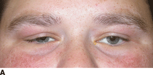
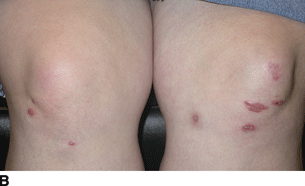
Figure 12.4 A: Right upper eyelid swelling, ptosis, and S-shaped deformity in this patient with systemic sarcoidosis and lacrimal gland involvement. B: Sarcoid skin nodules are present in this patient with systemic sarcoidosis.
Stay updated, free articles. Join our Telegram channel

Full access? Get Clinical Tree


