Fig. 3.1
Goniosynechiolysis technique with the trabectome tip. (a) The footplate (golden oval) is placed anterior to the tip of the peripheral anterior synechiae and gently pushed outward and down. (b) After access to the trabecular meshwork is achieved, the tip is inserted into Schlemm’s canal with a 45° upward and left movement and plasma-mediated ablation is initiated
The ablation is started at 0.8 mW and titrated up. The first 60° can usually be ablated continuously without many adjustments and up to 90° can be achieved by experienced surgeons. The tip is disengaged, rotated downward 180° (away from the cornea), and the ablation is started in the other direction. Again, the goal is to ablate for 90° in both directions. A total of 180° ablation will achieve near 240° of total outflow because a single opening through the TM typically provides access to about 60° of Schlemm’s canal [8]. Once ablation is complete, a viscoadaptive substance can be injected to fill about 20 % of the anterior chamber in order to leave a crescent of tamponading viscoelastic over the ablation arc to minimize postoperative hyphema from flowback. Preservative-free dexamethasone (100 μL, 10 mg/mL) is injected under the viscoelastic shell into the capsular sac. We used the described technique in a consecutive series of 192 patients. After 1 year, 81 % of patients had an IOP ≤ 18, while 52 % of patients had an IOP ≤ 15, and 27 % had an IOP ≤ 12 [9].
All glaucoma medications can be discontinued on the day of surgery. The patient is seen on postoperative day 1, week 1, month 1, 2, and 3. The postoperative regimen for the first week consists of a topical fluoroquinolone, prednisolone acetate 1 %, and pilocarpine 2 %, all of which are started four times daily on the day of surgery. The fluoroquinolone is continued for 7 days and prednisolone is tapered over 2–4 weeks. Pilocarpine can be maintained at four times daily for 4 weeks, then three times per day for 4 more weeks. Flattening the peripheral iris using pilocarpine is thought to reduce the chance of forming peripheral anterior synechiae (PAS) [10].
3.3.2 Goniosynechiolysis
In narrow angles with synechiae, the smooth base plate of the trabectome handpiece serves as an excellent instrument for goniosynechiolysis. Video 3.1 shows an example of goniosynechialysis and trabectome surgery in an eye with two failed tube shunts in a patient who had pressures around 25 mmHg and had a postoperative IOP of about 15 mmHg and needed one medication less at 1-year follow-up. For loosely connected synechiae, gentle posterior and outward pushing and sweeping motions with the base plate is often sufficient to displace them (Fig. 3.1). The pointed tip does not have to be used but more adherent synechiae can also be engaged directly between the tip and the baseplate and gently peeled away. Caution must be exercised to not injure the ciliary body band or create a cyclodialysis. The continuous irrigation under gonioscopic view offers direct visualization of the procedure while debris or blood is flushed away or actively aspirated and the anterior chamber stays well formed and pressurized, minimizing corneal striae.
3.4 Results
3.4.1 Overall Results
The initial clinical description of the trabectome was extended to 101 patients and demonstrated a 40 % IOP decrease [11]. Only 11 of those patients had follow-up for 30 months and that small group sustained a 33 % mean IOP decrease. The published results of trabectome surgery are divided into standalone trabectome cases below in Table 3.1 and combined phaco-trabectome in Table 3.2. Those results show that trabectome can be expected to decrease the IOP by an overall average of 8 mmHg (33 %) while concurrently lowering the number of medications by two. There are no published randomized trials involving trabectome. The largest case series, dating back to the first case, had 1,878 trabectome cases (alone or with phacoemulsification) recorded as of 2010 [24]. There was data on 5 patients who had been evaluated a full 6 years after the procedure and they still maintained a 38 % mean IOP decrease.
Table 3.1
Outcomes for trabectome as the only procedure
Study | Type of study | Number of subjects | Mean baseline IOP (mmHg) | Mean final IOP (mmHg) | % IOP decrease | Mean # decrease in medication | Length of study (months) |
|---|---|---|---|---|---|---|---|
Minckler et al. [2] | Prospective | 37 | 28 | 16 | 45 | 1.1 | 13 |
Minckler et al. [11] | Prospective | 101 | 28 | 16 | 41 | Not reported | 30 |
Minckler et al. [12] | Prospective | 738 | 26 | 18 | 32 | 2.43 | 60 |
POAG of Ting et al. [13] | Prospective | 450 | 26 | 17 | 34 | 0.57 | 12 |
PXG of Ting et al. [13] | Prospective | 67 | 29 | 16 | 44 | 0.88 | 12 |
Jea et al. [14] | Retrospective | 115 | 28 | 17 | 41 | 1 | 30 |
Minckler et al. [15] | Prospective | 1,151 | 26 | 17 | 36 | 1.7 | 60 |
Mosaed et al. [16] | Prospective | 538 | 26 | 17 | 37 | 0.79 | 12 |
Ahuja et al. [17] | Retrospective | 88 | 26 | 13 | 50 | 0.3 | 48 |
Maeda et al. [18] | Prospective | 80 | 27 | 18 | 33 | 1.7 | 12 |
Table 3.2
Combined phaco-trabectome outcomes
Authors | Type of study | Total n | Mean pre-op IOP (mmHg) | Mean final IOP (mmHg) | % IOP decrease | Mean # decrease in medication | Length of study (months) |
|---|---|---|---|---|---|---|---|
Minckler et al. [12] | Retrospective | 366 | 20 | 17 | 18 | 0.93 | 30 |
Francis et al. [19] | Prospective | 304 | 20 | 17 | 17 | 1.22 | 21 |
POAG of Ting et al. [13] | Prospective | 263 | 20 | 16 | 22 | 0.75 | 12 |
PXG of Ting et al. [13] | Prospective | 45 | 22 | 14 | 35 | 0.96 | 12 |
Minckler et al. [15] | Prospective | 681 | 20 | 16 | 21 | 0.87 | 36 |
Mosaed et al. [16] | Prospective | 290 | 20 | 16 | 23 | 0.85 | 12 |
Francis and Winarko [20] | Prospective | 89 | 22 | 15 | 30 | 1 | 12 |
Ahuja et al. [17] | Retrospective | 158 | 19 | 12 | 39 | 0.5 | 45 |
Klamann et al. [21] | Retrospective | 27 | 23 | 14 | 40 | 0.29 | 12 |
Klamann et al. [22] | Retrospective | 74 | 21 | 13 | 37.5 | 0.12 | 6 |
Francis [23] | Prospective | 114 | 22 | 15 | 31 | 1 | 24 |
Studies reporting on the success of the trabectome procedure have used varying definitions of success, making it difficult to compare results. The only common definition of success was final IOP < 21 mmHg with a 20 % decrease in IOP from baseline. For standalone trabectome cases, the success rate using this definition ranged from 65 % [13] to 71 % [13] after 1 year, but then fell as low as 22 % after 2 years [14]. For cases combined with phacoemulsification, the success rate ranged from 87 % [16] to 95 % [20] after 1 year and 80 % at 2 years [23]. A series of 1,127 patients (738 cases of standalone trabectome and 366 cases of phaco-trabectome) found a 45 % success rate after 4 years [12]. The same cohort was extended and analyzed success rates of 1,415 patients [25]. Using a less stringent definition of success as IOP < 21 or a 20 % IOP decrease from baseline, the success rate was approximately 95 % for combined cases after 3 years and approximately 52 % for standalone trabectome cases after 6 years.
3.4.2 Trabectome in Secondary Glaucomas
The realization that AIT with the trabectome works beyond primary open angle glaucoma (POAG) is relatively recent. Consequently, fewer results have been reported for trabectome in secondary glaucomas specifically, although they may be included in outcomes without detailed subtype analysis. In fact, of all the published papers with nonoverlapping cohorts that detail the subtype of glaucoma, POAG alone accounts for two-thirds of the total reported cases. Pseudoexfoliation glaucoma (PXG) is the second largest subgroup and is detailed below. Numerous secondary glaucomas have been treated but in numbers too small for subgroup analysis and often with overlapping pathomechanisms. In order of prevalence in the reported literature of trabectome treatment, they are: pigment dispersion glaucoma, uveitic, steroid-induced, anti-VEGF agent-induced, postsurgical, and traumatic.
It was thought that trabectome may be more effective in PXG because in addition to removing the obstructed and dysfunctional TM, the irrigation, ablation, or resulting mild inflammatory response may help clear pseudoexfoliation material from the anterior chamber [13, 21]. Also, trabectome may deepen the angle, which could decrease iridolenticular touch and lead to decreased exfoliative material release [26]. Combining trabectome with phacoemulsification may be the most meaningful way to avoid the accumulation of pseudoexfoliative material produced primarily by the lens. Further, the combined treatment often allows for bypassing of highly difficult surgeries and complications from zonular dehiscence seen later in the course of the disease while preventing the unexpected, aggressively worsening IOP that may occur in some individuals.
There are three publications that investigated trabectome in secondary glaucomas. The first was a prospective study designed to compare the IOP outcomes in POAG versus PXG [13]. In the group that received standalone trabectome, the PXG group started from a significantly higher baseline IOP, at 29 mmHg, versus 26 mmHg in the POAG group. The mean decrease in IOP at 1 year was better for PXG (44 % versus 34 % in POAG). The success rate at 1 year (defined as a 20 % decrease in IOP from baseline without reoperation) was superior in PXG at 79 % versus 63 % in POAG and the reoperation rate was also significantly lower in the PXG group. In the combined phaco-trabectome group, the baseline IOP of the two groups was statistically similar but the mean decrease in IOP was again superior for PXG at 35 % versus 22 % for POAG. The complication rates were non-visually threatening and similar in both groups.
The second, retrospective study investigated PXG only [21]. The study compared the results of 27 phaco-trabectomes to 28 cases of phacoemulsification combined with trabecular aspiration. The baseline IOP was statistically equivalent at 23 mmHg in the phaco-trabectome group and 22 mmHg in the phaco-aspiration group. After 1 year, the IOP was 40 % lower in the phaco-trabectome group versus 23 % after phaco-aspiration. These outcomes are difficult to compare to other studies because according to the author’s protocol, all of the patients had a target IOP below 16 mmHg and medications were used as needed to maintain the target IOP, even in the postoperative period. Strict adherence to this protocol would explain why there was no statistically significant decrease in medication in either group after 1 year.
The third study aimed to prospectively examine the outcome of trabectome in different types of glaucoma, although POAG alone still accounted for 45 % of the 557 cases [27]. The second largest subgroup was PXG, accounting for 30 % of cases, and was the only subtype besides POAG large enough for subgroup analysis with a short follow-up time of 7 months. Results were reported combining standalone trabectome with phaco-trabectome. The mean IOP in the POAG group decreased by 25 % from a baseline of 24 mmHg versus a 30 % decrease from a baseline of 25 mmHg in the PXG group. Although the final difference in IOP in PXG was not large (final IOP 17.6 mmHg in PXG versus 18.2 in POAG), the success rates were considerably higher in PXG. Defining success as a 20 % decrease in IOP, the rate after 2 years was 50 % for PXG and only 32 % for POAG. A fourth study found in their risk factor for failure analysis, that PXG was protective when compared to POAG since the hazard ratio was 0.43 [28].
However, there are several weaknesses in concluding that trabectome is more effective in PXG as compared to POAG. First, the number of studies is small. Second, in both comparative studies conducted to-date [13, 27], the POAG group had worse IOP outcomes and started from a lower baseline IOP. Lower baseline IOP has been consistently shown to have worse IOP outcomes with trabectome, including in multivariable analysis [14, 19, 29]. The lower baseline IOP in the POAG group would then be expected to have worse outcomes on that basis alone. Finally, in cases of pseudoexfoliation, simply increasing the volume of irrigation has been shown to be associated with a lower IOP for as long as 2 years [26]. Further, the possible IOP-lowering effect of washing out the exfoliation material (as may occur with almost any intraocular surgery) cannot be easily separated from the effects of TM ablation alone. Pigment dispersion glaucoma can continuously produce dispersed pigment until the burn-out phase (after an average of 10 years) [30] or resolution of the reverse pupillary block [31, 32]. We find that pigment dispersion glaucoma can be treated effectively, but patients have to be prepared to experience several months without compelling IOP reduction when massive pigment is liberated at the time of surgery. This freed material can obstruct collector intakes, but typically resolves eventually. Pigment dispersion eyes often have a high axial length that can lead to displacement of refluxed blood into the posterior chamber during abnormal iris lens diaphragm movement. It is plausible that POAG has a considerable outflow resistance component within the distal outflow tract [33], while PXG and possibly other types like steroid-induced glaucoma [34] primarily have a TM component and, therefore, have better results with ablation of the TM.
Several abstracts have also investigated the use of trabectome in secondary glaucomas. A small case series followed 15 eyes with complex glaucoma for one year after treatment with trabectome [35]. The IOP in five cases of retinal detachment repaired with scleral buckle fell 52 % from 31 ± 10 mmHg with a reduction of medications by 3.1. Three traumatic cases had a 55 % IOP decrease from a baseline of 32 ± 9 mmHg on four fewer medications. In three cases with inactive, regressed neovascularization of the iris and angle after panretinal photocoagulation, the IOP fell 37 % from 46 ± 12 mmHg on 2.3 fewer medications, and the IOP of three cases of uveitic glaucoma decreased 60 % from a baseline of 30 ± 6 mmHg on 2.4 fewer medications. Only one patient (7 %) needed further surgery within 1 year and received cyclophotocoagulation. No other complications or vision loss occurred. A case series of 61 patients with uveitic glaucoma found a 32 % decrease from a baseline IOP of 32 mmHg after 1 year on 0.5 fewer medications with a 26 % reoperation rate [36]. Another abstract analyzed 12 cases of isolated anterior uveitis over 2 months and found that the IOP fell 43 % from a baseline of 31 mmHg on two fewer medications [37]. A small series of 15 cases of steroid-induced glaucoma found that IOP fell 47 % from a baseline of 31 mmHg on 1.5 fewer medications after 1 year [38].
3.4.3 Trabectome in Glaucomas with Narrow Angle as an Atypical Indication
A narrow anterior chamber angle has, in the past, been considered a relative contraindication because it was feared that in addition to poor visualization of the target structure during surgery, PAS, descemetization of the angle, and fibrosis may form more readily and hasten failure [39]. This has prevented a large number of glaucoma patients from taking advantage of the highly favorable risk profile of trabectome surgery compared to traditional filtering glaucoma surgery as angle-closure glaucoma contributes to approximately 70 % of glaucoma cases in women and to 87 % of cases in Asians [40].
In our prospective study of trabectome combined with phacoemulsification and trabectome-only in patients with narrow angles of Shaffer grade ≤2 (SG ≤ 2) versus open angles with a Shaffer grade ≥3 (SG ≥ 3), we analyzed outcomes that included IOP, medications, complications, secondary surgery, and success (IOP < 21 mmHg and >20 % reduction without further surgery) [41]. Of 671 included cases with at least 1 year of follow-up, AIT patients with SG ≤ 2 (n = 43) had an IOP reduction of 42 % from 27.3 ± 7.4 to 15.7 ± 3.0 mmHg (p < 0.01) versus AIT SG ≥ 3 (n = 271) with an IOP reduction of 37 % from 26.1 ± 7.8 mmHg to 16.4 ± 3.9 (p < 0.01). In phaco-AIT with SG ≤ 2 (n = 48), IOP was reduced 24 % from 20.7 ± 7.0 mmHg to 15.7 ± 3.6 (p < 0.01) versus phaco-AIT with SG ≥ 3 (n = 309) with an IOP reduction of 25 % from 22.6 ± 6.4 mmHg to 17.0 ± 3.4 (p < 0.01). There was no significant difference between SG ≤ 2 and SG ≥ 3 in reduction of IOP or medications, complications, secondary surgery, and success rates (Fig. 3.2, p > 0.05).
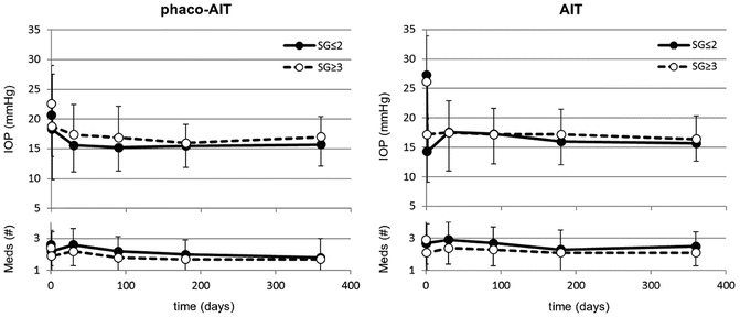
Fig. 3.2
In phaco-AIT (trabectome combined with phacoemulsification, left), no significant differences were found between IOP and glaucoma medication use in patients with a narrow anterior chamber angle (SG ≤ 2, n = 48) compared to those with an open angle (SG ≥ 3, n = 309) over a 1-year period. Similarly, in AIT (trabectome, right), no significant differences were found in IOP and glaucoma medication use between patients with a narrow anterior chamber angle (SG ≤ 2, n = 43) and a wide angle (SG ≥ 3, n = 271) during 1-year period (all p > 0.05)
These results indicate that both phaco-trabectome and standalone trabectome can significantly reduce the IOP and number of medications regardless of degree of angle opening. This suggests that indications for trabectome should be expanded to include narrow angles. The results show that trabectome can be considered in the relatively large population of patients with narrow angles who might have mixed mechanisms that lead to increased IOP. Phacoemulsification in itself can decrease IOP by 1.5–3 mmHg [42–44], via decompression or mechanical stretch of the TM and Schlemm’s canal [45] or activation of a stress response pathway by ultrasound [46]. However, after TM ablation, this mechanism would not achieve additional IOP reduction via these mechanisms, disaffirming cataract surgery as a significant contributor.
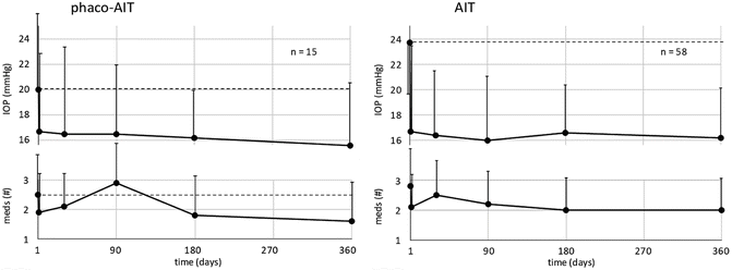
3.4.4 Trabectome as a Secondary Procedure After Failed Trabeculectomy
Trabectome can be successfully used in cases following failed incisional surgery. We analyzed 73 patients with a minimum of 1 year of follow-up who were treated with trabectome following failed trabeculectomy (Fig. 3.3) [47]. In the standalone trabectome group, the mean IOP was reduced from 23.7 ± 5.5 mmHg to 16.2 ± 3.9 mmHg (28 % mean reduction, p < 0.01) on 0.8 fewer medications (p < 0.01) at 1 year. In the phaco-trabectome group, the mean IOP was reduced from 20.0 ± 5.9 mmHg to 15.6 ± 5.1 mmHg (19 % mean reduction, p = 0.11) on 0.9 fewer medications (p = 0.24). While 89 % of standalone trabectome and 92 % of phaco-trabectome cases finished with an IOP < 21 mmHg, defining success as IOP < 21 mmHg with a 20 % IOP decrease meant only 62 % of patients in both groups were successful at 1 year. There were no visually threatening complications. A recent abstract reported on 24 patients who had trabectome after failed aqueous shunt surgery [48]. The IOP fell 30 % from a baseline of 23 mmHg on 0.8 fewer medications after 1 year. 83 % of patients maintained an IOP < 21 mmHg with a 20 % decrease at 1 year. There were no visually threatening complications.