Fig. 15.1
Anterior segment OCT showing malignant glaucoma with anterior displacement of IOL and anterior chamber shallowing (top) and resolution of malignant glaucoma and deepening of anterior chamber after treatment in the same eye (bottom)
Malignant glaucoma is a diagnosis of exclusion and it is important to rule out other causes of postoperative anterior chamber shallowing, including incision leakage, pupil block, capsular block syndrome, plateau iris, ciliary body cysts, zonulopathy-induced lens subluxation, and supraciliary or choroidal effusion/hemorrhage [3].
In some cases, malignant glaucoma may present intraoperatively and manifest as acute and marked anterior migration of capsule, lens, and iris following an intraoperative anterior chamber shallowing or decompression event. Iris often prolapses and it may not be possible to reposit despite decompressing the anterior chamber. The condition does not improve with usual maneuvers done for “positive pressure” such as relieving tension on the lid speculum or encouraging the patient to avoid valsalva/strain. As well, unlike intraoperative suprachoroidal hemorrhage, pain is not a hallmark of this condition.
15.3 Pathophysiology
Decompression, or sudden drop in IOP, is thought to be the inciting event for malignant glaucoma. It is usually accompanied by anterior chamber shallowing. During surgery, decompression may occur during incision creation, while moving instruments in and out of the eye particularly larger ones like hand-pieces for phacoemulsification (phaco) or irrigation-aspiration, when creating openings for glaucoma filtration surgery or during wound closure if tissue apposition is insufficient. Following surgery, decompression can occur due to wound leak or excessive filtration from glaucoma surgery.
Reduced IOP from decompression may result in choroidal expansion as increased blood volume rushes into the potential vascular space. The choroid covers a large surface area and a small increase in its thickness can add significant volume to the intraocular contents pushing other structures anteriorly. Normally, aqueous outflow either via trans-scleral drainage or anteriorly through the trabecular meshwork would compensate for this event. However, this may not occur in the susceptible eye due to a combination of factors. These include a reduced ability for fluid to transmit through vitreous due to poor conductivity, limited lens-iris channel area for anterior migration of aqueous, and reduced trans-scleral outflow due to thicker sclera. In eyes with these characteristics, a decompression event may result in anterior lens-iris movement and significant anterior chamber shallowing [4].
15.4 Prevention
The risk of malignant glaucoma may be reduced, though not eliminated, by avoiding decompression during surgery.
During cataract surgery, when withdrawing the phaco hand piece after lens removal or when withdrawing the irrigation-aspiration hand piece after cortex or viscoelastic removal, the anterior chamber often shallows, signaling decompression. To avoid this, one can inject balanced salt solution (BSS) or viscoelastic through the paracentesis to maintain the anterior chamber pressure. The choice between BSS or viscoelastic would depend on the next step planned in surgery. Typically, the authors would inject BSS following phacoemulsification to prepare for cortex removal, viscoelastic following cortex removal to prepare for lens insertion, and BSS after viscoelastic removal to prepare for wound hydration. To reduce tremor, the surgeon can depress the plunger using the index finger while holding the syringe between the thumb and middle finger, rather than the traditional method of depressing the plunger with one’s thumb. This technique takes practice and one can do so on routine cases so that the skill is honed for use when needed.
During filtration surgery, decompression can occur when creating an ostium as well as during closure. Viscoelastic can be used in the anterior chamber during ostium creation to avoid shallowing. As well, during closure of a trabeculectomy flap, periods of excessive aqueous egress can occur resulting in anterior chamber shallowing. With the microscope centered on the flap and the surgeon’s attention focused on suturing, this may go unnoticed. By periodically checking the anterior chamber status during this step, one can inject BSS or viscoelastic to help avoid excessive shallowing and decompression. Similarly, during tube shunt entry and manipulations, care in avoiding excessive anterior chamber shallowing may help reduce the risk of subsequent malignant glaucoma.
15.5 Medical Treatment
Definitive treatment for malignant glaucoma aims to restore the normal relationship between the anterior and posterior segments by establishing a unicameral eye. This can be achieved through a stepwise approach which begins with less invasive approaches and reserves more invasive strategies only for cases that do not resolve with earlier steps (Fig. 15.2).
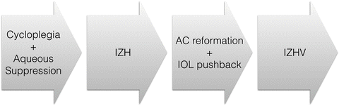
Fig. 15.2
Stepwise treatment approach beginning with medical therapy using cycloplegia and aqueous suppressants, then adding iridozonulohyaloidotomy (IZH) followed by anterior chamber (AC) reformation and IOL pushback and ultimately moving to iridozonulohyaloidotomy with vitrectomy in those cases that do not resolve with the previous steps
Medical management consists of cycloplegics to help pull the lens-iris diaphragm posteriorly and aqueous suppressants to reduce intraocular volume as well as control pressure. Although traditionally this has been reported to be successful in up to 50 % of patients [7], more recent reports suggest this number may be as low as 10–14 % [8, 9]. Furthermore, since medical management does not address the underlying anatomic issue, cycloplegia often cannot be discontinued [2], suggesting a role for medications to help control but not necessarily cure malignant glaucoma.
15.6 Surgical Treatment
15.6.1 Iridozonulohyaloidotomy
Iridozonulohyaloidotomy (IZH) aims to create a channel for aqueous to pass through the iris, zonules, and anterior hyaloid effectively allowing aqueous to pass freely between the posterior and anterior segments. This is intended to equalize pressure between the two segments of the eye and if successful should result in a deepening of the anterior chamber within 24 h [10]. IZH can resolve malignant glaucoma in 35 % of cases [9].
An IZH may be performed using a YAG laser aimed at iris peripheral to the lens. A preexisting iridotomy is often a good starting point for this procedure and settings would mirror those used for a YAG laser iridotomy. Once through the iris, the laser is focused posteriorly to penetrate the zonules and hyaloid respectively. If necessary, the aperture can be enlarged to ensure adequate visualization of deeper structures. Often in malignant glaucoma, because the vitreous is compressed anteriorly, an IZH may pass through both anterior and posterior hyaloid.
In pseudophakic eyes with widely dilated pupils, one can aim the laser through anterior and posterior capsule leaflets peripheral to the lens edge rather than through iris performing a capsulohyaloidotomy rather than the traditional IZH.
15.6.2 Anterior Chamber Reformation and Intraocular Lens Pushback
In pseudophakic patients, once a patent IZH has been created, if malignant glaucoma persists or cycloplegia cannot be discontinued, it may help to reform the anterior chamber (AC) with viscoelastic and manually push the intraocular lens (IOL) posteriorly. The surgeon may note a palpable sense as anteriorly rotated ciliary processes “pop” back into place. As well, the posterior movement of the IOL can force fluid trapped in the posterior segment to migrate through the patent IZH into the anterior segment. This technique may resolve a further 30 % of malignant glaucoma cases [9].
This procedure can be done at the slit lamp or in an operating suite depending on the surgeon’s comfort level. When performing this at the slit lamp, it is advisable to use iodine as well as a lid speculum to ensure sterility. A 30-gauge needle is placed on a viscoelastic syringe and introduced into the anterior chamber at the limbus. Viscoelastic is injected to maintain the anterior chamber and within this stable environment, the needle can be advanced over the IOL optic avoiding the visual axis. The needle is used to push the IOL posteriorly.
Stay updated, free articles. Join our Telegram channel

Full access? Get Clinical Tree


