Fig. 13.1
The drops and gel to be applied before needling, proparacaine, apraclonidine, povidone iodide, and 3.5 % xylocaine gel
The half-inch 30 g needle, mounted on a TB syringe, is bent into a “Z” shape (Fig. 13.2) [16]. This is done by holding the syringe upright with the needle bevel facing you, grasping the needle 3 mm from the hub with a blade-breaker or needle driver, bending the needle backwards at the needle hub and then twisting it forwards where it is being held by the blade-breaker. This makes an instrument that is held comfortably with the thumb and second and third fingers, and in which the tip of the needle can approach the eye parallel to the corneal surface.
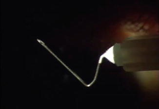
Fig. 13.2
Bent 30-gauge needle mounted on a tuberculin syringe
After the patient has been seated comfortably at the slit lamp (sitting upright with good support of the chin and forehead) a solid bladed lid speculum is placed to isolate the lashes. The surgeon likewise must be seated comfortably in an upright position, have the elbows resting on either the slit lamp table or elbow supports, and the hand holding the TB syringe with needle stabilized by contact of the fourth and fifth fingers with the patient’s cheek.
The needle is held over its intended path to be sure that there is sufficient length beyond the distal bend to pass through the internal ostium, out through the episcleral margin of the scleral wound and to the outer margins of the bleb (in case needling of the bleb itself is required) (Fig. 13.3).
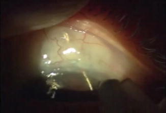
Fig. 13.3
Checking that the final portion of the needle will reach the bleb margins
The cornea is entered parallel to the surface and passed vertically and somewhat obliquely (so that the needle entry is not in the visual axis) aimed towards the internal ostium, with the bevel up, and until just the bevel is within the cornea. Then the syringe is tilted up so that the needle tip passes on a somewhat more perpendicular path to enter the anterior chamber. This is done so that the path in the cornea is not so long as to impede side to side and up and down manipulations of the needle tip. The needle is then advanced slowly by gentle wiggling of the syringe, so that it does not plunge too rapidly forwards. The needle tip must then pass through the internal wound of the trabeculectomy. This region is determined preoperatively with gonioscopy and noting any landmarks, such as an iridectomy or iris details. The needle should slide without significant resistance through to the scleral surface. This may require some gentle probing with retreat if resistance is encountered. One does not want to penetrate the sclera outside of the path of the trabeculectomy, as that does not restore aqueous flow and can produce pain and bleeding. The procedure is far easier to perform in the eyes in which a short scleral tunnel or short scleral flap, with a wide internal entry, had been used in the trabeculectomy procedure [16].
Once the needle advances through the episcleral tissue to reach the interior of the bleb, the bleb may expand dramatically and that would indicate that the major unwanted resistance has been eliminated and that one may withdraw the needle. However, if the bleb does not expand, the needle may be advanced along the scleral surface to penetrate Tenons adhesions at the bleb margin, and the side of the 30 g needle used to slice such adhesions in one or a few places, until the bleb expands in size beyond its original margins.
13.4 Advantages of the Ab-interno Technique
There are several advantages of this procedure over trans-conjunctival needling, especially if the latter is done in an outpatient or hospital operating room. First, there are logistic advantages, as it is performed in the clinic at the time the patient is seen, with little cost and inconvenience, since no scheduling of the procedure and no hospital or outpatient facility paperwork and expense are involved. This is particularly advantageous to the practitioner within the global period, in which any postoperative interventions are not reimbursed, and in which returns to the hospital within the global period may be monitored. Second are the psychological advantages, in that one can explain to the patient that the operation they recently had has not “failed”, but just needs adjustment, and deal with any anxiety by promptly dealing with the problem, rather than have it weigh on their mind until a revision could be scheduled and performed. Third, the procedure avoids several risks of trans-conjunctival needling.
With trans-corneal needling, the only entry into the eye is a self-sealing 30 g needle entry. To achieve a self-sealing wound, it is important that the bevel be parallel to the cornea so that a tunnel, and not a groove, is made. It is also important that the needle path be sufficiently long, and this is assured by having the bevel of the needle fully in the cornea before turning the needle tip more perpendicularly for entry into the anterior chamber. In contrast, a trans-conjunctival needling by definition makes a hole in the conjunctiva, which can cause hypotony and can serve as an entry site for bacteria. For that reason, trans-conjunctival needling entry sites must always be Seidel tested and closed, if positive, with a suture or cautery.
Stay updated, free articles. Join our Telegram channel

Full access? Get Clinical Tree


