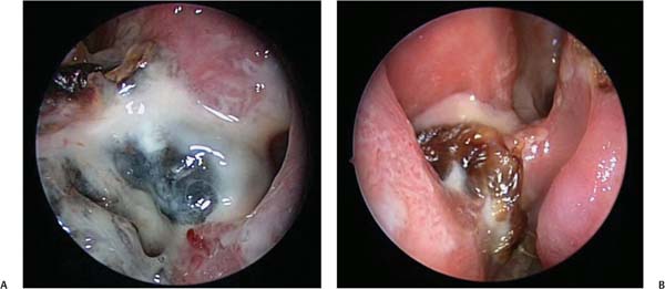
28 Three Difficult Cases in the Management of Maxillary Sinus Disease The maxillary sinus, as we have seen in this book, is without a doubt the most difficult sinus to cure medically or surgically. The endoscopic sinus surgeon can perform a flawless endoscopic antrostomy and yet fail to improve the diseased state of the sinus. Similarly, the endoscopic sinus surgeon can revise a scarred maxillary antrostomy and fail to improve the ability of the sinus to drain properly. Finally, the revision endoscopic sinus surgeon can remove the diseased lining in a failed maxillary antrostomy case and still find that the postoperative maxillary sinus is in no way improved than its preoperative state. Our intent in this concluding chapter is to present three cases of maxillary sinusitis, in which the patients have failed to improve their symptoms following attempted surgical treatment. The authors of previous chapters have elucidated how to perform a maxillary antrostomy successfully. They have also illustrated how to identify and correct technical reasons for failure. In this chapter, we would like to discuss the most difficult cases that we have encountered in the previous 23 years at Vanderbilt University Medical Center (Nashville, TN). The following discussion assumes that the patient has undergone a previous maxillary antrostomy, which has failed, but that no abnormalities in the surgical technique for the antrostomy have been found. Before presenting you with the three cases, we will outline the treatment philosophy that has been used to evaluate and treat our maxillary sinus patients: • The first treatment point is to adequately manage the patient medically, once maxillary sinusitis has been diagnosed via computed tomography (CT) scan or directly by nasal endoscopy. In my practice, adequate management includes the use of systemic steroids for a minimum of 10 days and oral antibiotics for a minimum of 3 weeks. All attempts are made to obtain a culture endoscopically, from the middle meatus or directly from the maxillary sinus. • A second point concerns the management of biofilms. In the case of an isolated maxillary sinus infection, irrigation via the maxillary antrostomy site is utilized to break up and flush out the infected materials. All irrigations are cultured. If Staphylococcus is isolated on culture, mupirocin is instilled directly into the maxillary sinus and the patient is placed on culture-directed antibiotics. Further treatments of the biofilms are initiated by weekly office visits to irrigate and instill mupirocin. • If bacteria are isolated on culture that is not amenable to oral antibiotics, either due to allergy or to the lack of a suitable oral antibiotic, an infectious disease consult is obtained. Once the plan for intravenous (IV) antibiotics is initiated, plans are made either to see the patient weekly for biofilm treatment, or to see the patient at 3 weeks to evaluate the effectiveness of the IV antibiotic. If at 3 weeks there is observable improvement, then the patient proceeds on to a complete course of IV treatment, usually lasting 6 weeks. In our experience, one of two results will occur with the treatments outlined above. The patient will improve and become healthy, or the sinus will not improve and the patient will relapse. The usual case is that the sinus will relapse quickly after the weekly treatments or after the cessation of the IV antibiotics. The patients in our three cases were all treated in the manner just described. When the maxillary sinus fails to improve, our diagnosis is failure of the ciliated respiratory epithelium to regenerate. Given that the maxillary sinus must drain against gravity, an intact ciliary clearance mechanism must be present and functional. The data we have used to guide our management of the failed cilia are based on our experience with the use of Caldwell–Luc surgery to manage this group of patients. Our review of this group of maxillary sinus patients found a 93% success rate to manage the maxillary sinus after removal of the maxillary sinus lining via a Caldwell–Luc approach.1 The first case is a 67-year-old man with a chief complaint of chronic nasal crusting and nasal drainage. He had a history of two previous sinus surgeries and was on Coumadin following an aortic valve replacement. Nasal endoscopy revealed metaplastic changes in both maxillary sinuses. In addition, the nasal cavities were filled with crust and purulent material. A CT scan of the sinuses revealed bilateral maxillary sinus opacification. A culture with sensitivities was obtained from both maxillary sinuses. The patient was then treated with numerous culture-directed antibiotics without improvement. The recommended plan of treatment was a bilateral Caldwell-Luc operation. The clinical findings at the time of surgery revealed avascular bone along the medial wall of the left maxillary sinus and purulent debris within both maxillary sinuses. The sinus mucosa was also found to be edematous. Microscopically, the left maxillary sinus lining was found to be polypoid and edematous with markedly inflamed respiratory mucosa and submucosa with severe chronic inflammation and mild acute inflammation. There were areas of necrotic inflammatory debris, fragmented fungal hyphal forms, and benign reactive bone. On the right side, the mucosa was likewise found to be polypoid and the submucosa also revealed severe chronic and mild acute inflammation, along with fragments of benign bone, but no definite fungal organisms. Part of the pathology report stated that “the mucosa exhibits relatively prominent plasmacytic and lymphocytic infiltrates. A few areas of ulceration and acute inflammation are present.” The follow-up at one month noted no signs of infection in either maxillary sinus. The patient returned at 2 months with complaints of globus sensation and postnasal drip. The endoscopic findings were crusting in the bilateral maxillary sinuses. Topical Pulmicort (AstraZeneca, Pharmaceuticals, LP, London, UK) was instituted. The patient returned 2 months later with complaint of hoarseness and nasal crusting. A culture was taken, the Pulmicort was stopped, and Bactroban (GlaxoSmithKline, Mississauga, Ontario, Canada) was started topically. The culture was positive for Klebsiella. Appropriate antibiotic management was initiated. The patient returned 3 months later. His complaint at that time was persistent postnasal drip. Physical exam revealed scarring of the bilateral maxillary sinuses. A CT scan of the sinuses was obtained. It noted significant opacification within the bilateral maxillary sinuses. Given the CT findings, exploratory surgery was recommended with a possibility of bilateral Caldwell–Luc approaches. One month later the patient was taken to the operating room. The surgical findings were a small area of loculated purulence within the right maxillary sinus and no purulence within the left maxillary sinus (Fig. 28.1). The postoperative visit at one week found the patient well healed and much improved. He was continued on Flonase (GlaxoSmithKline, Mississauga, Ontario, Canada) and Bactroban topically. The patient was followed regularly and was taken back to the operating room about a year later for continued chronic maxillary sinusitis unresponsive to medical management. He underwent bilateral revision endoscopic maxillary antrostomies with tissue removal and bilateral maxillary sinoscopies. Intraoperative findings at that time included loculated areas of purulence involving the right maxillary and no loculated areas on the left, but scarring of the antrostomies bilaterally. He returned to the operating room about 4 months later for nasal endoscopy given the persistent opacification of the bilateral maxillary sinuses. The findings at that time included scarring of left and right maxillary openings and his maxillary sinuses were filled with scar tissue. No pockets of purulent material were noted. The patient continues his use of Flonase and Bactroban topically. He continues to be followed regularly in our clinic and has had no new problems. This case illustrates the following important points: 1. The maxillary sinus respiratory lining can become irreversibly damaged as exemplified by the first pathology report. The treatment of the diseased lining in these cases may require its removal. 2. Once the diseased lining is removed, the assessment of the status of the sinus may be limited on endoscopic exam. The use of the CT scan may also be limited due to the development of scarring that can obliterate the interior of the maxillary sinus. The inability to assess the sinus in the office may require a second look in the operating room. 3. The need to get pre- and postoperative CT scans. Fig. 28.1 (A,B) Intraoperative findings looking into the patient’s left maxillary sinus with a 30-degree endoscope. The second case is a 43-year-old woman who presented with recurrent episodes of maxillary sinusitis causing foul-smelling nasal drainage. She had undergone a previous endoscopic sinus surgery with bilateral maxillary antrostomies in January of 1999, and a septoplasty in 1988. The endoscopic exam revealed scarring at both maxillary antrostomies. Cultures were obtained in clinic, but were nondiagnostic. A sinus CT scan was read as bilateral maxillary mucopurulent thickening, as well as opacification in the frontal sinuses and left ethmoid cells. The patient was taken to the operating room on December 31, 2001, and underwent correction of the scarring at both maxillary antrostomies. Scar tissue was found between the natural maxillary ostium and the surgical ostium. Pathologically, the specimen was unremarkable, only revealing chronic inflammation. The patient continued to have recurrent episodes of maxillary sinusitis, which were culture-positive for Staphylococcus. She underwent numerous treatments with antibiotics and endoscopic debridement of the bilateral maxillary sinuses. Her maxillary sinus cultures began to grow Serratia along with Staphylococcus. After failure of numerous antibiotics, bilateral Caldwell–Luc operations were performed on March 16, 2006. Crusting, edema, and purulent debris were found in both maxillary sinuses. The pathology report found respiratory-lined mucosa with marked edema, acute and chronic inflammation, and occasional eosinophils (~5 per high-powered field). The culture was positive for Serratia. The patient eventually improved over the course of one year. She then relapsed with recurrent maxillary sinus infection, which was culture-positive for Pseudomonas. Oral antibiotics and debridements failed to control the infection. The infectious disease team was consulted and the patient was started on 6 weeks of IV antibiotics with weekly debridements. The medical management failed to control the maxillary sinusitis and the symptoms of malodorous nasal drainage. A second Caldwell–Luc procedure was performed on the left maxillary sinus on September 15, 2008. The findings were very hyperplastic, edematous membrane with an abscess pocket in the medial inferior aspect of the sinus. Pseudomonas was found on culture. The pathology report found respiratory mucosa with granulation tissue and chronic inflammation, but no increase in eosinophils (see Video 28.1). The patient’s postoperative clinical course was remarkable for persistent pseudomonal maxillary sinusitis. Immunologic and genetic testing revealed that her immunoglobulin levels were within normal limits (immunoglobulin G [IGG] level was 1131 mg/dL with reference range of 694–1618, IGA level was 202 mg/dL with reference range of 68–378, and IGE level was 44 IU/mL with reference range of 0–99). Cystic fibrosis testing revealed that the patient was a carrier of the ΔF508 mutation, detected in one allele (Poly dT tract in intron 8–5/9). Her seat chloride test was 32 mEq/L. (Reference range: less than 40 mEq/L sweat chloride = normal; 40–60 mEq/L sweat chloride = borderline; greater than 60 mEq/L sweat chloride = abnormal.) The third patient is a 76-year-old man who presented with a chief complaint of odor from his nose along with crusting and nasal congestion. His history was remarkable for three previous sinus surgeries, and type II diabetes. Nasal endoscopy revealed marked mucosal edema involving both maxillary sinuses with a question of scarring at both natural maxillary sinus openings. There was a large amount of nasal crusting. A CT scan found circumferential moderate mucosal thickening within the bilateral maxillary sinuses. A culture was taken from the maxillary sinuses and reported light growth of upper respiratory bacteria. He was started on ciprofloxacin and scheduled for surgery for possible Caldwell–Luc procedures. The patient was taken to the operating room approximately one month later and found to have purulence in the maxillary sinuses with discrete abscesses within the maxillary sinus cavities, loculated purulence in the posterior ethmoid sinuses, and purulence in the right sphenoid. The patient underwent removal of scarring at both maxillary sinus openings and removal of diseased maxillary mucosa via a sinoscopy approach. A culture taken during surgery found a light growth of coagulase-positive Staphylococcus. The pathology report found respiratory mucosa with chronic inflammation and up to 20 eosinophils per high-power field. Postoperatively, the patient did not show improvement in his maxillary sinus disease. Approximately 2 months later, he underwent bilateral Caldwell–Luc procedures. The surgical findings were massive edema and purulence pus within the maxillary sinuses. Intraoperative culture was again positive for heavy growth of coagulase-positive Staphylococcus. The pathology report found edematous respiratory mucosa and submucosa with extensive lymphoplasmacytic inflammation. An unofficial hematology consult revealed that the report findings were consistent with chronic inflammation. Since the last operative intervention, the patient has had weekly maxillary sinus debridements and is using a saline nasal wash. The crusting and odor have diminished and although they are still present, they no longer affect the patient’s quality of life. The maxillary sinus lining continues to be swollen and inflamed. A sweat chloride was performed and found to be 53 mEq/L. (Reference range: less than 40 mEq/L sweat chloride = normal; 40–60 mEq/L sweat chlo-ride = borderline.) The patient is scheduled for genetic testing to determine if he has a variant of cystic fibrosis. The two patients discussed in Cases 2 and 3 have required multiple medical and surgical treatments of their maxillary sinus disease as a result of unhealthy maxillary sinus mucosa. They both show evidence of a symptomatic carrier state for the cystic fibrosis (CF) gene. The patient in Case 2 has been shown to be a carrier for ΔF508 mutation, which is the most common CF mutation worldwide, although she had a sweat chloride test within normal limits. Conversely, the patient discussed in Case 3 had a borderline sweat chloride test, although thus far genetic testing has not revealed him to carry a known CF mutation. Furthermore, this patient’s pathology report noted up to 20 eosinophils per high-power field, a known marker for inflammation. Studies have shown that chronic rhinosinusitis (CRS) patients with hypereosinophilia have a worse prognosis when compared with controls.2 Treatment paradigms for hypereosinophilic CRS have shifted to involve control of the inflammation and away from antimicrobial and surgical treatments.3 Cystic fibrosis has been known to be associated with CRS, but the story does not stop there.4 The majority of patients with CF suffer from CRS and 20% of patients will eventually require surgical treatment of their sinuses.5 More recent data suggest that there is a higher prevalence of CRS in carriers for the CF gene as compared with the general population. More specifically, 36% of CF carriers were noted to have signs and symptoms of CF as compared with 13 to 14% in the general population.6 The patient discussed in Case 2 is known to be a carrier for the CF gene. Furthermore, the bacteriology of the maxillary sinusitis in this patient is characteristic for that found in airway infections of CF. Staphylococcus aureus and Pseudomonas aeruginosa have long been considered of primary importance with respect to virulence.7 This would explain the propensity for developing CRS in the patient in Case 2 as well as the difficulty in managing her maxillary sinus disease. The difficulty in establishing the existence of a CF mutation in the patient discussed in Case 3, in the setting of difficult to treat sinus disease and intermediate sweat chloride test, is not surprising when one bears in mind that there are more than 1600 CF mutations.8 Mild cystic fibrosis without pulmonary symptoms has also been previously described with some CF mutations. There are many cases reported in the literature of patients with adult-onset or atypical presentations of CF.9–11 This may delay confirmation of the diagnosis in a patient with a borderline sweat chloride test as studies have shown that a borderline sweat chloride test can be associated with a CF phenotype or it can exist in patients who do not exhibit the CF phenotype.12 Some authors advocate looking for CF and ciliary dyskinesia mutations in all patients displaying signs and symptoms of atypical or severe and persistent CRS.13 A borderline sweat chloride test can therefore be thought of as predictor of CF, but not the basis for a definitive diagnosis. Maxillary sinusitis and sinusitis in general have become common clinical entities affecting ~15% of adults.14 Patent ostia, mucus of the proper viscosity, and actively beating cilia are necessary to protect against infection of the paranasal sinuses. Sinusitis that does not respond to appropriate medical therapy is often treated surgically.15 Underlying disease processes that affect the upper airway such as asthma, cystic fibrosis, ciliary dyskinesia, and allergic rhinitis complicate the treatment of maxillary sinusitis, both medically and surgically.16,17 Some authors have suggested that patients with chronically diseased maxillary sinuses and poor mucociliary clearance from long-standing inflammation or scarring from previous surgery, may benefit from an endoscopic maxillary mega-antrostomy and avoid persistent sinus mucosa stripping.18 Others have suggested that endoscopic revision of the maxillary sinus yields comparable outcomes to a repeat Caldwell–Luc procedure in patients with a history of previous failed Caldwell–Luc surgery, and thus, endoscopic revision surgery is a viable alternative for surgical rehabilitation of the post-Caldwell–Luc maxillary sinus.19 The authors believe that even though endoscopic sinus surgery techniques have proven to be safe and effective in the vast majority of patients requiring surgical management, the Caldwell-Luc procedure is safe and effective as described, and should remain in the repertoire of surgeons managing the maxillary sinus.20 We have presented three cases from our experience, which illustrate the difficulty that can be encountered in treating maxillary sinus disease despite careful medical and surgical management. Whether the maxillary sinus mucosa is irreversibly damaged during a prior procedure, as the first case exemplifies, or whether the lining is diseased as a result of a genetic predisposition, as illustrated in the last two cases, a difficult problem is posed to the treating physician and sinus surgeon. When treating these patients, one must keep in mind that the focus of management, both medical and surgical, should be on maintaining the health of the sinus mucosa, which ensures good ciliary function and patent sinus ostia, thus achieving adequate mucociliary flow. • Always take an endoscopic-directed culture of the maxillary sinus. • Always do an endoscopic debridement during antibiotic therapy to break up the biofilm • Weekly endoscopic debridements may be necessary. • Use of systemic steroids may be helpful. • Endoscopic surgical exploration may be indicated to determine the nature and extent of disease. • Always send tissue to the laboratory when in the operating room. • In cases of persistent sinusitis, consider immune workup and infectious disease consultations. • In cases of persistent sinusitis, consider sweat chloride/genetic testing.
 Case 1
 Case 2
 Case 3
 Conclusion
Three Difficult Cases in the Management of Maxillary Sinus Disease
Pearls
< div class='tao-gold-member'>Only gold members can continue reading. Log In or Register to continue

Full access? Get Clinical Tree


