Otolaryngologists frequently encounter patients with diseases of the upper airways. Patients with upper airway disease present a unique challenge. In addition to having diseases like allergic rhinitis (AR) or chronic rhinosinusitis (CRS) that negatively impact their quality of life, they are at risk of concurrently having or subsequently developing lower airway disease, such as asthma. Although upper airway disease has significant cost and morbidity in and of itself, asthma may have even more associated morbidity and socioeconomic burden. As such, recognition of concomitant lower airway disease and effective management of upper and lower airway disease is paramount.
Recent evidence has shown that the upper and lower airways behave as a single functional unit, with similar histologic characteristics and concurrent inflammation. Worsening of disease in one part of the airway negatively impacts other parts of the airway. Furthermore, effectively managing disease in one part of the airway seems to improve disease in other parts of the airway. This has led to the concept of the unified airway and it has revolutionized the understanding of the relationship between the upper and lower airways.
The concept of unified airway has solidified the need for multidisciplinary care for these patients involving primary care physicians, otolaryngologist, allergists, immunologists, and pulmonologists. Historically, otolaryngologists have less experience in recognizing and managing lower airway disease. Thus it is important for otolaryngologists to understand the concept of the unified airway and apply this to their practice to best manage these patients.
The concept of a unified airway is based on a centuryold observation that patients with lower airway disease had a high incidence of upper airway disease as well. In the last 15 years, interest has increased in the relationship between the lower and the upper airways and the shared inflammation between the two. Krouse has proposed three criteria in support of the theory of the unified airway and they are as follows:
Patients with upper airway disease such as rhinitis and rhinosinusitis should have a higher prevalence of lower respiratory diseases such as asthma; the corollary, increased prevalence of upper respiratory disease among patients with lower respiratory diseases, also should be present.
Interrelated pathophysiologic mechanisms between upper and lower airway diseases should exist to explain the interaction of these two disease processes.
Treatment of one portion of the unified airway should improve symptoms in a separate portion of the respiratory system (1).
Asthma may represent the most important aspect of this concept insofar as it carries the most morbidity of all the airway diseases. The presence of AR and CRS are clues to the development of asthma, and effective management of these diseases can positively impact the management of asthma. In this chapter, the evidence and literature that has helped support the concept of a unified airway is reviewed. Furthermore, the disease processes that affect the upper and lower airways are discussed. Finally, treatment modalities and the impact of treatment of one part of the airway on the rest of the airway are reviewed.
ALLERGIC RHINITIS
AR is a common disease affecting 20% of adults and up to 40% children in United States. Its importance lies in its direct and indirect costs to society and its negative impact on quality of life (2,3). AR is characterized by an immunoglobulinE (IgE)-mediated, type 1 hypersensitivity reaction that is triggered by an inhaled antigen. The immunologic response to this inhaled antigen produces the classic symptoms of AR, which are characterized in Table 36.1 (4).
TABLE 36.1 NASAL AND NONNASAL SYMPTOMS OF ALLERGIC RHINITIS
Nasal
Sneezing
Rhinorrhea
Pruritus
Congestion
Smell impairment
Postnasal drip
Eustachian tube dysfunction
Nonnasal
Lacrimation
Conjunctivitis
Itching eyes
Fatigue
Sleep disturbances
Depression
Headache
Palatal pruritus
Ear fullness/otalgia
Midface pressure
Cognitive impairment
Reprinted from Ahmad N, Zacharek MA. Allergic rhinitis and rhinosinusitis. Otolaryngol Clin North Am 2008;41:267-281.
With regards to the underlying mechanisms of AR, the first phase is known as the priming phase during which one’s mast cells are sensitized to a specific antigen. The first exposure to an antigen leads to processing by an antigen presenting cell (APC) or a macrophage. After processing the antigen, the APC interacts with a CD4+ Th2 helper T cell, leading to release of Th2 type cytokines like interleukin (IL)-4 and IL-13, as well as inducing B cells of the immune system to differentiate into plasma cells. The plasma cells produce IgE specific to the initial inciting antigen and attach them to the surface of mast cells rendering the mast cells sensitized. The patient is now primed to have a mast cell degranulation of inflammatory mediators upon subsequent allergen exposure.
The next exposure to the specific allergen will result in the degranulation of the mast cell and release of preformed mediators such as histamine, kinins, and proteases, which lead to the classic symptoms of AR, listed in Table 36.1. This “early phase response” takes place 10 to 30 minutes after exposure to the allergen. This is in contrast with the late phase of the allergic response, which occurs 4 to 8 hours after exposure. This delayed reaction is the result of chemotaxis and migration of neutrophils, basophils, eosinophils, T-lymphocytes, and macrophages across the mucosal endothelium into the nasal submucosa (4).
Supporting the concept of a unified airway, there is a clear relationship between AR and asthma. Over 80% of asthmatics have rhinitis and 10% to 40% of patients with rhinitis have asthma (5). Although there are distinct differences between the two diseases, there are many similarities, especially the hallmark inflammatory response. Later in this chapter, more evidence supporting the link between AR and asthma and how treating AR can retard the progression to asthma is reviewed. The link between AR and CRS is controversial and is examined later in this chapter.
CHRONIC RHINOSINUSITIS
Adult CRS has been defined as a group of disorders characterized by inflammation of the mucosa of the nose and paranasal sinuses of at least 12 weeks duration. It affects more than 30 million Americans and has a significant burden on society from a cost and quality of life standpoint (6). Symptoms of CRS have been divided into major and minor symptoms and are listed in Table 36.2.
For over 70 years, there has been a recognized coexistence and suspected association between asthma and sinusitis (7,8,9). Much like asthma and AR, the hallmark of CRS is inflammation. Patients who have CRS have a 20% prevalence of asthma, approximately three to four times greater than the 5% to 8% prevalence of asthma in the general population. Multiple mechanisms have been proposed to explain this link. The nasobronchial reflex, the pharyngobronchial reflex, and posterior nasal drainage of inflammatory mediators have been postulated to link CRS with asthma and a lower airway inflammatory response, but none of these hypotheses provides a complete explanation of the relationship between CRS and asthma (10). Multiple studies have demonstrated that a local inflammatory reaction in one portion of the airway can reach the systemic circulation and potentially affect distant airway sites. These are discussed in more detail below.
TABLE 36.2 SIGNS AND SYMPTOMS IN CRS
Major
Nasal obstruction
Facial pressure
Nasal discharge/postnasal drainage
Purulence
Anosmia/hyposmia
Minor
Cough
Headache
Dental pain
Ear pressure
Fatigue
Halitosis
Reprinted from Joe SA, Thakkar K. Chronic rhinosinusitis and asthma. Otolaryngol Clin North Am 2008;41:297-309. Original data from Lanza DC, Kennedy DW. Adult rhinosinusitis defined. Otolaryngol Head NeckSurg 1997;117:S1-S7.
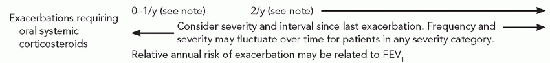
TABLE 36.3 CLASSIFICATION OF ASTHMA SEVERITY FOR PATIENTS ≥ 12 OF AGE
Components of Severity
Classification of Asthma Severity (Youths ≥ 12 y of age and adults)
Persistent
Intermittent
Mild
Moderate
Severe
Symptoms
≤2 d/wk
>2 d/wk but not daily
Daily
Throughout the day
Nighttime awakenings
Less than or equal to two times per month
Three to four times per month
Less than one time per week but not nightly
Often seven times per week
Impairment
Short-acting beta2-against use for symptom control (not prevention of EIB)
Expert Panel Report 3: Guidelines for the Diagnosis and Management of Asthma. National Heart, Lung, and Blood Institute, Aug 2007.
ASTHMA
Despite the marked heterogeneity of the asthma phenotype, a consensus definition for asthma has been developed that recognizes this condition to be a chronic inflammatory disorder of the airways in which many cells and cellular elements play a role, in particular, mast cells, eosinophils, T lymphocytes, neutrophils, and epithelial cells. The classic symptoms of asthma are recurrent episodes of wheezing, breathlessness, chest tightness, and cough, particularly at night and/or in the early morning. These episodes are typically characterized by widespread but variable airflow obstruction that is often reversible either spontaneously or with treatment. The inflammation also causes an associated increase in the existing bronchial hyperresponsiveness to a variety of stimuli. It is the association of asthma with AR and CRS that is one of the central points to the idea of the unified airway, as effective management of AR and CRS may prevent the development of asthma or beneficially impact the severity of asthma and its resultant morbidity.
Diagnostic tests for asthma include pulmonary function tests, such as forced expiratory volume (FEV), peak expiratory flow, and the methacholine challenge test, which provokes bronchial hyperresponsiveness. Furthermore, a diagnosis of asthma is established when a reversible airflow obstruction is confirmed with the above-mentioned tests as well as a response to an inhaled beta-agonist. Asthma severity can be classified clinically as mild intermittent, mild persistent, moderate persistent, and severe persistent. This classification scheme is detailed in Table 36.3, and is useful in guiding management. Management typically consists of anti-inflammatory modulators such as inhaled or systemic corticosteroids as well as short or long acting beta-agonist bronchodilator agents. Other options include leukotrieneinhibitors, methylxanthines like theophylline, and mast cell stabilizers like cromolyn or nedocromil. Anti-IgE monoclonal antibodies are available but reserved for severe cases due to their significant cost (11). Furthermore, pulmonary function tests may be repeated to assess response to therapy, and treatments can be adjusted based on these results.
Only gold members can continue reading. Log In or Register to continue