The Role of Genetic and Environmental Factors in Age-Related Macular Degeneration


The Role of Genetic and Environmental Factors in Age-Related Macular Degeneration


Philip Storey

Larry A. Donoso


INTRODUCTION

Both genetic and environmental factors have been implicated in the pathogenesis of age-related macular degeneration (AMD).1,2 These factors have been difficult to differentiate and characterize in part because AMD is an adult onset condition. Genetic studies are challenging because affected individuals are typically 65-years of age, and in most cases, have children too young to manifest the disorder and parents who are deceased. The finding that AMD increases rapidly with advancing age implies that environmental risk factors may contribute to or modify the disease as well. Of the risk factors that have been investigated, cigarette smoking has been clearly and most consistently identified.3,4,5,6,7,8,9,10

The last decade has seen an explosion of knowledge related to human genetics. The Human Genome Project has provided an entire map of the human genome as well as another map, the HapMap.11 The HapMap, in which the entire genome of 16 “normal” humans was sequenced, revealed that only 0.1% of the 3 billion nucleic acids are responsible for genetic diversity. These nonconserved sites are “normal” sequence variants known as single nucleotide polymorphisms, or SNPs. One percent of these SNPs, the nonsynonymous SNPs, cause single amino acid changes in their translated proteins. Unlike mutations, which are “abnormal” variations in the DNA sequence that result in gene products with significantly altered structure and function, nonsynonymous SNPs may be understood as “normal” variations which alter susceptibility to rather than cause disease.

Several SNPs have been identified and implicated as either predisposing or protective, or in conjunction with specific environmental factors, able to modify the risk of developing advanced AMD.12,13,14,15,16,17,18,19 This chapter will review the latest information regarding several chromosomal regions which have been implicated in the pathogenesis of AMD. These chromosomal regions code for several proteins involved in the inflammatory process including CX3CR1 (C3p21.3), complement factor H (C1q25-31), complement factor B (C6 MHC), complement component 2 (C2) (C6 MHC), complement component 3 (C3) (C19p13.3), and complement factor I (C4q25). In addition, the LOC387715 gene (C10q26), a gene of unknown function, appears to modify further the smokers’ risk of developing AMD. Taken together these studies provide insight into the genetic and environmental factors that play a prominent role in the pathogenesis of AMD.

The following discussion will present a brief overview of the complement system, summarize the recent reports regarding these SNPs and their effect on AMD, and discuss their importance in reshaping our understanding of the pathophysiology of AMD and other seemingly unrelated systemic disorders that may share the same underlying mechanism, specifically, a chronic localized inflammatory process.


GENERAL FEATURES OF AMD


Clinical Features

The clinical and angiographic features of AMD have been well described. In particular, Gass20,21,22,23 helped to characterize the “dry” form of AMD including drusen, the extracellular deposits associated with the condition, and also described the contribution of the choroidal vasculature to the “wet” form (Figs. 23C.1, 23C.2 and 23C.3). The clinical features of this condition are not a subject of this Chapter and the reader is referred to Foundations, Volume 2, Chapter 12 for a more detailed discussion.


Histopathological Features

Drusen, the hallmark of AMD, are extracellular deposits of degraded normal cellular materials which accumulate between the basal lamina of the retinal pigment epithelium (RPE) and the inner collagenous layer of Bruch’s membrane24,25(Fig. 23C.4). Drusen are predominantly composed of cholesterol, vitronectin, and apolipoproteins B and E (Table 23C.1).24,26,27,28 In addition, amyloid,25,29 a substance not normally found in healthy cells, as well as complement proteins (C5 and C5b-9), immunoglobulin light chains, and Factor X and their RNA transcripts have been identified in drusen and the RPE respectively.25,30,31 Similar material has been identified the glomerular deposits of poststreptococcal and membranoproliferative glomerulonephritis type II (MPGN II) and in the neuritic plaques which characterize Alzheimer’s disease (AD)32 as well as in other diseases of aging25 including atherosclerosis,33,34 amyloidosis,35 dense deposit disease,25 and elastosis36 (Table 23C.2). This suggests that these systemic disorders may have a similar pathophysiology. The association with MPGN II is particularly notable. In the majority of cases, MPGN II is characterized by an autoantibody against the C3bBb complex, which leads to uncontrolled activation of the alternative complement pathway. Renal lesions in MPGN II have been shown to be identical in composition and structure to drusen in AMD, supporting the hypothesis of the central importance of the complement system in AMD.37 Recent studies have also implicated allelic variations in factor H in MPGN II, AMD, coronary artery disease, and atherosclerosis.38,39,40,41 A common denominator in these seemingly diverse and unrelated ocular and systemic diseases may be the co-localization of factor H, C-reactive protein (CRP), and heparin on the arterial endothelium. This indicates that factor H may play an important role in the protection against complement mediated vascular damage with associated secondary exudation or neovascularization.16,41


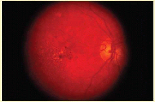
FIG. 23C.1 Drusen. Fundus photograph demonstrates numerous small drusen and RPE pigment clumping in an eye with “dry” AMD.


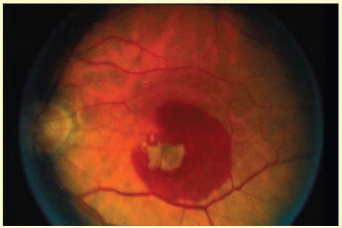
FIG. 23C.2 Exudative AMD. A large neovascular membrane with associated subretinal hemorrhage consistent with “wet” or exudative AMD is present in the macular region.


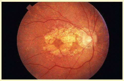
FIG. 23C.3 Atrophic AMD. Choroidal and RPE atrophy surround the optic disc and spare the foveal region in this example of atrophic AMD.


GENETIC APPROACHES TO THE STUDY OF AMD

Several approaches have been utilized to study AMD including investigations of genes known to be causative for juvenile onset macular dystrophies, twin and linkage studies, and more recently, genome wide scans in related and unrelated adults.


Juvenile Onset Macular Dystrophies

The juvenile onset macular dystrophies consist of a number of disorders which in most cases become manifest between the ages of 10 and 20 years. These conditions have been well characterized, in part because large families consisting of multiple generations have been available for study. Many specific genes42,43,44,45,46,47,48,49,50,51,52,53,54,55,56,57,58,59,60,61,62,63 or chromosomal regions64,65,66,67,68 have been identified and their discoveries are contributing to a better understanding of the basic underlying mechanisms of degenerative macular disease. In general these genes have not been implicated in a significant percentage of adult onset AMD cases. For more information regarding juvenile onset macular dystrophies the reader is referred to several excellent reviews on these important scientific developments.69,70,71


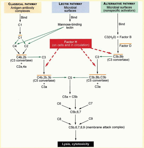
FIG. 23C.4 An Overview of Inflammation and the Complement System. Green arrows indicate proteolytic cleavage of the molecule at the tip of the arrow. Orange lines over complexes indicate that each is enzymatically active. Dashed red arrows indicate the roles of factor H in binding to C3b, accelerating the decay of the alternative pathway C3-convertase (C3bBb) and acting as a cofactor in proteolytic inactivation of C3b. (Modified from Levinson W, Jawetz E. Medical Microbiology and Immunology. 7th ed. New York, NY: The McGraw-Hill Companies; 2002:401.)

The complement system is composed of a group of inactive plasma enzymes (zymogens) and cell membrane regulatory proteins that interact to maintain a balance between activation (with phagocytosis of pathogens) and inactivation (with protection of self). In response to initiating stimuli, the complement cascade produces peptide mediators of inflammation, phagocyte recruitment, opsonization, and cell lysis.

The three pathways lead to complement activation. They differ in their initiating stimuli. Once activated, the complement reaction in most cases takes place on the cell surface on which it was initiated. The three pathways converge on complement C3b, the primary effector of the complement system.

The classical pathway is activated by antigen-antibody complexes on the surface of invading pathogens. The mannan-binding lectin pathway is activated by pathogen mannose and is important in infancy when adaptive immune system is not yet able to produce antibody.

The alternative pathway is initiated spontaneously and continuously in the plasma where complement C3 undergoes spontaneous hydrolysis. As factors D and B cleave and activate the C3 convertase, complement C3b is produced and deposited on both pathogens and host cells. Complement C2 is an activator of the classical pathway analogous to factor B.

C3b opsonizes cells marking them for phagocytosis by cells with complement receptor Cr1. Other C3 cleavage products, C3a and C5a, are potent mediators of both a local inflammatory response and phagocyte recruitment and in addition, have been shown to promote choroidal neovascularization by increasing VEGF expression in mice.205 The terminal peptides, C5b, C6, C7, C8, and C9 interact to form the membrane attack complex (MAC) and induce cell lysis.

Excessive amplification of the complement system is regulated by additional proteins. In the alternative pathway, MCP (membrane cofactor protein) and DAF (decay accelerating factor) are the cell-bound proteins that prevent complement activation on host cells, and plasma proteins factor H and factor I down-regulate inflammation by coating host cells marked with C3b and inactivating plasma C3b respectively.

The CX3CR1 receptor for the chemokine, fractalkine, a membrane bound protein is expressed by endothelial cells, neurons, and glia. Its soluble form, released by TNF-alpha-converting enzyme (TACE), is important in the adhesion and recruitment of monocytes, natural killer cells (NK), and activated T cells which bear the CX3CR1 receptor. Fractalkine blocks TNF-alpha secretion and thereby participates in the downregulation of inflammation.


TABLE 23C-1 Components of Drusen


Cholesterol


Complement protein C5


Vitronection


Complement protein C5b-9


Apolipoprotein B


Factor X


Apolipoprotein E


Immunoglobulin light chains


Amyloid


Malondialdehyde


Familial Studies

Evidence supporting a genetic contribution is derived from studies of twins72 and first-degree relatives73 of persons with AMD. These studies indicated that at least 25% of AMD may be genetically determined and that genetic factors play an important role in determining the onset and severity of the disease. Several families with AMD spanning multiple generations have been studied which have provided significant information regarding the genetics of AMD. Using the technique of linkage analysis and genome wide scanning two chromosomal regions 1q25-31 and 10q26 have been consistently associated with AMD.17,74,75,76,77,78,79,80,81,82


TABLE 23C-2 Nonocular Diseases with Drusen-like Deposits


Alzheimer’s disease


Elastosis


Atherosclerosis


Poststreptococcal glomerulonephritis


Amyloidosis


Membranoproliferative glomerulonephritis


Dense deposit disease


Genome Wide Scans, SNPs and Susceptibility to AMD

Modern molecular biological techniques have made it possible to scan the entire genome for putative disease causing genes. Genome wide scans in AMD have implicated chromosomal regions1q25-31 and 10q26 as well as four genes associated with the inflammatory process: the chemokine receptor CX3CR1,12,19 factor H,13,15,16,17 factor B, and complement factors C2,14 C3,83,84 and I85 (Fig. 23C.1). SNPs identified within these genes are associated either with a predisposing (at risk) or protective effect for AMD. LOC387715, a gene of unknown function, has also been linked to AMD especially in association with a history of smoking.18,86 Overall these studies have identified SNPs or haplotypes which are associated with at risk, protection, or modifying effects in upwards of 75% of AMD cases. What has emerged is the strong association of AMD with inflammation, the complement system, and smoking as summarized in Table 23C.3.


Chemokine Receptor CX3CR1

CX3CR1 is a macrophage chemokine receptor which may play a role in clearing age-related deposits. Two SNPs of CX3CR1, V249I, and T280M, are known to be associated with reduced chemoattraction of macrophages. The affected CX3CR1 receptors are fewer in number and have lower binding affinity with their substrate fractralkine. Tuo et al.19 showed that there was an association between these two SNPs and AMD. They proposed that a lower number of receptor binding sites and reduced binding affinity resulted in reduced macrophage recruitment and altered macrophage clearing of age-related sub-RPE deposits. In support of this theory, microdissection of retinal cells detected the M280 allele in 29.7% of eyes with AMD as well as reduced levels of CX3CR1 in the macular region of these eyes.12 It is interesting to note 249I and 280M were protective against atherogenesis,87,88 but I249 “not balanced” by M280 was associated with increased risk of CAD and re-stenosis following cardiac artery stenting.89,90 Also, the haplotype 249I/280M increased progression to AIDS in HIV+ individuals,91,92 but also increased favorable response to HAART93 while 249I/280T was associated with long-term nonprogression.94


TABLE 23C-3 SNPs Associated with AMD


Gene Product


SNP*


Haplotype


Type


P value


OR*


Reference


CX3CR1


V249I


At risk


4 × 10⁻²


1.86


Tuo


T280M


At risk


4 × 10⁻²


1.94


Tuo


Factor H


I62V


3.2 × 10⁻⁷


1.95


Hageman


Factor H


Y402H


At risk


1.6 × 10⁻¹³


1.79


Hageman


H1


At risk


1 × 10⁻⁵


2.46


Hageman


H2


Protective


13 × 10-5


0.54


Hageman


Factor H


Y402H


At risk


4.1 × 10⁻⁸


7.4


Klein


Factor H


Y402H


At risk


6 × 10⁻⁵


2.45


Haines


Factor H


Y402H


At risk


4.95 × 10⁻¹⁰


2.7


Edwards


Factor B and C2


H1


At risk


1.3 × 10⁻³


1.32


Gold


Factor B and C2


R32Q/intron 10


H7


Protective


1 × 10⁻⁴


0.36


Gold


Factor B and C2


L9H/E318D


H10


Protective


1 × 10⁻⁴


0.45


Gold


LOC387715


Ala69Ser


Modifying


3.13 × 10⁻⁸


5.73


Schmidt


Factor C3


rs2230199


At risk


5.9 × 10⁻⁵


2.6


Yates


Factor C3


rs2230199


At risk


4.5 × 10⁻¹²


1.61


Maller


Factor I


rs10033900


H2


Protective


9.11 × 10⁻⁸


0.70


Fagerness


* OR (odds ratio) is a statistical measure of relative risk. If the OR is 1, then the likelihood of an event in either the first or second group is the same. OR < 1 indicates reduced risk, OR > 1 indicates increased risk.


Complement Factor H

The complement system is composed of a group of inactive plasma enzymes (zymogens) and cell membrane regulatory proteins that interact to maintain a balance between activation with phagocytosis of pathogens and inactivation with protection of self. In response to initiating stimuli, the complement cascade results in the production of peptide mediators of inflammation, phagocyte recruitment, opsonization, and cell lysis. The complement cascade is summarized in Figure 23C.4.

Complement factor H is a plasma protein and is one of the primary regulators of the alternative complement pathway.40 Factor H binds with C3b on host cells which it is able to distinguish from C3b on pathogens through an interaction with host cell sialic acid. This prevents inadvertent host cell damage. Although it preferentially binds with membrane bound C3b, factor H is also able to modulate plasma C3b.

Various data support the hypothesis that particular DNA sequence variations of complement factor H may be associated with AMD. First, factor H is located in chromosome 1q31, a region known to link to a large family with AMD (ARMD1 locus).74,75,76,77,78,80 Second, complement has been identified in drusen, Bruch’s membrane, and the intercapillary pillars of the choriocapillaris.25,95 In addition, drusen of persons with MPGN II, a renal condition known to be caused by a deficiency of factor H, have complement components similar to those associated with AMD.32 Third, risk factors associated with AMD, age, and smoking, affect levels of plasma factor H.96

Four independent studies (Table 23C.3) tested this hypothesis and consistently identified a SNP in the factor H gene, Y402H, which significantly increases the likelihood of developing AMD. As a predisposing factor, this defect may explain up to 50% of the cases of AMD. Y402H is significantly and similarly associated with both drusen and advanced AMD, indicating that other genetic or environmental factors may be responsible for progression to advanced neovascular or atrophic AMD.97 The results of these studies are summarized below.

Klein and associates16 performed a whole-genome case-control association study of over 100,000 SNPs in 96 white, nonhispanic patients and 50 similar controls and identified two SNPs which were significantly associated with AMD. Both are located on chromosome 1q31. Reconstruction of inferred haplotypes revealed that six SNPs within the factor H gene region associated in four distinct haplotypes or allelic variations. Homozygosity for one of these haplotypes increased the risk of disease by a factor of 7.4. Resequencing of the exons in this haplotype revealed 50 SNPs. One nonsynonymous SNP in exon 9, DNA sequence T1277C (substitution of cysteine for tyrosine at position 1277) caused an amino acid sequence change Y402H (substitution of histidine for tyrosine at position 402) in complement factor H which was strongly associated with AMD. The authors found this SNP was present in 97% of the chromosomes with the high-risk haplotype indicating this was a polymorphism underlying susceptibility to AMD.

Haines and coworkers16 studied 44 SNPs located on chromosome 1q32 in the ARMD1 locus. In two independent data sets (family-based and AMD cases and controls) the authors found multiple SNPs strongly associated with AMD. However, one haplotype composed of five of the SNPs was present in 46% of cases and 33% of controls. This haplotype was more frequent in diseased than nonaffected family members. DNA resequencing revealed only one sequencing variant, T1277C (DNA) or Y402H (factor H protein), which increased odds of AMD 2.45 to 5.57 times in heterozygotes with any form of AMD and homozygous with neovascular disease, respectively. Because residue 402 is located in the heparin and CRP binding site of factor H, the authors postulated the amino acid substitution could affect factor H function by altering its ability to bind CRP and heparin, and as a consequence, its affinity for C3b and its ability to prevent host damage.

Edwards et al.13 tested nonsynonymous SNPs within the ARMD1 locus for association with AMD in two independent case-control populations. Heterozygosity for histidine at 402 (Y402H) increased risk of AMD by a factor of 2.7. Genotype frequencies among cases with and without family history of AMD were similar, indicating that the Y402H polymorphism is a more general predisposing factor for AMD.

Hageman and associates15,25 identified factor H and the membrane attack complex (MAC) in drusen, RPE basement membrane, and the choroidal capillary walls in the macular rather than the extramacular region.15 The authors also showed that Factor H was present in the intercapillary pillars and was synthesized locally by the RPE. Case-control allele association analysis of the factor H gene revealed several SNPs with highly significant associations with AMD. Haplotype analysis revealed one haplotype which increased risk, and two which decreased risk. Increased and decreased risk was more pronounced in homozygotes. These associations were observed to be stronger in individuals with drusen and neovascular AMD, rather than geographic atrophy. Half of individuals with AMD carried the same at-risk haplotype.

Others have confirmed the association of Y402H with AMD17,98 and additional studies of various ethnic populations including Japanese,99 French,100 and British101 individuals have confirmed the association in groups with disparate heredity. Of interest is that the frequency of Y402H was much lower in Japanese, as is the incidence of advanced AMD. Additional risk haplotypes were present that appeared to be responsible for disease susceptibility in this population.99 Allelic variations among other populations may contribute to the understanding of observed racial/ethnic variability in AMD prevalence not attributable to other known risk factors.102

Only gold members can continue reading. Log In or Register to continue

Stay updated, free articles. Join our Telegram channel

Jul 11, 2016 | Posted by in OPHTHALMOLOGY | Comments Off on The Role of Genetic and Environmental Factors in Age-Related Macular Degeneration

Full access? Get Clinical Tree

Get Clinical Tree app for offline access