The Lacrimal System
Edward H. Bedrossian Jr.
The precorneal tear film is essential for maintaining normal function of the cornea as well as the refractive optical integrity of the eye. It is composed of three layers: (a) the outer or superficial oily layer; (b) the middle aqueous layer; and (c) the inner mucoid layer. The outer superficial oily layer is produced by the meibomian and Zeis glands. The meibomian glands are found closely packed within the tarsal plates and are arranged in a parallel fashion, extending the entire height of the tarsal plate. They are taller and more numerous in the larger superior tarsus than in the smaller inferior tarsus. The oily layer prevents the escape of aqueous tears over the edge of the eyelid margin and retards evaporation. The glands of Zeis are found along the roots of the eyelashes, to which their contribution is less significant.
The middle aqueous layer is the thickest layer of the precorneal tear film and is produced by the main lacrimal gland and the accessory lacrimal glands (Wolfring and Krause); these glands are the subject of this chapter.
The inner mucoid layer is produced by the goblet cells and the conjunctival epithelial cells. These goblet cells are found in greatest concentrations along the eyelid margins, conjunctival fornices, antimarginal tarsal borders (crypts of Henle), and corneal-scleral limbus (glands of Manz).
The lacrimal apparatus is divided into two components. Those structures that contribute to the formation of the middle (aqueous) layer of the precorneal tear film are known as the secretory system. Those that form the conduit by which the tears pass from the conjunctival fornices into the nasal cavity are known as the excretory system (Fig. 30.1).
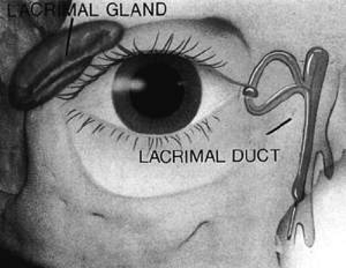 Figure 30.1. The lacrimal apparatus is divided into two components: the secretory system and the excretory system. |
The Secretory System
The lacrimal glands are exocrine glands. They are divided into two groups: the main lacrimal gland and the accessory lacrimal glands.
Gross Anatomy of the Secretory System
The Main Lacrimal Gland
The main lacrimal gland resides in the superotemporal orbit, partially within a shallow bony fossa in the lateral angular process of the frontal bone (fossa glandula lacrimalis). The gland is situated between the eyeball below and the curved orbital wall above, giving it a somewhat compressed and curved shape (Fig. 30.2). It may extend inferiorly to the lateral canthal tendon. The lateral horn of the levator aponeurosis crosses the gland anteriorly, separating it into a larger superior or orbital lobe and a smaller inferior or palpebral lobe (Fig. 30.3). The division is incomplete because the larger orbital lobe is connected to the smaller palpebral lobe posteriorly by a bridge of glandular tissue, draining tubules, and Müller muscle, which is attached to the underside of the levator muscle and aponeurosis. The lacrimal gland is surrounded by fibrous tissue that is attached superiorly to the periosteum of the frontal bone and inferiorly to the orbital portion of the zygomatic bone.1
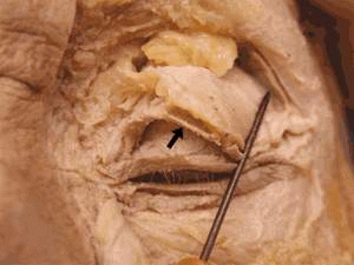 Figure 30.2. Orbital lobe of the lacrimal gland (pointer). The insertion of the levator aponeurosis (arrow) is seen dissected from the anterior surface of the tarsus. |
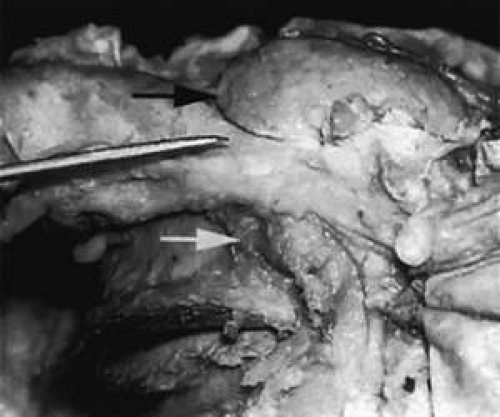 Figure 30.3. With the roof and lateral wall of the left orbit removed, this cadaver dissection demonstrates the orbital lobe (black arrow) and the palpebral lobe (white arrow) separated by the lateral horn of the levator aponeurosis (pointer). |
These attachments may become attenuated in older persons, allowing the gland to herniate through a weakened orbital septum to give a temporal bulge in the upper eyelid (Fig. 30.4). The lacrimal gland tissue is usually grayer and pinker than the surrounding yellow adipose tissue.
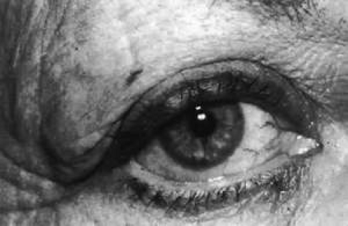 Figure 30.4. Disinsertion or attenuation of the suspensory fibrous attachments of the lacrimal gland will allow the orbital portion to herniate through a weakened orbital septum, producing a temporal bulge in the upper eyelid. |
The almond-shaped orbital lobe (see Fig. 30.3) represents approximately 65% to 75% of the gland and measures 20 mm long × 5 mm thick × 12 mm wide.2 Its sharp anterior border rests behind the superior orbital rim and is covered by the orbital septum (Fig. 30.5) and a portion of the temporal aspect of the central preaponeurotic fat pad (Fig. 30.6). Posteriorly, its rounded border is supported by a large superotemporal fat pad at the plane of the posterior pole of the globe.3 The convex superior surface is suspended from the periorbita of the lacrimal gland fossa of the frontal bone. The inferior border is convex and attached to the sheath of the levator aponeurosis. Its lateral border is smooth and convex in contour with the bony fossa.
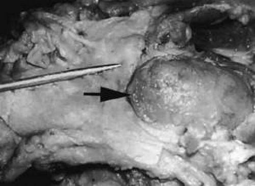 Figure 30.5. The sharp anterior border of the left lacrimal gland (black arrow) rests behind the orbital septum (pointer), which in this cadaver has been reflected anteriorly. |
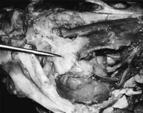 Figure 30.6. The temporal portion of the central preaponeurotic fat pad (pointer) rests near the anterior border of the orbital portion of the lacrimal gland. The left eyelids are seen below and to the left. |
The inferior or palpebral lobe of the lacrimal gland (see Fig. 30.3) represents approximately 25% to 35% of the gland and lies beneath the levator aponeurosis in the subaponeurotic space (Fig. 30.7). It extends anteriorly beyond the orbital margin to lie in the lateral portion of the superior fornix. The palpebral lobe can be seen through the conjunctiva when the eyelid is elevated or everted (Fig. 30.8).
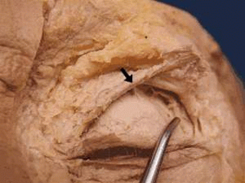 Figure 30.7. The palpebral lobe at the lacrimal gland (pointer) is seen posterior to the reflected levator aponeurosis (arrow). |
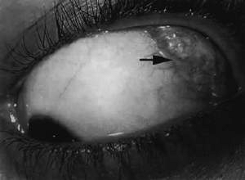 Figure 30.8. The palpebral lobe (arrow) is seen through the conjunctiva when the eyelid is elevated (left eye). |
The palpebral lobe is attached primarily on its underside to conjunctiva and to the intermuscular membrane between the superior and lateral rectus muscles. Only medially is it separated from conjunctiva by the superior tarsal muscle of Müller (Müller muscle).
The parenchyma of the gland is made up of small lobules separated by a fine connective tissue network. The lacrimal gland has approximately 12 secretory ducts (Fig. 30.9), which average 0.66 mm in diameter and 2.31 mm in length.4 Two to five of them originate from the orbital lobe and six to eight from the palpebral lobe. The ductules from the orbital portion of the lacrimal gland pass through the parenchyma of the palpebral lobe before exiting into the superotemporal portion of the conjunctival fornix, 4 or 5 mm above the upper border of the tarsus. One or two may open near the lateral canthus.5 Excision of the palpebral lobe may therefore interrupt drainage from the orbital lobe as well.
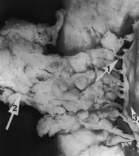 Figure 30.9. Secretory ducts (arrow 1) of the right lacrimal gland (arrow 2) are seen passing to the palpebral conjunctiva (arrow 3). (Courtesy of James Sanderson, M.D., Orkan Stasior, M.D., and George Stasior, M.D.) |
Sanderson and Stasior found islands of “peripheral” lacrimal glandular tissue below the lateral canthus unassociated with the main gland in 60% of cadaver specimens (Fig. 30.10).4 These peripheral islands of glandular tissue were found less commonly in or above the lateral canthal tendon.
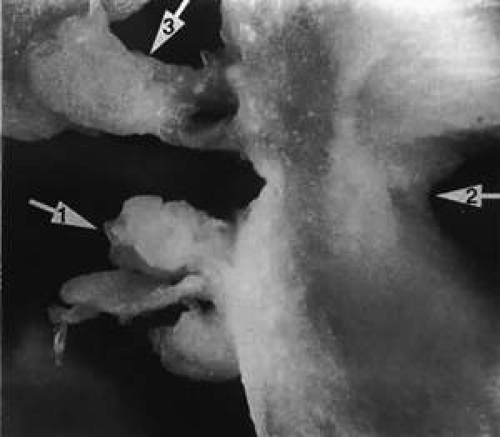 Figure 30.10. “peripheral” lacrimal gland lobules (arrow 1) are seen below the right lateral canthus (arrow 2) and separate from the palpebral lobe of the lacrimal gland (arrow 3). (Courtesy of James Sanderson, M.D., Orkan Stasior, M.D., and George Stasior, M.D.) |
The Accessory Lacrimal Glands
The accessory lacrimal exocrine glands of Wolfring and Krause structurally resemble the main lacrimal gland but on a lesser scale. Between 20 and 40 glands of Krause are located in the superior conjunctival fornix; two to eight are located in the inferior conjunctival fornix. Between three and 20 glands of Wolfring can be found along the upper border of the superior tarsus, one to four below the lower tarsus, and an occasional gland in the caruncle and in the plica semilunaris. The glands of Wolfring are larger in size than the glands of Krause.4,5,6
Jones termed the accessory lacrimal glands the basal secretors because they do not possess direct secretory motor fibers.7 The other basal secretors are the sebaceous glands (meibomian and Zeis) and the mucous glands (goblet cells). The reflex secretors are the main lacrimal glands. Reflex secretion provides additional secretion by peripheral sensory (fifth nerve efferent, seventh nerve afferent), retinal, or psychogenic stimulation.
Vascular Supply to the Secretory Apparatus
The lacrimal gland receives its arterial supply from the lacrimal branch of the ophthalmic artery, which enters the gland near its posterior margin. An additional arterial supply often is seen from the infraorbital branch of the internal maxillary artery. The lacrimal artery divides within the gland and continues anteriorly to form the lateral palpebral pretarsal arterial arcades of the eyelid.
Venules form a lacrimal vein (Fig. 30.11), which exits the gland near its posterior margin. The lacrimal vein follows an intraorbital course similar to that of the lacrimal artery-superior to the lateral rectus muscle and lateral to the superior rectus muscle-before it drains into the superior ophthalmic vein and eventually into the cavernous sinus.
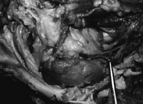 Figure 30.11. In this dissection, the roof of the orbit and the lateral orbital wall have been removed. The lacrimal vein (pointer) is seen near the posterior margin of the orbital portion of the lacrimal gland. The eyelids are seen below and to the left. |
Lymphatic drainage from the lacrimal gland enters the conjunctival and palpebral systems to drain into the preauricular nodes.
Nerve Supply to the Secretory Apparatus
Tear secretion is under complex neural control. This provides protection against environmental conditions such as cold, noxious chemicals, foreign bodies, and various organisms. Tears also provide nutrients and immunologic protection, and remove waste from the cells of the ocular surface.8
The lacrimal gland is richly innervated by the lacrimal nerve (sensory), the facial nerve (parasympathetic), and the sympathetic nervous system.
The lacrimal nerve carries sensory fibers from the lacrimal gland, and it is one of the three branches of the ophthalmic division of the trigeminal nerve (cranial nerve V1). Afferent sensory fibers of the lacrimal nerve leave the lacrimal gland posteriorly, near the lacrimal vessels (Fig. 30.12). Sensory fibers of the lacrimal nerve traverse the orbit superotemporally, along the superior border of the lateral rectus muscle, remaining outside the muscle cone. They leave the orbit through the superior orbital fissure, then pass through the cavernous sinus. Their cell bodies are found in the gasserian ganglion.
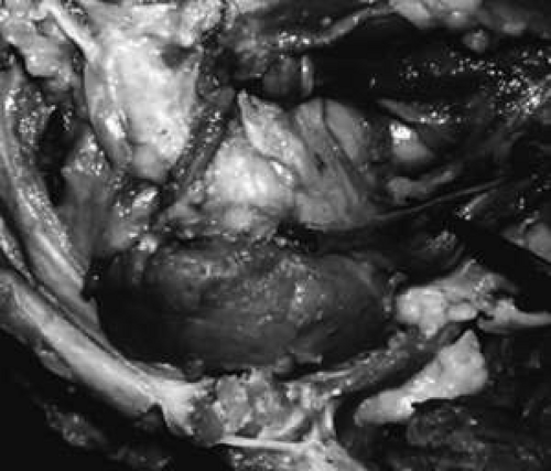 Figure 30.12. The lacrimal nerve (above pointer) is seen entering the posterior aspect of the lacrimal gland with the vessels (below pointer). |
The facial nerve supplies parasympathetic secretory fibers to the lacrimal gland. They arise from the lacrimal nucleus near the facial nucleus in the caudal pons. They emerge from the pons-medullary junction between cranial nerves VI and VIII. The facial nerve passes into the internal auditory meatus and extends to the geniculate ganglion, where the nerve cells of the preganglionic parasympathetic fibers lie. The greater superficial petrosal nerve arises from the preganglionic parasympathetic fibers of the geniculate ganglion and courses back into the middle cranial fossa to join with the great deep petrosal sympathetic nerve from the internal carotid plexus, forming the nerve of the pterygoid canal (Vidian nerve). Parasympathetic fibers pass into the sphenopalatine ganglion and synapse. From there, postganglionic parasympathetic fibers can pass either directly to the lacrimal gland or, more commonly, along with the communicating branch of the zygomaticotemporal nerve (a branch of the maxillary nerve) to anastomose with the lacrimal nerve.
The postganglionic cervical sympathetic fibers rise from the superior cervical ganglion and travel, along with the internal carotid arteries (carotid plexus), to reach the lacrimal gland by way of the deep petrosal nerve, the nerve of the pterygoid canal, the sphenopalatine ganglion, and then the zygomaticotemporal nerve. A communicating branch to the lacrimal nerve from the zygomaticotemporal nerve provides sympathetic and parasympathetic innervation. Other sympathetic fibers may pass directly from the carotid plexus to the trigeminal nerve and then to the lacrimal nerve. Other sympathetic nerve fibers are contiguous with the lacrimal artery.
It appears that the trigeminal nerve forms the sensory (afferent) pathway and the facial nerve the parasympathetic (efferent) pathway for reflex secretions of the main lacrimal glands.9
Histology of the Lacrimal Glands
Arrangement
Each lobe of the lacrimal gland is separated into numerous lobules by interlobular fibrovascular connective tissue (Fig. 30.13). Each lobule, as seen by light microscopy, is composed of two units (Fig. 30.14): the acinar unit (or secretory unit) and the ductal system. The acinar units are further separated by intralobular fibrovascular connective tissue.
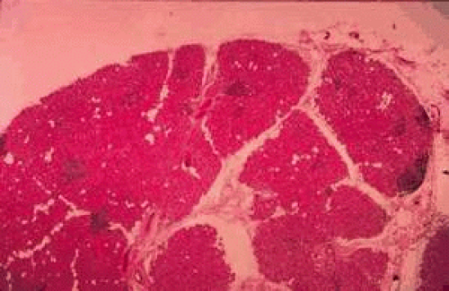 Figure 30.13. Lacrimal gland architecture. Each lobe of the lacrimal gland is separated into numerous lobules by interlobular fibrovascular connective tissue. (H & E, original magnification ×5; Courtesy of Ralph Eagle, M.D., Philadelphia, PA.) |
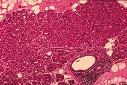 Figure 30.14. Lacrimal gland. Each lobule is composed of acinar secretory units (above) and a ductal system. An interlobular duct is seen (lower right). (H & E, original magnification ×25; Courtesy of Ralph Eagle, M.D., Philadelphia, PA.) |
The Acinar Unit
The acinar unit is illustrated in Figure 30.15. Each lobule consists of a complicated grape-like (Fig. 30.16) arrangement of acini. Each acinar unit (Fig. 30.17) consists of a central lumen, a continuous inner layer of columnar secretory epithelial cells, and a surrounding interrupted outer layer of irregularly shaped myoepithelial (basket) cells.10
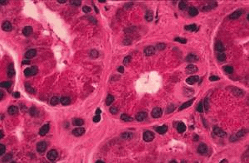 Figure 30.15. The acinar unit is composed of a central lumen, a continuous inner epithelial layer of secretory cells, and an interrupted outer layer of myoepithelial cells. (H & E, original magnification ×250; Courtesy of Ralph Eagle, M.D., Philadelphia, PA.) |
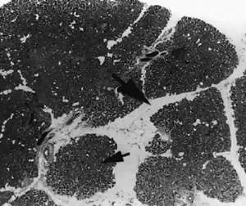 Figure 30.16. Tubuloracemose architecture of the lacrimal gland. Interlobular fibrovascular connective tissue (large arrow) separates the lobules of acinar tissue (small arrow). (H & E, original magnification ×5; Courtesy of Ralph Eagle, M.D., Philadelphia, PA.) |
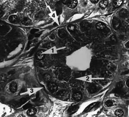 Figure 30.17. The acinar unit. The columnar secretory cell has a basally located nucleus (arrow 1). Zymogenic secretory granules (arrow 2) are seen in the midportion and apical cytoplasm. A myoepithelial cell (arrow 3) is interspersed between the secretory cell and the basement membrane (arrow 4). (H & E, original magnification ×250; Courtesy of Ralph Eagle, M.D., Philadelphia, PA.) |
The acinar secretory cell typically has a basally located nucleus with one or two nucleoli, as seen with electron microscopy. The predominant structures in its cytoplasm are the numerous zymogenic secretory granules located mainly in the apical or midportions of the cell (see Fig. 30.17).10
The secretory granules vary in shape from round to oval, and have a surrounding membrane. The secretory granules vary in electron density from homogeneous (electron-dense) to finely granular (less electron-dense).11 The number of granules in the cytoplasm varies from cell to cell. The size of the secretory granules also varies: The larger ones are located in the more peripheral portion of the lobule. Although chemical studies show that the secretory granules of the lacrimal gland contain both serous proteins and mucous polysaccharides, the predominance of dense granules containing protein suggest that the serous type of secretion is the primary function of the lacrimal glands.12 In addition, IgA immunoglobulins have been detected in human tear fluid, implicating the role of the lacrimal gland as part of the immune system.13
The mechanism by which the zymogenic granules are discharged into the lumen of the acinus is thought to be emiocytosis,14 a process in which the limiting membrane of the granules fuses with the apical cell membrane, after which the granular contents are released into the lumen. Other cytoplasmic structures include cisternae of rough-surfaced endoplasmic reticulum and the Golgi complex. The cytoplasm also has mitochondria, free ribosomes, lipid droplets, and dispersed tono-filaments.12,13,14,15
The luminal surface of the secretory cell has microvilli, whereas the basal surface of the secretory cell is in direct contact with the surrounding basement membrane or in contact with an interspersed myoepithelial cell.11,16,17
Stay updated, free articles. Join our Telegram channel

Full access? Get Clinical Tree


