Systemic Hamartomatoses (“Phakomatoses”)


Systemic Hamartomatoses (“Phakomatoses”)


Carol L. Shields

Jerry A. Shields


THE SYSTEMIC HAMARTOMATOSES (phakomatoses) comprise a group of syndromes with variable clinical manifestations that primarily affect the ocular region, central nervous system (CNS), skin, and occasionally, the viscera (1,2,3,4,5,6,7). The recognition of singular ocular or cutaneous features should prompt a referral to specialists familiar with the variety of manifestations of these conditions for diagnostic purposes and to provide patient management. In this chapter, we review the hamartomatoses, otherwise termed phakomatoses or oculoneurocutaneous syndromes. We emphasize the clinical spectrum of findings and illustrate the salient features.


GENERAL CONSIDERATIONS


Historical Aspects

The term “phakoma” was coined by Van der Hoeve in 1932 to indicate a mother spot or birthmark, a characteristic finding in many of these conditions (1). At that time, retinal and cerebellar hemangiomatosis (von Hippel-Lindau syndrome), neurofibromatosis (von Recklinghausen syndrome), and tuberous sclerosis (Bourneville syndrome) were grouped under this heading. Later, encephalofacial hemangiomatosis (EFH; Sturge-Weber syndrome), racemose hemangiomatosis (Wyburn-Mason syndrome), and cavernous hemangioma of the retina with cutaneous and CNS involvement were grouped with these other conditions.


Terminology

To better understand these syndromes, the clinician should be familiar with certain terms such as hamartia, hamartoma, chorista, and choristoma.

Hamartia and hamartoma are terms that refer to malformations that are composed of tissues that are ordinarily present at the location where they occur. A hamartia is a nontumorous anomaly composed of tissues that are normally present at the involved site, while a hamartoma is a tumorous malformation composed of tissues that are normally present at the involved site. Most of the entities discussed in this chapter are characterized by the presence of hamartomas. The term systemic hamartomatosis is used to designate multiple-organ involvement. Examples of hamartomas include the vascular tumors that occur in patients with either retinocerebellar hemangiomatosis or EFH, and the glial and peripheral nerve tumors are those that occur with tuberous sclerosis or neurofibromatosis. These tumors develop in areas where vascular and neural tissues are normally present.

Chorista and choristoma, on the other hand, are terms that refer to malformations composed of elements that are not ordinarily present at the location where they occur. A chorista is a nontumorous anomaly composed of tissues which are not normally present at the involved site. The microscopic rests of ectopic lacrimal gland tissue that sometimes occur in the anterior chamber and deep orbit are examples of choristas. A choristoma is a tumorous malformation composed of tissues that are not normally present at the involved site. The classic example of a choristoma is the limbal dermoid, a tumor composed of dermal elements that are not normally present in the bulbar conjunctiva or cornea.

A particular lesion can be classified as either a hamartoma or a choristoma depending on the organ involved. For example, a 5 mm nodule of mature bone would be classified as a hamartoma if it occurred on the superior orbital rim, but a similar mass of osseous tissue in the liver would be classified as a choristoma.


Heredity

Most of the hamartomatoses have an autosomal dominant mode of inheritance, often with incomplete penetrance. Specific chromosomal abnormalities have been recognized in association with these entities. Notable exceptions are EFH (Sturge-Weber syndrome) and racemose hemangiomatosis (Wyburn-Mason syndrome) in which heredity does not
appear to play a role and genetic abnormalities are not yet delineated. In Sturge-Weber syndrome, there is some evidence that the condition is a mosaic mutation with only segmental involvement. Those that receive the full mutation die in utero. Although these conditions are generally hereditary, many do not become clinically apparent until the teenage years or young adulthood.


Benign and Malignant Tumors

In general, the tumors that develop in these syndromes are benign. They differ from true neoplasms by virtue of the fact that they are anomalies of tissue formation, rather than tumors that arise from fully developed tissues. Furthermore, they are usually stationary or slowly progressive lesions that generally lack the capacity for the limitless proliferation seen with malignant neoplasms (5,6). Some of these syndromes, however, can be associated with malignant neoplasms. For example, there is an increased incidence of malignant schwannomas of the peripheral nerves in patients with neurofibromatosis. Hypernephroma occurs with greater frequency in patients with retinal capillary hemangiomatosis.


Formes Frustes and Combined Phakomatoses

It is common for patients with systemic hamartomatoses to only manifest some of the clinical features of a particular syndrome. This lack of complete expressivity is referred to as a forme fruste. Furthermore, patients can occasionally exhibit certain lesions characteristic of one entity and other lesions characteristic of another. For example, the café au lait spots seen in patients with neurofibromatosis can occasionally be seen in patients with tuberous sclerosis. Some patients with features of Sturge-Weber syndrome also manifest oculodermal melanocytosis, a condition termed phakomatosis pigmentovascularis. This coincidence of two conditions has been attributed to a genetic phenomenon of twin spotting (8,9,10).


TUBEROUS SCLEROSIS COMPLEX (BOURNEVILLE SYNDROME)


Ophthalmologic Features

The retinal astrocytic hamartoma is the characteristic fundus lesion of TSC (17,18,19,20,21). The smaller noncalcified tumor can be extremely subtle and appear only as ill-defined translucent thickening of the nerve fiber layer. A slightly larger tumor is more opaque and appears as a sessile white lesion at the level of the nerve fiber layer of the retina (Fig. 20.1). It may contain characteristic dense yellow, refractile foci of calcification that resemble fish eggs or tapioca (Fig. 20.2). In some cases, this tumor can develop a central cavity and occasionally, there can be localized overlying vitreous seeds (22,23). The retinal astrocytic hamartoma is generally stable but can rarely enlarge and produce retinal detachment and neovascular glaucoma.

A fairly common but often overlooked fundus feature of TSC is the retinal pigment epithelial (RPE) depigmented (punched out) lesion (19,20). This finding can be tiny at only 50 to 100 µm diameter or it can be obvious at 3 to 4 mm diameter. Multiple RPE depigmented lesions should be suggestive of TSC.

Ancillary studies, such as fluorescein angiography, ultrasonography, and optical coherence tomography, can assist in the diagnosis (24). Fine-needle aspiration has been used to make a diagnosis in atypical cases.

The retinal astrocytic hamartoma shows rather typical pathologic features. It is located in the nerve fiber layer of the retina and is composed of fibrillary astrocytes. Foci of dystrophic calcification, sometimes resembling psammoma
bodies, are frequently present in the tumor. The uveal tract is rarely affected in tuberous sclerosis. A depigmented iris sector, seen in some affected patients, is believed to be the equivalent of depigmented cutaneous lesions.


Table 20.1 DIAGNOSTIC CRITERIA (REVISED) FOR ESTABLISHING THE DIAGNOSIS OF TUBEROUS SCLEROSIS COMPLEX (TSC)


Category


Feature


Major features


  • Facial angiofibromas or forehead plaque


  • Ungual or periungual fibroma


  • Hypomelanotic macules (>3) (ash leaf macule)


  • Shagreen patch (connective tissue nevus)


  • Multiple retinal nodular hamartomas


  • Cortical tuber


  • Subependymal nodule


  • Subependymal giant cell astrocytoma


  • Cardiac rhabdomyoma


  • Lymphangiomyomatosis


  • Renal angiomyolipoma


Minor features


  • Multiple pits in dental enamel


  • Hamartomatous rectal polyps


  • Bone cysts


  • Cerebral white matter migration lines


  • Gingival fibromas


  • Non-renal hamartoma


  • Retinal achromic patch


  • Confetti skin lesions


  • Multiple renal cysts


Classification


  • Definite TSC: Either 2 major or 1 major with two minor features


  • Probable TSC: 1 major and 1 minor features


  • Possible TSC: Either 1 major or ≥2 minor features


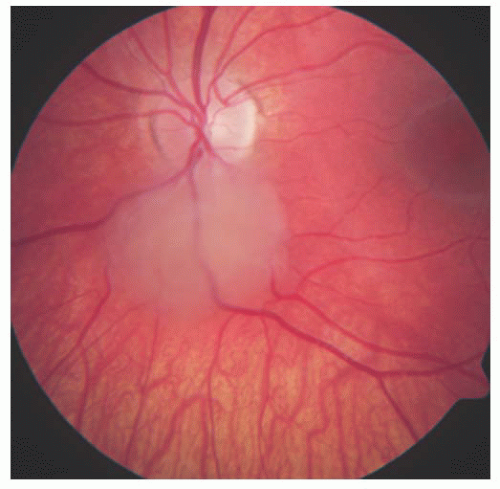
FIGURE 20.1. Tuberous sclerosis complex: non-calcified retinal astrocytic hamartoma.


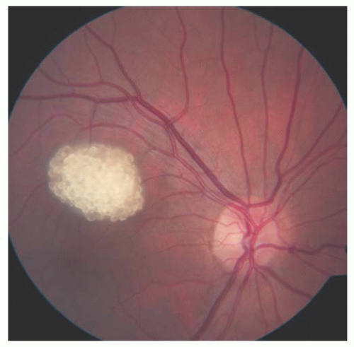
FIGURE 20.2. Tuberous sclerosis complex: calcified retinal astrocytic hamartoma.


Dermatologic Features

The main cutaneous manifestations of TSC include adenoma sebaceum, depigmented macules, and café au lait spots. Adenoma sebaceum is characterized clinically by multiple slightly elevated, rubbery, yellow-red papules. They are most often found on the face, frequently in a butterfly-shaped distribution (Fig. 20.3). They are composed microscopically of a benign proliferation of fibrous tissue and blood vessels. In reality, these tumors are angiofibromas, and the sebaceous gland hyperplasia appears to be a secondary change and is not part of the primary process. Consequently, the term “sebaceous adenoma” is a misnomer. Similar angiofibromas can occur beneath or adjacent to the fingernails or toenails in patients with tuberous sclerosis. These subungual fibromas, when present, are highly suggestive of tuberous sclerosis.

Depigmented macules resembling vitiligo are commonly present on the skin of patients with TSC (25). Because these patches frequently assume a configuration of a leaf from the mountain ash tree, the characteristic lesion is often referred to as the ash leaf sign (Fig. 20.4). These lesions are considered to be highly characteristic or even pathognomonic of tuberous sclerosis.


Other Features

The characteristic brain findings in patients with TSC include subependymal and cortical astrocytomas, sometimes with giant tumor cells (13,14,26) (Fig. 20.5). Both can demonstrate cystic and calcific changes, which account for the name tuberous sclerosis (potato-like masses). These lesions contribute to the seizures and mental deficiency. Contrary to early reports, mental deficiency is not necessarily a part of this syndrome. Many of the early reported patients were recruited from mental institutions where individuals with mental derangement were hospitalized. It is now recognized that many patients have only mild symptoms and signs and are of normal or near-normal intelligence.


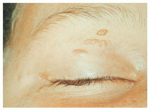
FIGURE 20.3. Tuberous sclerosis complex: adenoma sebaceum in the periocular region.


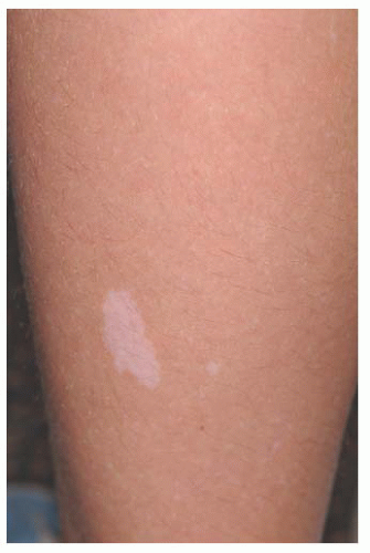
FIGURE 20.4. Tuberous sclerosis complex: ash leaf sign.


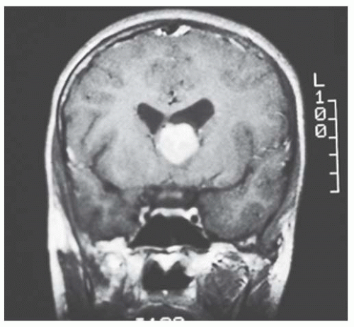
FIGURE 20.5. Tuberous sclerosis complex: brain astrocytoma.

The renal lesions commonly predispose the patient to recurrent nephritis and elevated blood urea nitrogen. They have been shown histologically to be benign angiomyolipomas, with no tendency to undergo malignant transformation
or to metastasize (26). The cardiac lesions have been shown to be rhabdomyomas, composed of large spider cells with prominent vacuoles containing glycogen. Some patients with tuberous sclerosis develop slowly progressive subpleural cysts that result from anomalous development of pulmonary tissue. These cysts can rupture, leading to spontaneous pneumothorax. Irregular cortical thickenings of bones, particularly the metatarsals and metacarpals, as well as hamartomas of the liver, thyroid, pancreas, testes, and other organs, have been reported.


Management

The retinal astrocytic hamartoma and RPE depigmented lesions are typically asymptomatic, nonprogressive, and do not require treatment. Ocular examination should be performed yearly, and the patient followed for other manifestations of tuberous sclerosis. If there should be an associated retinal detachment that extends into the foveal area, then laser photocoagulation, photodynamic therapy, or plaque radiotherapy can be employed to bring about resolution of the subretinal fluid. The astrocytic hamartoma of the retina has an extremely low tendency to undergo malignant change and has no tendency to metastasize. The visual prognosis is also excellent, except in the rare instances in which exudation, retinal detachment, or vitreous hemorrhage occur.

Most of the cutaneous lesions of TSC require no treatment. Larger facial angiofibromas may require surgical excision for cosmetic purposes. In recent years, the brain lesions have been treated with mTOR inhibitors (27).


NEUROFIBROMATOSIS (VON RECKLINGHAUSEN SYNDROME)


Neurofibromatosis Type 1


General Considerations

Neurofibromatosis type 1 occurs at a rate of 1 in 3,000 persons, but it is estimated that the frequency could be higher as some individuals manifest only mild features. Approximately one-half of affected patients represent a new mutation. This condition is caused by an autosomal dominant mutation in the NF1 gene that leads to decreased production of the protein neurofibromin, which has a tumor suppressor function. Only one deleted is necessary to manifest this condition. The NF1 gene is chromosome 17q. There have been more than 250 mutations identified. Complete gene deletion leads to severe phenotype. This highly penetrant phenotype has a wide variety of manifestations and can vary within families. Another locus, the SPRED1 gene, has been found in patients with “mild neurofibromatosis” and this represents Legius syndrome.

The criteria for diagnosis of neurofibromatosis type 1 are listed in Table 20.2 (29,30). This condition more often affects patients of the Caucasian race and equally in boys and girls. Scoliosis can be a prominent feature.


Ophthalmologic Features

Neurofibromatosis has the most diversified ocular findings among the phakomatoses. This condition can involve the eyelid, conjunctiva, aqueous outflow channels, uveal tract, retina, orbit, and optic nerve (31,32).

Eyelid involvement is characterized by nodular or plexiform neurofibroma. Nodular neurofibroma appears as a solitary or multifocal painless, smooth-surfaced and well-defined mass, often the size of a pea, and without color change. Plexiform neurofibroma presents as a diffuse thickening of the eyelid that can produce the typical S-shaped curvature to the eyelid, a finding highly characteristic of neurofibromatosis. The conjunctiva can be involved by diffuse or localized neurofibromas. Patients with neurofibromatosis have an increased incidence of congenital glaucoma, which can be secondary to several mechanisms. There is a high association of neurofibromatosis type 1 with optic nerve glioma.

Iris Lisch nodules are the most common ophthalmic abnormality of neurofibromatosis type 1. These characteristically orange-tan nodules appear in early childhood (usually by age of 5 to 6 years) as discrete, multiple, bilateral tumors of the anterior border layer of the iris, classically measuring less than 1 mm diameter, and best detected by slit lamp biomicroscopy (Fig. 20.6). Histopathologically, iris Lisch nodules are hamartomas composed of aggregates of melanocytes on the anterior border layer of the iris.

The choroidal findings in patients with neurofibromatosis type 1 include unifocal or multifocal choroidal nevus, diffuse plexiform neurofibroma, neurilemoma, and melanoma. Multiple bilateral, choroidal nevi are highly suggestive of neurofibromatosis type 1. They are usually small, illdefined, and randomly distributed. Choroidal neurofibroma usually appears a diffuse thickening of the uveal tract from an increased number of neurofibromatous and melanocytic
elements. There appears to be a higher incidence of uveal melanoma in patients with neurofibromatosis.


Table 20.2 DIAGNOSTIC CRITERIA FOR NEUROFIBROMATOSIS TYPE 1 (NF1)


Feature


Number


Café au lait


≥6 café au lait spots larger than 5 mm in diameter in prepubertal children (<10 y) or ≥6 café au lait spots larger than 15 mm in diameter in postpubertal individuals (adults)


Freckles in axilla or inguinal region


Crowe sign


Skin neurofibroma


≥2 typical neurofibroma or ≥1 plexiform neurofibroma


Optic nerve glioma


Iris Lisch nodules


≥2 lesions


Osseous lesion


Sphenoid dysplasia or long bone abnormalities (cortex thinning or pseudoarthrosis)


Relative (first degree) with NF1 by above criteria


Parent, sibling, or offspring


At least two of the seven criteria should be present for diagnosis. Some manifestations do not appear until later life, delaying diagnosis. Adapted from: Stumpf DA, Alksne JF, Annegers JF. Neurofibromatosis. Conference Statement. National Institute of Health Consensus Development Conference. Arch Neurol 1988;45:575-578; Gutmann DH, Aylsworth A, Carey JC, et al. The diagnostic evaluation and multidisciplinary management of neurofibromatosis 1 and neurofibromatosis 2. JAMA 1997;278:51-57.


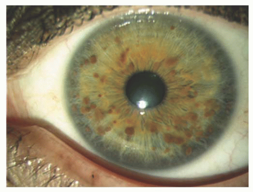
FIGURE 20.6. Neurofibromatosis type 1: Iris Lisch nodules.

Several retinal and optic disk lesions can occur with neurofibromatosis type 1. Retinal astrocytic hamartoma is a manifestation of neurofibromatosis, but is more often found with TSC. Retinal vasoproliferative tumor can occur with neurofibromatosis, leading to exudative retinopathy and risk for blindness (33). Congenital hypertrophy of the RPE is believed to be more common in patients with neurofibromatosis type 1. Fundus changes can occur secondary to optic nerve glioma including optic disk edema, optic atrophy, opticociliary shunt vessels, and central retinal vein obstruction.


Dermatologic Features

The most important cutaneous manifestations of neurofibromatosis include café au lait spots (pigmented macules), freckles in the axillary or inguinal region, and urticarial pigmentosa (Fig. 20.7). Café au lait spots are found in 95% of patients with neurofibromatosis type 1, but they can be seen in patients with other conditions such as McCune-Albright syndrome, TSC, and Fanconi anemia. Café au lait spots are also found in persons without neurofibromatosis type 1. They can become more noticeable over time with sun exposure.

Subcutaneous or cutaneous benign neurofibromas are an important finding but typically appear in older children or later. Deep neurofibromas might not be visible and are only detected by palpation. Puberty and pregnancy can increase number and growth of neurofibromas. Plexiform neurofibromas can be invasive and ill defined, occasionally associated with pain. Rapid growth of neurofibroma is suggestive of malignant degeneration.


Central Nervous System Features

The most important CNS feature of neurofibromatosis type 1 is the optic nerve glioma (juvenile pilocytic astrocytoma). Optic nerve glioma can present with painless proptosis
or subtle features of color or visual acuity abnormalities (Figs. 20.8 and 20.9). Magnetic resonance imaging shows enlargement of the optic nerve, often so large that it develops a fold (kink) within its substance to accommodate the orbit, leading to down-and-out proptosis. This mass shows enhancement on T1-weighted, gadolinium contrast images, particularly notable in the axial and coronal views. It is important to differentiate this tumor from optic nerve sheath meningioma as the systemic implications and therapy differ. This is best determined using gadolinium-enhanced, orbital fat suppressed, T1-weighted coronal views. With glioma, the central substance of the nerve enhances, whereas with meningioma the peripheral encircling arachnoid sheath enhances. Glioma is more often associated with neurofibromatosis type 1, whereas meningioma with neurofibromatosis type 2.


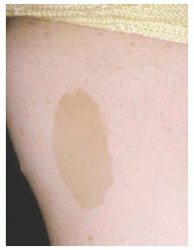
FIGURE 20.7. Neurofibromatosis type 1: cutaneous café au lait spot.


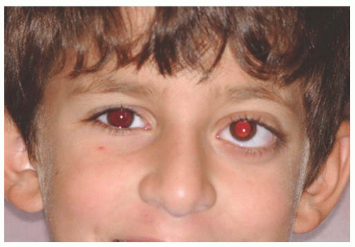
FIGURE 20.8. Neurofibromatosis type 1: mild proptosis from optic nerve glioma.

Only gold members can continue reading. Log In or Register to continue

Stay updated, free articles. Join our Telegram channel

Jun 20, 2016 | Posted by in OPHTHALMOLOGY | Comments Off on Systemic Hamartomatoses (“Phakomatoses”)

Full access? Get Clinical Tree

Get Clinical Tree app for offline access