Supraorbital Nerve Nuances/Dissections from Above
1 The supraorbital foramen/notch varies from 2.3 to 2.7 cm from the midline in men and 2.2 to 2.5 cm from the midline in women.
2 The “medial” or superficial branch of the supraorbital nerve pierces the frontalis muscle at varying levels to ascend cephalad on the surface of the frontalis muscle with many separate fibers. These nerves supply sensation to the off-midline forehead into the scalp.
3 Present always is a deep or “lateral” branch of the supraorbital nerve that runs obliquely up the forehead toward the lateral brow. This branch may exit with or from a separate foramen above or lateral to the superficial branch of the supraorbital nerve.
4 The exit foramen for this branch may be 1 cm above the rim, or along a path 3 to 4 cm lateral to the medial nerve’s exit. Because the lateral or deep branch may exit above or lateral to the usual notch area, anesthetic block to the supraorbital nerve foramen region may require augmentation to anesthetize the upper lateral forehead. The needle should be passed along the periosteum above the rim from the level of the foramen and laterally for 3 to 4 cm.
5 The nerve’s lateral branch usually runs between the galea and periosteum; the nerve may bifurcate or trifurcate into two or three branches as it courses cephalad. Sometimes there are small branches that may travel subperiosteally and groove the skull. These probably supply the periosteum itself. The lateral branches can always be found in the midforehead to lie 0.5 to 1.5 cm medial to the superior temporal line.
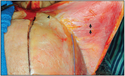 Figure 6.1 Supraorbit from above. A cut is made just to the right of the midline. A supraperiosteal dissection is done. The blue strip marks the superior temporal line. Note deep supraorbital nerve branches (arrows) and the corrugator origin (dart). |
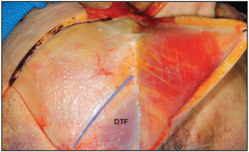 Figure 6.2 Side view. The blue strip is on the superior temporal line. The deep temporal fascia (DTF) is marked. The deep nerves would lie medial to the superior temporal line. |
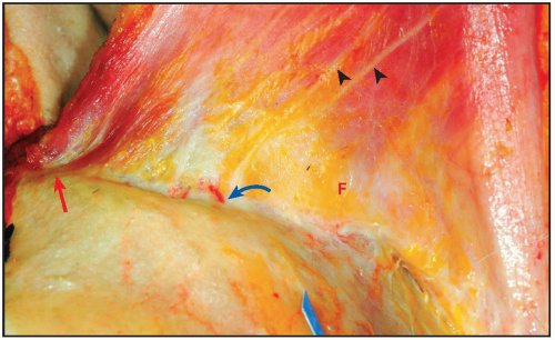 Figure 6.3 Dissection is taken almost to the rim. Note the galeal fat pad (F). There is a vessel (arrow) commonly seen here that goes from the supratrochlear nerve toward the retro-orbicularis oculi fat (ROOF). Deep branches of the supraorbital are noted with darts. Note the corrugator origin (red arrow). |
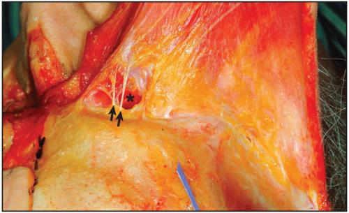 Figure 6.4 The galeal fat pad is removed from under the corrugator from medial to lateral, exposing supratrochlear nerves (arrows). The underside of the corrugator can be seen coming into view (*).
Stay updated, free articles. Join our Telegram channel
Full access? Get Clinical Tree
 Get Clinical Tree app for offline access

|