Superomedial Zone
1 The corrugators, proceri, and frontalis muscles are innervated by the frontal (temporal) branches of the facial nerve on each side. The depressor supercilii and medial orbicularis oculi are not; the zygomatic branch of cranial nerve VII innervates those.
2 The corrugator on one side may be of varying size, but two specific heads do not exist. The supratrochlear nerves always pass through these muscles. The nerves do not really separate the corrugator into two heads. The corrugators are panel-like muscles that require surgical diligence for full extirpation.
3 The supraorbital nerve passes under the corrugator and then through them sometimes, but usually through the frontalis to provide multiple branches vertically on the surface of the frontalis.
4 The supraorbital nerve separates into a medial (superficial) and a lateral (deeper) part, which will be presented in depth. The deep branch may exit together with the former, or separately as one to three branches, proceeding cephalad and always medial to the superior temporal line.
5 The corrugator insertion into the underside of the frontalis is 4.2 to 4.5 cm from the midline and over a large area. The origin is about 1 cm wide.
6 The muscles of the lower forehead are surrounded by fat, which accounts for their mobility in the lower forehead. This fat is all part of the galeal fat pad, which extends across the entire lower forehead on each side.
7 Distances of key nerves and arteries from the midline are presented for memorization to help in surgery, such as paramedian forehead flaps, surgical dissections, etc.
8 Vessels also course horizontally across the lower forehead; the key vessels for the supratrochlear region are arteries, but veins predominate about the supraorbital zone.
9 The procerus muscles are separate from one another in the midline. Under each edge, fat protrudes to assist in motion. Between the proceri in the forehead, fascia, but no muscle, runs vertically in the midline.
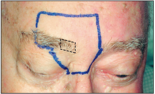 Figure 5.1 The superomedial zone. This region concentrates on the corrugators, procerus, supraorbital, and supratrochlear nerves, and some smaller muscles. Starter fact: The medial orbicularis oculi and depressor supercilii muscles have insertions under the medial brow in area noted. |
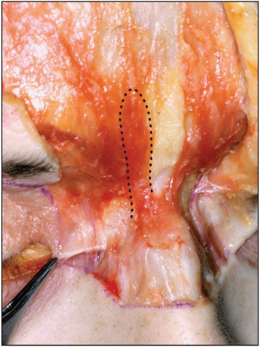 Figure 5.2 New specimen, skin off, shows the medial canthal tendon in forceps. The dotted area denotes the right procerus. Note the fascial insertion over the nasal bones in this case. Sometimes the procerus continues as muscle. The right and left always are separated in the middle. Also note that fat always can be found under the medial procerus edge, which is part of the galeal fat pad and facilitates gliding. |
 Figure 5.3 The midline is marked (black line). A supratrochlear vessel can be noted (red) to supply the paramedian forehead flaps 1.6 to 2.3 cm from the midline. Note that the fat under the medial procerus edge (X) is part of the fat glide plane. |
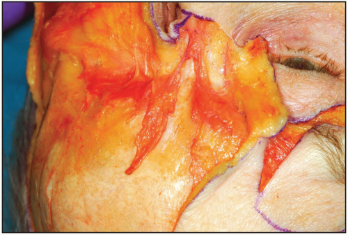 Figure 5.4 The procerus muscles are free on the left, still part of the frontalis on the right. They always tend to meet above or below the nasofrontal groove region. |
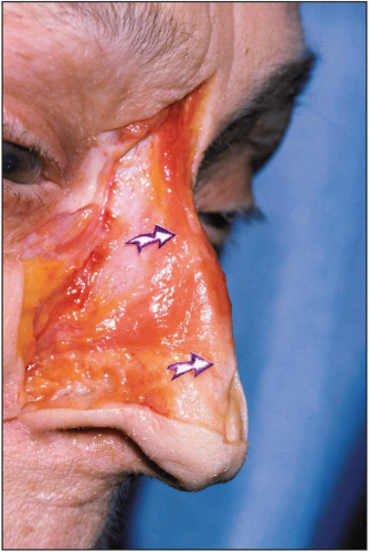 Figure 5.5 In this case, the procerus muscles proceed all the way down to meet the nasalis transverses on both sides. The arrows show the nasalis transverses insertion broadly over the mid-dorsum. The Corrugator(s), Depressor Supercilii, and Key Nerves of the Superomedial Zone 1. The corrugator muscles vary greatly in size. 2. Distinct oblique and transverse heads (as suggested) are not present, although at times two muscles can appear separated by the nerve branches. 3. The corrugators may be quite large; therefore, residual movement, despite so-called “total removal,” occurs frequently, due to partial excision. 4. The origin(s) of the corrugators on the supraorbital ridge of the frontal bone start just over 0.5 cm from the midline and cover an area of 1.0 ×2.5 cm. The vertical height may be considerable. 5. The lateral extent of the corrugators into the underside of the frontalis and beyond occurs from 4.25 to 4.5 cm from the midline. As they insert here, the muscle panels blend all the way to the lateral third of the brow. 6. The corrugators are innervated by the frontal (temporal) branch laterally, and transection or ablation of these nerves may reduce the vertical glabellar lines, but will not totally eliminate them. Nor will this prevent brow depression, as other muscles not innervated by this branch also depress the brow. 7. From the broad-based origin to the lateral third of the eyebrow, the corrugator muscle(s) courses in “panel-like” fashion in an oblique parallel direction. The medial muscle is pierced by supratrochlear branches, which may seem to separate the muscle(s) into separate compartments. 8. The supratrochlear nerve intrusion into the muscle varies in position. Although the supratrochlear nerves come around the rim 1.6 to 2.3 cm (average: 1.9 cm) from the midline, they penetrate the muscle with multiple small branches. The supraorbital nerve, which lies 22 to -32 mm (average: 2.6 cm) from midline, starts with a medial branch that runs posterior to the muscle bundle, and usually one branch penetrates the muscle 1 to -2 cm above the notch.
Stay updated, free articles. Join our Telegram channel
Full access? Get Clinical Tree
 Get Clinical Tree app for offline access

|