Stomatitis


Stomatitis


Richard O. Wein

Miriam O’Leary


This chapter covers the broad category of diagnoses that fall under the diagnosis of stomatitis. The term “stomatitis” indicates an inflammation and potential breakdown of the oral cavity mucosal surfaces and is typically symptomatic. The clinical presentation may extend to the oropharynx or the external perioral region. The etiologies are multiple and include treatment-related, infectious, immunologic, systemic, nutritional, and idiopathic causes.

The diagnoses that fall under this classification can be challenging to identify and require a detailed assessment of a patient’s history of presentation and associated conditions. Additionally, these presentations are not solely seen in the adult population and some forms of stomatitis are unique to the pediatric population.

Given that many of these diagnoses are localized manifestations of systemic diseases, collaborative management of patients with our Dermatology, Infectious Disease, Oral Medicine, and/or Rheumatology colleagues is expected.

Interestingly, despite being encountered daily in general practice, the etiology of many of these oral lesions remains unclear. Many of the advocated treatment options vary dramatically within the medical literature and may only offer symptom control and not affect cure of the underlying diagnosis. As such, this chapter attempts to summarize some of the common perspectives relative to these diagnoses; however, for those seeking more detail on any one condition, the reader is encouraged to review the articles cited within the chapter’s reference list.


CLINICAL ASSESSMENT

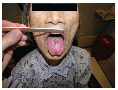
The clinical appearance of stomatitis is variable. Presentations can differ from simple localized to fulminant manifestations and may include components of mucosal erythema, hyperkeratosis, ulceration, pseudomembrane, and/or vesicle formation.

The rate of epithelial turnover in the oral cavity ranges from 3 to 7 days. As a result, this anatomic location is quick to demonstrate visible signs of a metabolic disturbance from a systemic condition or in relation to treatment (1).

Throughout this chapter, a theme should develop relative to the importance of a thorough history of presentation and the assessment for associated diagnoses. Some of the conditions that are discussed are diagnoses of exclusion, while others may represent the earliest presentation of a more significant systemic process.

Pertinent issues that should be addressed in the history include the circumstances and the time course surrounding the onset of symptoms, recent changes in medications, aggressiveness of oral hygiene and product use, palliating/provoking factors, quality of pain and any prodromal symptoms.

A full head and neck examination is necessary in these cases in an effort to assess for synchronous findings beyond the oral presentation. Dentures should be removed and assessed for quality of fit while asking the patient about the original date of manufacture in addition to visits for modification. In cases of buccal mucosal lesions, the relationship of the ipsilateral dentition and any amalgam that contacts the mucosa in a closed mouth position should be identified. Additionally, the quality of salivary production should be assessed.

Exudative lesions should be considered for gram stain and culture (with fungal cultures additionally requested). Tzanck smear and viral cultures should also be considered in patients with vesicular lesions.

Laboratory assay, in cases where a wide differential diagnosis continues to exist after history and physical examination, can include a complete blood count (CBC) with differential, erythrocyte sedimentation rate (ESR), antinuclear antibody (ANA), and cytoplasmic-antineutrophil cytoplasmic antibodies (c-ANCA) in addition to levels
for iron, ferritin, total iron-binding capacity, folate, thiamine (B1), riboflavin (B2), pyridoxine (B6), cobalamin (B12), zinc, and magnesium (2). In xerostomic patients, anti-SSA and anti-SSB can be drawn to assess for Sjogren disease.

Biopsy remains a powerful tool for establishing a definitive diagnosis in patients with stomatitis of unclear etiology. Patients with vesiculobullous lesions should have perilesional tissue sent for direct immunofluorescence (DIF) for deposition of IgG, IgA, and/or complement C3. Biopsy should be considered in patients with suspected recurrent aphthous stomatitis (RAS), oral lichen planus (OLP), or graft-versus-host disease (GVHD) to rule out oral squamous cell carcinoma because of the potential for similarity in appearance.

Patients with a history and findings highly suggestive of an inflammatory bowel condition, such as Crohn disease or ulcerative colitis (UC), should be considered candidates for colonoscopy.


ETIOLOGY

Our understanding of the etiology of treatment-related mucositis at the cellular level continues to evolve. Some aspects may be genetically predetermined and explain the heterogeneity of response between patients to different treatments.

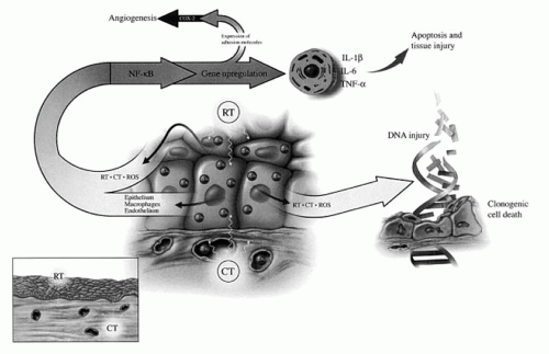
Theories on the development of mucositis have traditionally stated that it was the result of direct damage to the basal epithelial cell layer leading to an impairment of the renewal capacity of the surface epithelium. The resultant clonogenic cell death would result in atrophy and consequent ulceration with the clinical presentation of mucositis.

Additional evidence has suggested that an acute microvascular injury resulting from endothelial apoptosis may play a more significant role in the etiology of mucositis than previously considered. Electron microscopy has demonstrated that endothelial and connective tissue damage precede epithelial changes in irradiated oral mucosa. This finding suggests that the development of treatment-related mucositis is not just an epithelial process.

Sonis et al. (3) describe the five phases of mucosal barrier injury as a result of chemotherapy and/or radiation therapy as:


  • Initiation with generation of reactive oxygen species


  • Up-regulation with generation of messengers including nuclear factor-κB


  • Signaling and amplification with proinflammatory cytokines including tumor necrosis factor-alpha (TNF-α)


  • Ulceration with inflammation with bacterial colonization and pain


  • Healing with epithelial proliferation and the return of local microbial flora


Diagram 51.1 Mucositis as a result of radiation therapy or chemotherapy occurs as a result the creation of reactive oxygen species (ROS) and the stimulation of transcription factors (such as nuclear factor-κB). Resultant double-stranded DNA breaks leads to clonogenic cell death. Up-regulation of TNF-α and the interleukins (IL-6 and IL-1β) leads to apoptosis of cells in the basal epithelium. Additional expression of factors, cyclooxygenase-2, stimulates reactive angiogenesis. (From Sonis ST, Elting LS, Keefe D, et al. Perspectives on cancer therapy-induced mucosal injury: pathogenesis, measurement, epidemiology, and consequences for patients. Cancer 2004;100(9 Suppl):1995-2005, with permission.)


TREATMENT-RELATED STOMATITIS

Stomatitis can occur as a result of multiple forms of therapy; however, it is most evident and has the greatest impact in the treatment of cancer. It is expected that with chemotherapy and radiation therapy some degree of stomatitis, also known as mucositis, development will be experienced. Strategies for prevention, assessment, and treatment of treatment-related mucositis have been well documented; however, variation in findings and recommendations exist.

Assessment tools typically categorize the severity of a presentation by assessing a patient’s well-being, utilizing mouth and throat soreness (MTS) scores, and defining the patient’s functional capacity. Oral function can be assessed through dietary capacity, the ability to eat in public, and overall speech quality. An example of a quality-of-life instrument used in this setting is The Oral Mucositis Weekly Questionnaire—Head and Neck Cancer. It has been shown to be a reliable and valid means for assessing the impact of mucositis on patients undergoing treatment (4).

Administration of 5-fluorouracil, with or without leucovorin, is associated with oral mucositis in as many as 40% of patients with grade 3 and 4 presentations occurring in 10% to 15% of patients (5). In a double-blind randomized study by Sorensen et al., patients undergoing chemotherapy with 5-fluorouracil and leucovorin received prophylactic chlorhexidine or cryotherapy in attempt to decrease treatment-associated mucositis. Prophylactic chlorhexidine (10 mL at 0.1% concentration) was utilized three times per day with 1-minute oral rinses. Cryotherapy was performed with crushed oral ice 10 minutes before and
35 minutes after administration of chemotherapy. The frequency and duration of oral mucositis were significantly improved with both treatment options. Combined use of cryotherapy and chlorhexidine were not examined during this study (6).

Another preventative treatment option, frequently reported in patients with hematologic malignancies, is palifermin. Palifermin is a recombinant human keratinocyte growth factor-1 (hKGF-1) that is considered to offer mucosal protection by inducing epithelial hyperplasia. Typical administration is in multiple pre- and posttreatment administrations of 60 µg/kg. Use of single-dose palifermin was studied in sarcoma patients receiving doxorubicin chemotherapy in a randomized, double-blind, placebocontrolled trial. Single-dose intravenous palifermin (180 µg/kg body weight), administered before each cycle, was noted to significantly reduce the incidence and severity of mucositis experienced. During the trial, the drug was well tolerated and majority of patients experienced thickening of oral mucosa and tongue. Biopsies of the affected areas noted increased levels of proliferative marker Ki-67 suggesting a palifermin-associated etiology (7).

The medications associated with treatment-associated mucositis are not limited to the conventional chemotherapeutic options but also include agents like hydroxyurea (8) and the newer targeted therapies such as imatinib mesylate (9), cetuximab, panitumumab, gefitinib, erlotinib, rapamycin, sorafenib tosylate, and lapatinib (10).

Mucositis associated with radiation therapy is a common finding that tends to be self-limited after the completion of treatment. Mucositis typically peaks between weeks 5 to 6 during treatment as assessed by MTS scores (11). Radiation-associated mucositis is worse in patients undergoing radiation with accelerated regimens (5). Grade 3 and 4 mucositis requiring gastrotomy tube placement is more significant with chemoradiation protocols (38%) than compared to radiation alone (18%). The risk of developing mucositis is the same for patients undergoing treatment of an oral cavity and oropharyngeal cancer as those treated for a laryngeal and hypopharyngeal cancer.

Prophylactic pilocarpine has been shown to decrease the incidence and severity of treatment-associated mucositis that is presumed through the up-regulation of salivary function during treatment (12). However in contrast, in a randomized phase III prospective trial of bethanechol, which has a similar ability to stimulate salivary function, prophylactic treatment did not prevent mucositis or candidiasis in head and neck cancer patients undergoing radiotherapy (13).

In 2004, the Multinational Association of Supportive Care in Cancer and the International Society for Oral Oncology (MASCC/ISOO) offered a consensus statement with recommendations on treatment-associated mucositis.

In the setting of radiation therapy, the MASCC/ISOO advocated the use of benzydamine, a nonsteroidal antiinflammatory that inhibits TNF-α (not available in the United States), and did not support the use of chlorhexidine as a prophylactic for mucositis (5).

In patients treated with chemotherapy, the prophylactic use of acyclovir was not recommended. Cryotherapy was not advocated in the setting of treatment with 5-fluorouracil, with or without leucovorin. Additionally, the panel did not support the prophylactic use of pentoxifylline or low-level laser therapy in patients preparing for stem cell transplant (5).

Novel agents requiring additional evaluation include Gelclair, AES-14, and human keratinocyte growth factor-1 (hKGF-1) and hKGF-2. Gelclair, a mixture of polyvinylpyrrolidone, sodium hyaluronate, and glycyrrhetinic acid, has demonstrated improved pain scores during treatment yet clinical trial data were lacking. AES-14, a proprietary form of administering L-glutamine, demonstrated the capacity to decrease grade 2 or greater mucositis when compared to placebo. Repifermin (hKGF-2) was shown to reduce grade 2 to 4 mucositis in clinical trials, and head and neck cancer patients receiving palifermin (hKGF-1) experienced a lower incidence and shorter duration of mucositis when compared to placebo (5).

The Cochrane Collaboration reviewed 131 studies with over 10,000 randomized patients in assessing treatment options for the prevention of oral mucositis in patients receiving cancer treatment. Less than 10% of the studies were considered to have a low risk of bias associated with their performance. The therapies shown to offer some benefit compared to placebo included: aloe vera, amifostine, cryotherapy, granulocyte-colony stimulating factor, intravenous glutamine, honey, keratinocyte growth factor, polymyxin/amphotericin antibiotic paste, and sucralfate. The strength of the evidence was described as variable (14).


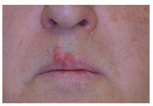
Graft-Versus-Host Disease

Acute GVHD is dependent upon type I cytokine-driven CD8 effectors, while chronic graft-versus-host disease (cGVHD) has traditionally been associated with type II CD4 T cells (
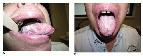
15). The median onset of GVHD is 6 months after transplantation with oral manifestations seen in up to 80% of patients.

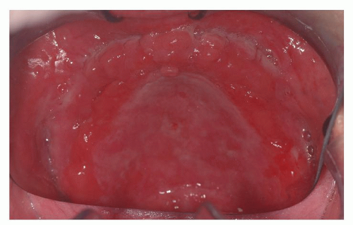
The clinical presentation includes xerostomia, oral ulceration, altered capacity for taste, and recurrent candidal infections (16) (Fig. 51.1A and B). The clinical appearance can mimic oral squamous carcinoma. Of note, patients who have undergone hematopoietic stem cell transplant are at increased risk of developing oral squamous cell carcinoma and should have regular surveillance exams with biopsy performed for suspicious lesions (
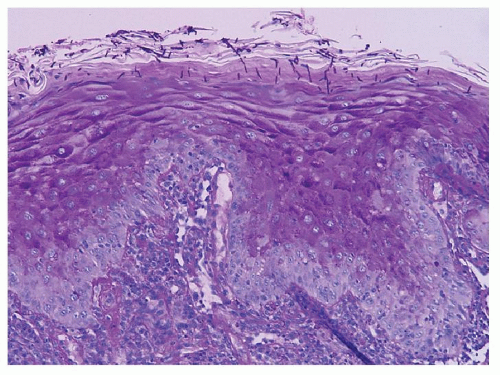
17). Histologic findings of GVHD on biopsy include hydropic degeneration of the basal layer of the epithelium, intracellular edema, apoptotic bodies, fibrosis, and lymphocytic infiltrate in the submucosa (18).

The current therapy for cGVHD is nonspecific immunosuppression.


Figure 51.1 GVHD of the lateral (A) and dorsal (B) tongue in a patient with prior history of bone marrow transplant.


Contrary to conventional belief, Imanguli et al. believe that cGVHD is not a type II cytokine driven disorder. They advocate a model that oral cGVHD results from type I interferon (IFN)-driven proliferation and differentiation of Th1/Tc1 differentiated effectors. This concept was supported by the finding of T cells in cGVHD infiltrates expressing T-bet, a transcription factor critical for Th1/Tc1 polarization and the production of IFN-γ and granzyme B. With these new findings, the authors suggest that new treatment approaches for cGVHD should focus on affecting the IFN-mediated inflammatory axis (15).


Lichenoid Reactions

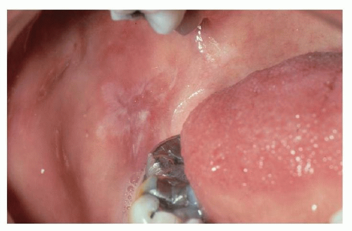
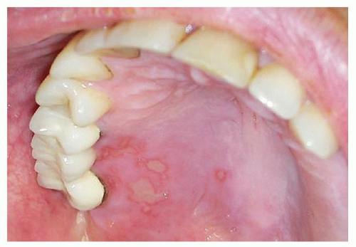
Lichenoid reactions of the oral cavity represent an immunologic response to a systemic or local stimulus. Reactions can present as a contact stomatitis, as seen in the buccal mucosa opposing a dental amalgam (Fig. 51.2), or may represent a drug reaction from the initiation of a common medication. An extensive number of oral medications have been associated with lichenoid drug reactions (Table 51.1). The most common agents responsible for oral lichenoid drug reactions are nonsteroidal anti-inflammatories, antihypertensives, and HIV antiretrovirals. Presentation of a lichenoid reaction can vary widely but typically occurs within 2 to 3 months after initiation of a medication (19).


Figure 51.2 Lichenoid contact reaction from local amalgam.

In addition to dental amalgam, contact lichenoid stomatitis has been reported with cinnamon flavoring (as a result of cinnamic aldehyde) and other flavorings commonly found in mints and mouthwashes. A history of a recent change in the use of oral hygiene products or variation in the habitual use of a mint or gum brand may help elucidate this risk factor.


TABLE 51.1 MEDICATIONS ASSOCIATED WITH ORAL LICHENOID DRUG REACTION


Tetracycline


Chloroquine


Carbamazepine


Quinine


Phenytoin


Quinidine


Valproic acid


Isoniazid


Chlorpropamide


Rifampin


Glipizide


Streptomycin


Insulin


Zidovudine


Tolbutamide


Imatinib


Bismuth


Dactinomycin


Amphotericin B


INF-α


Ketoconazole


Gold salts


Atenolol


Penicillamine


Enalapril


Aspirin


Hydrochlorothiazide


Ibuprofen


Methyldopa


Naproxen


Metoprolol


Benzodiazepines


Lithium


Tricyclic antidepressants


Indomethacin


Allopurinol


Adapted from Schlosser BJ. Lichen planus and lichenoid reactions of the oral mucosa. Dermatol Ther 2010;23(3):251-267.


Individuals with contact allergy to inorganic mercury amalgam present with an oral mucosal lesion within 1.0 cm of the suspected offending restoration, and they lack associated cutaneous lichen planus (CLP). Patch testing to inorganic mercury has been advocated for recalcitrant presentations and should be read on days 7, 10, and 14 in an effort to identify late reactions. In the setting of a positive patch test, subsequent improvement of oral lesions is highly likely with removal and replacement of the restoration. Contact lichenoid reactions are not unique to mercury and may also occur to gold and palladium (20,21).


INFECTIOUS


Candidiasis

Oral candidiasis is the most common form of fungal stomatitis. Approximately 30% to 60% of individuals carry candidal species as part of their normal oral flora (22,23). Candida albicans is the most common candidal species in the oral cavity and accounts for up to 80% of oral isolates.

Multiple local and systemic factors can lead to candidal overgrowth. Recurrent oral candidiasis is typically seen in patients with poorly controlled diabetes, HIV-positive patients (with decreased CD4 counts), at the extremes of age (premature and elderly), and in patients with xerostomia. Use of broad-spectrum antibiotics and topical or inhalational steroids can also predispose to candidal overgrowth (22,23,24).

The most recognized clinical presentation of Candida is readily diagnosed but other forms of oral candidal infection may be more difficult to identify. The category of erythematous candidiasis is actually the most common form of oral candidiasis and presentations under this subheading are varied. Erythematous candidiasis includes acute and chronic atrophic candidiasis, angular cheilitis, median rhomboid glossitis, and chronic multifocal candidiasis (22,23).

Multiple classifications are reported for oral candidal infections. Table 51.2 presents a system that delineates candidal infections into five categories (24).

Pseudomembranous candidiasis, also known as thrush, is common in immunocompromised populations and may be diagnosed in all age groups. White creamy plaques on an erythematous mucosal base may be seen throughout the oral cavity with various degrees of associated symptoms. Extension of thrush to involve the pharynx and esophagus may also occur and present with dysphagia and odynophagia (23,24).

Acute atrophic candidiasis is also known as “antibiotic sore mouth.” It occurs as a result of use of broad-spectrum antibiotics. Patients complain of oral burning and present with an erythematous patches on the buccal mucosa or dorsal tongue with papillary atrophy noted on examination. Chronic atrophic candidiasis is seen with denture use and is discussed in greater detail below.

Chronic hyperplastic candidiasis, also known as candidal leukoplakia, is the least common form of oral candidal infection. It presents on the buccal mucosa as a superficial white lesion that is incapable of being wiped away from the underlying mucosa. If the lesion does not resolve with use of antifungal therapy, leukoplakia should be viewed as more likely representing the diagnosis and biopsy considered (23,24). Candidal-associated diagnoses include actinic cheilitis, median rhomboid glossitis, and linear gingival erythema.


TABLE 51.2 CLASSIFICATIONS OF VARIOUS TYPES OF ORAL CANDIDIASIS


Pseudomembranous (exudative) candidiasis


(Exudative candidiasis)


Acute atrophic (erythematous) candidiasis


Median rhomboid glossitis


Oral candidiasis


Chronic atrophic candidiasis


(Denture stomatitis)


Chronic hyperplastic candidiasis


(Candidal leukoplakia)


Candida-associated diagnoses


Actinic cheilitis


Median rhomboid glossitis


Linear gingival erythema


Parentheses indicate additional names used to describe the above-listed entity.


Adapted from Sharon V, Fazel N. Oral candidiasis and angular cheilitis.


Dermatol Ther 2010;23(3):230-242.


Angular cheilitis, also know as perleche (Fig. 51.3), represents a combination of fungal (C. albicans) and bacterial (Staphylococcus aureus) infections. Pain and erythema with fissuring at oral commissure are noted. This finding is common in denture wearers and is the result of the loss of alveolar bone height over time with the pooling of secretions at the oral commissure. In young dentate patients, infection with HIV should be considered as a potential diagnosis in recurrent presentations (23,24).

Median rhomboid glossitis, also known as central papillary atrophy, presents at the midline dorsal oral tongue.
The smooth or lobular demarcated region anterior to the circumvallate papillae is typically asymptomatic in presentation (Fig. 51.4). A mirrored palate lesion may be seen from direct contact with the tongue (23,24).


Figure 51.3 Angular cheilitis.


Figure 51.4 Median rhomboid glossitis in a patient with a history of Sjogren disease and immunosuppression.

The diagnosis of chronic multifocal candidiasis, separate from the classification above, is assigned to those patients with more than one form of candidal infection (e.g., angular cheilitis and denture stomatitis) that is present for greater than 4 weeks with the lack of predisposing risk factors (immunosuppression, xerostomia, medications) for candidal infection (23).

The diagnosis of oral candidiasis can be established solely on clinical exam or may be augmented with the use of exfoliative cytology, culture, or biopsy. On smear, budding oval yeast forms with nonbranching septate pseudohyphae with blastocysts are noted. On biopsy specimens, fungal elements invading the parakeratin layer of the epithelium can be seen (Fig. 51.5) (23,24).


Figure 51.5 Periodic acid-Schiff staining of a biopsy specimen with candidal invasion of the parakeratin layer of epithelium.

A wide variety of topical and systemic treatment options are available. For the limited common forms of acute infection, topical agents are typically employed prior to use of to systemic antifungal agents. Topical therapies come in the form of lozenges (clotrimazole), tablets (miconazole), suspensions (nystatin and amphotericin B), and creams (ketoconazole). Oral agents typically include the azole class of antifungals (fluconazole, ketoconazole, and itraconazole) (23,24).

The length and type of treatment is dependent upon the presentation (acute vs. chronic), patient characteristics (immunocompromised), and response to the initiation of therapy. Although the length of treatment varies with respective medication, treatment for at least 2 to 3 weeks is common with most agents. Drug resistant forms of Candida may be seen in patients requiring longer courses of antifungal therapy. Oral agents are typically used in patients at recalcitrant presentations or when a significant risk of developing systemic infection exists (22,23,24).


Denture-Related

Denture-related stomatitis occurs in up to 70% of denture wearers and is highest in the elderly, women, smokers, and individuals with compromised immune function. Causes include includes poor hygiene and continual (night-time) denture use. Colonization with C. albicans and with the formation of resistant biofilms on the denture result in a presentation that is resistant to use of nystatin oral rinses. Culture of the denture can help confirm the diagnosis (24,25).

Denture stomatitis, also known as chronic atrophic candidiasis, has been described to have three subtypes of presentation that include:


Type I—localized inflammation

Type II—diffuse erythema involving the denture-bearing mucosa

Type III—papillary hyperplasia on mucosa (Fig. 51.6) (23,24).


Figure 51.6 Erythema and papillary hyperplasia seen with denture stomatitis.


Poor fitting dentures exacerbate oral trauma through rough edges and inadequate contact spots and should be addressed with routine maintenance. The importance of sanitization with commercially available denture cleaners needs to be emphasized to address the formation of biofilms. Treatment with oral fluconazole or topical clotrimazole cream and nystatin (to the mouth and denture) may require 1 month of treatment for resolution (25).


Noncandidal Fungal Etiologies

Noncandidal etiologies of fungal infections of the oral cavity that can present as stomatitis include paracoccidioidomycosis, histoplasmosis, and mucormycosis.

Paracoccidioidomycosis is primarily seen in South America (Brazil, Colombia, and Venezuela) and is associated with smoking, poor nutrition, and alcohol abuse. It affects the mucous membranes, lymph nodes, lungs, and bones. Chronic presentations are more common than subacute and more likely to be associated with mucosal lesions. Painful oral cavity lesions are seen in about 60% of patients. The gingiva is the most commonly affected site with involvement also seen in the soft palate, lower lip, and tongue (26). The classical clinical presentation is a superficial ulcer with granular appearance and hemorrhagic points (Table 51.3). Treatment involves use of antibacterials (sulfamethoxazole) and antifungals (itraconazole and amphotericin B) and is dependent upon the clinical presentation (27).

Histoplasmosis is caused by the organism Histoplasma capsulatum. It is most commonly found in the Ohio and Mississippi River Valley regions of the United States and in South America (Argentina and Brazil). Infection results from inhalation of fungal spores that can be found in caves, as a result of birds and bats, or from working around chickens. Infection is most problematic in immunocompromised patients. Mucocutaneous lesions are more common in South America than is seen with the North American HIV-infected patients (27). Oral lesions vary and may present as papules, patches, ulcers, or vegetations. Patients may present with only oral ulceration without the finding of extraoral manifestations of histoplasmosis (28). The clinical appearance of the oral findings may mimic squamous cell carcinoma. Treatment involves antifungals, such as itraconazole and amphotericin B, and may require lifetime suppressive therapy in immunocompromised populations (27).


TABLE 51.3 LIST OF THE MOST COMMON CAUSES OF GRANULOMATOUS ORAL/PERIORAL CONDITIONS


Foreign body reaction (suture, amalgam)


Tuberculosis


Leprosy


Cat-scratch disease


Tertiary syphilis


Histoplasmosis


Cryptococcosis


Blastomycosis


Paracoccidioidomycosis


Sarcoidosis


Crohn disease


ORG


Adapted from Alawi F. Granulomatous diseases of the oral tissues: differential diagnosis and update. Dent Clin North Am 2005;49: 203-221.


Mucormycosis is a life-threatening invasive fungal infection that occurs predominantly in patients with diabetes who are prone to ketoacidosis. It may also present in neutropenic and posttransplant patients or associated with malignancy (leukemia). Palatal ulceration and necrosis are classic for the rhinocerebral presentation of mucormycosis (29). Vascular invasion by hyphae with thrombosis and infarction of tissue is seen on biopsy specimens. A mortality rate of greater than 40% exists. Treatment requires the removal of the underlying risk factor, aggressive surgical intervention, and systemic antifungal therapy (27).


Viral


Herpes Simplex Virus

Primary herpetic gingivostomatitis is predominantly the result of herpes simplex virus-1 (HSV-1) and occurs in 25% to 30% of children (30). Clear yellowish pinhead vesicles develop after a prodrome of fever, anorexia, and malaise. Vesicles appear on keratinized tissue such as the attached gingiva, hard palate (Fig. 51.7), and the lip at the vermillion border. The vesicles contain active virus and are considered very contagious; however, the contagion potential drops rapidly after rupture. Rupture occurs within hours revealing irregular ulcers covered by a yellow-gray membrane. The oral presentation is variable in extent but is typically asymptomatic. Lymphadenitis and odynophagia may accompany the presentation. Healing typically occurs in 7 to 10 days. Primary herpetic gingivostomatitis in an adult may present very similar to mononucleosis with predominantly pharyngotonsillar symptoms (22,30).


Figure 51.7 Herpes ulceration of the hard palate.


Figure 51.8 Herpes labialis.


During the asymptomatic latent state, the virus resides within the sensory ganglion of the trigeminal nerve. Exposure to a stressor leads to viral replication and reexpression with a subsequent vesicle formation. Stressors may include fever, radiation, chemotherapy, HIV infection, UV light exposure, pregnancy, and surgery (22,30).

Secondary or recurrent herpes affects labial and perilabial surfaces (cold sores) in the majority of cases and is typically unilateral (Fig. 51.8). Secondary presentations within oral cavity are uncommon yet when present occur on gingiva or palate mucosa and do not occur on mobile soft tissues. Multiple small vesicles may coalesce to larger lesions. Treatment is dictated by symptoms. Lubrication may reduce crusting of overlying lesions and enhance healing.

Treatment options include the use of topical agents and systemic antivirals; however, therapy primarily directed to symptom control. When treatment is initiated at the time of prodromal symptoms, reduction in healing time and pain reduction has been reported. Antiviral therapy is unnecessary in the setting of primary infection unless concern over progression to encephalitis arises. Long-term clinical ulceration should prompt investigation for other vesiculobullous conditions rather than an HSV-associated diagnosis (30).


HIV-Associated Stomatitis

HIV-positive patients presenting opportunistic oral lesions are likely to have higher viral loads and lower CD4 counts. Stomatitis may be the first manifestation of an undiagnosed HIV infection. Awareness of this association can prompt HIV testing and facilitate early diagnosis and treatment (31). In the HIV-positive patient undergoing treatment, emergence of stomatitis can signify the failure of an existing antiretroviral regimen.

Oral presentations seen in the HIV-positive population includes candidiasis, oral hairy leukoplakia, recurrent aphthous-like ulceration, orolabial HSV, oral warts, and HIV-associated periodontal disease such as linear gingival erythema (32).

Pseudomembranous candidiasis is the most common form of stomatitis seen in HIV-positive patients (31). Prophylactic fluconazole is not recommended except for in severe or recurrent cases because of concerns it could lead to multidrug resistance. Topical agents are recommended for patients with a CD4 counts greater than 200 cells/mm3. Culture and sensitivity is advised if the candidiasis does not respond to initial therapy (32,33).

For RAS, initial treatment approaches with topical and systemic steroids is the same for HIV-positive as for HIVnegative patients. Thalidomide has been used in severe refractory aphthous stomatitis in two randomized, doubleblind, placebo-controlled trials; however, drug toxicity can limit treatment. Cytomegalovirus (CMV) may also present with painful oral ulcerations and can be associated with the subsequent development of CMV-associated retinitis and encephalitis (32).

Only gold members can continue reading. Log In or Register to continue

Stay updated, free articles. Join our Telegram channel

May 24, 2016 | Posted by in OTOLARYNGOLOGY | Comments Off on Stomatitis

Full access? Get Clinical Tree

Get Clinical Tree app for offline access