Sinonasal Anatomy and Physiology
Randy M. Leung
William E. Walsh
Robert C. Kern
EMBRYOLOGY
Embryologic development of the nasal cavity and sinuses leads to the intricate features of sinonasal anatomy and can be divided into two ongoing processes. First, the embryo’s head develops into a structure with two distinct nasal cavities; second, the lateral nasal walls then invaginate to create complex folds, known as turbinates, and spaces, known as sinuses. During the fourth to eighth gestational weeks, the embryo develops separate nasal cavities as the frontonasal and maxillary processes join. The frontonasal process grows over the developing forebrain, contributing to the formation of the nasal olfactory placodes. Medial and lateral nasal prominences develop on either side of the placode eventually becoming the nares. The nasal placode invaginates to form the nasal pit and eventually the nasal sac. Fusion of the medial nasal prominence with the maxillary process forms the upper maxilla and the philtrum of the upper lip (Fig. 23.1). The septum arises from the posterior midline growth of the frontonasal process and midline extensions of mesoderm from the maxillary processes. The primary and secondary palatal shelves join in an axial plane to separate the nasal cavity and nasopharynx from the oral cavity and oropharynx. The descending septum merges with the fused palate to create two distinct nasal cavities (Fig. 23.1). Fusion failure of the medial nasal prominence with the maxillary process or fusion failure of the palatal shelves results in a cleft lip or palate deformity. Because clefting can extend into the nose, rhinoplasty to correct an associated nasal deformity is often technically difficult.
During the sixth gestational week, mesenchyme forms a simple lateral nasal wall. During the seventh week, three axial furrows form, giving rise to the three turbinates (Fig. 23.1). During the 10th week, development of the maxillary sinus begins with the invagination of the middle meatus. At the same time, the uncinate process and the bulla ethmoidalis form a narrow groove known as the hiatus semilunaris. During the 14th week, the anterior ethmoidal cells appear as several invaginations from the upper middle meatus and the posterior ethmoidal cells from the floor of the superior meatus. Finally, by the 36th week the lateral nasal wall is well developed and the turbinates are at adult proportions. All paranasal sinuses are present to varying degrees in the newborn, but sinuses have specific periods of significant growth. The ethmoid sinuses are the first to fully develop, followed in order by maxillary, sphenoid, and frontal sinuses.
ANATOMY
Refer to Figure 23.2 for illustrations of sinus anatomy discussed in the text.
Ethmoidal Sinuses and Lateral Nasal Wall
The ethmoid sinuses are the central structures of the nose with complex anatomy; they are best visualized as a boxlike structure with open anterior and inferior faces. The lateral portions form the medial walls of the orbits, the sphenoid establishes the posterior face, the superior surface is formed by the skull base of the anterior cranial fossa, and many of the key structures of the lateral nasal wall, derived from basal lamellas, extend posteroinferiorly from the skull base.
The lateral wall of the ethmoid sinus, or lamina papyracea, forms the paper-thin medial wall of the orbit. The midline vertical plate of the ethmoid bone is composed of a superior portion in the anterior cranial fossa called the crista galli and an inferior portion in the nasal cavity called the perpendicular plate of the ethmoid bone that contributes to the nasal septum. The anterior cranial fossa is separated from the ethmoid air cells superiorly by the horizontal plate of the ethmoid bone, which is composed of the thin medial cribriform plate and the thicker, more
lateral ethmoid roof. The ethmoid roof articulates with the cribriform plate at the lateral lamella of the cribriform plate, which is the thinnest bone in the entire skull base. The length of the lateral lamella depends on the position of the cribriform plate with respect to the ethmoid roof. In a Keros type 1 skull base, the plate is located 1 to 3 mm below the roof of the ethmoid, making the lateral lamella short or nonexistent (Fig. 23.2B). In Keros type 2, the distance is 4 to 7 mm. In Keros type 3, it is 8 to 16 mm, thus giving rise to a long vertical lateral lamella (Fig. 23.2C). Patients with a low cribriform plate, Keros III in particular, are believed to be at greatest risk for cerebrospinal fluid leak during endoscopic sinus surgery.
lateral ethmoid roof. The ethmoid roof articulates with the cribriform plate at the lateral lamella of the cribriform plate, which is the thinnest bone in the entire skull base. The length of the lateral lamella depends on the position of the cribriform plate with respect to the ethmoid roof. In a Keros type 1 skull base, the plate is located 1 to 3 mm below the roof of the ethmoid, making the lateral lamella short or nonexistent (Fig. 23.2B). In Keros type 2, the distance is 4 to 7 mm. In Keros type 3, it is 8 to 16 mm, thus giving rise to a long vertical lateral lamella (Fig. 23.2C). Patients with a low cribriform plate, Keros III in particular, are believed to be at greatest risk for cerebrospinal fluid leak during endoscopic sinus surgery.
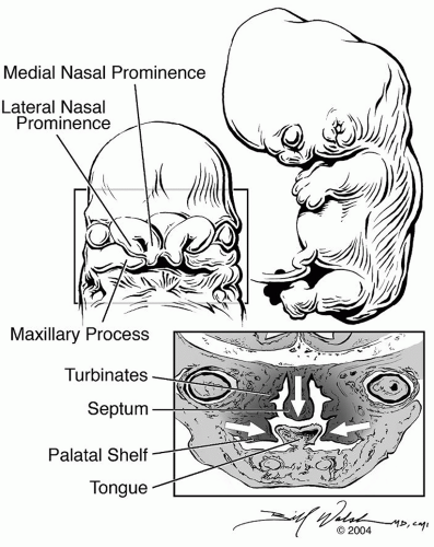 Figure 23.1 A 7-week gestational age embryo. Cross-section in bottom right shows the formation of turbinates and the separation of the nasal cavities by fusion of the nasal septum with the palatal shelves. Illustration by William E. Walsh, MD, CMI ©2004, used with permission. |
The ethmoid sinuses are separated by a series of recesses demarcated by five bony partitions or lamellae. These lamellae are named from the most anterior to posterior: first (uncinate process), second (bulla ethmoidalis), third (basal lamella), fourth (superior turbinate), and fifth (supreme turbinate). These partitions aerate during development forming the ethmoid air cells. If the aeration projects anterior to the attachment of the middle turbinate, the air cells are termed agger nasi cells. Continuing posteriorly, the uncinate process is an L-shaped bone that runs in an anterosuperior to posteroinferior direction. The posterosuperior margin of the uncinate runs parallel to the anterior border of the bulla ethmoidalis and the posterior end attaches to the palatine bone and the inferior turbinate. The superior portion of the uncinate most commonly attaches to the lamina papyracea but can also attach to the posteromedial wall of the agger nasi cell, the skull base, or the middle turbinate. The bulla ethmoidalis, or second lamella, gives rise to the most constant and usually largest anterior ethmoid air cell. It attaches laterally to the lamina papyracea and to a varying degree posteriorly to the ground lamella. Superiorly, the bulla may reach the roof of the ethmoid and form the posterior wall of the frontal recess. Minimal or absent pneumatization of the ethmoid bulla occurs in 8% of individuals (1). The basal lamella marks the dividing line between the anterior and posterior ethmoid sinuses. The inferior portion of the basal lamella connects the middle turbinate to the lateral nasal wall obliquely transitioning from a coronal plane anteriorly to an axial plane posteriorly. Preservation of the lower portion of the basal lamella during endoscopic sinus surgery provides stability to the middle turbinate. Posterior ethmoidal cells are generally larger and can pneumatize laterally and superiorly to the sphenoid sinus. This developmental variant is known as an Onodi cell, a risk factor for dehiscence and potential injury to the optic nerve during surgery.
The lamellae of the ethmoid sinuses are separated by a series of four recesses: the frontal recess, the infundibulum, the sinus lateralis, and the sphenoethmoidal recess. The frontal recess drains the frontal sinus; the anatomy is highly variable depending on the pneumatization patterns of the bulla ethmoidalis and agger nasi air cells (2). The ethmoidal infundibulum is a three-dimensional space lateral to the uncinate process. Between the free concave posterior margin of the uncinate process and the convex anterior face of the bulla ethmoidalis is a two-dimensional cleft called the hiatus semilunaris that serves as the door that leads anteriorly into the infundibulum. The ostium of the maxillary sinus lies deep within the ethmoidal infundibulum lateral to the uncinate process. The ethmoid sinuses anterior to the basal lamella, the maxillary sinus, and the frontal sinus all drain directly into or near the infundibulum. The ostiomeatal complex (OMC) refers to the area bounded by the middle turbinate medially, the lamina papyracea laterally, the basal lamella posteriorly, and the ethmoid roof superiorly (1). A sinus lateralis or retrobullar recess exists if the posterior wall of the bulla ethmoidalis aerates to become distinct from the basal lamella. The sphenoethmoidal recess is located at the posterior end of the superior meatus draining the posterior ethmoid and sphenoid sinuses separately, outside of the OMC.
The anterior ethmoid artery originates from the ophthalmic artery in the orbit and passes through the anterior ethmoidal foramen to enter the anterior ethmoidal cells. The artery typically crosses the ethmoids very near the skull base at the junction of the ethmoid roof and marks the posterior border of the frontal recess; the artery travels within a bony canal that may be partially or completely dehiscent in 40% of cases (1). The area where the anterior ethmoid
artery enters the anterior cranial fossa through the lateral lamella is the weakest portion of the skull base, being only one-tenth as strong as the roof of the ethmoid (1).
artery enters the anterior cranial fossa through the lateral lamella is the weakest portion of the skull base, being only one-tenth as strong as the roof of the ethmoid (1).
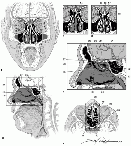 Figure 23.2 Sinus anatomy. A: Coronal section through the ostiomeatal complex with the left uncinate attaching medially to the septum. 1, right uncinate process; 2, maxillary sinus ostium; 3, ethmoidal infundibulum; 4, hiatus semilunaris; 5, bulla ethmoidalis; 6, perpendicular plate of the ethmoid bone; 7, crista galli; 8, lamina papyracea; 9, left uncinate process attaching medially to the septum; 10, middle turbinate; 11, Haller cell; 12, maxillary sinus; 13, inferior turbinate. B: Keros 1 skull base with the uncinate processes attaching superiorly to the skull base. 14, left uncinate process attaching superiorly to the skull base. C: Keros 3 skull base with uncinate processes attaching laterally to the lamina papyracea. 15, cribriform plate; 16, lateral lamella; 17, ethmoid roof; 18, left uncinate process attaching laterally to the lamina papyracea. D: Sagittal view of the lateral nasal wall. 19, inferior turbinate; 20, middle turbinate; 21, frontal sinus; 22, crista galli; 23, superior turbinate; 24, sphenoid sinus. E: Close-up sagittal view of the lateral nasal wall with the middle turbinate removed. 25, uncinate process; 26, agger nasi cell; 27, frontal ostium; 28, bulla ethmoidalis; 29, middle turbinate cut edge; 30, superior turbinate cut edge; 31, optic nerve prominence in the sphenoid sinus; 32, carotid artery prominence in the sphenoid sinus; 33, sphenoid sinus ostium; 34, inferior turbinate cut edge; 35, nasolacrimal duct. F: Axial view. 36, septum; 37, ethmoid cell; 38, Onodi cell; 39, optic nerve; 40, carotid artery; 41, sphenoid sinus. Illustration by William E. Walsh, MD, CMI ©2004, used with permission. |
Maxillary Sinus
The maxillary sinus is the pneumatized space within the maxillary bone and is the largest of the paranasal sinuses. The anterior wall derives from the facial surface of the maxilla, the posterior wall borders the pterygopalatine fossa, the medial wall constitutes the lateral wall of the nasal cavity, the floor of the sinus is the alveolar process, and the superior wall serves as the orbital floor. The infraorbital nerve crosses the orbital floor to exit the anterior portion of the maxilla via the infraorbital foramen. The canal for the infraorbital nerve is dehiscent into the maxillary sinus in 14% of cases and may be at risk during endoscopic sinus surgery. The first and second molar tooth roots are dehiscent into the maxillary sinus occurring in 2% of cases. These patients are at risk for development of an oroantral fistula following dental extraction at these sites.
The natural ostium of the maxillary sinus opens into the superior aspect of the medial wall to drain into the ethmoidal infundibulum. Accessory maxillary sinus ostia are found in 15% to 40% of subjects, most commonly superior and posterior to the uncinate process above the insertion of the inferior turbinate. Occasionally a Haller cell, or ethmoidal cell that pneumatizes laterally between the maxillary sinus and the floor of the orbit, may be present. The presence of Haller cells can potentially narrow the maxillary infundibulum and impair sinus drainage (3).
Frontal Sinus
The size of the frontal sinus varies depending on the degree of pneumatization, may be completely absent (5%), and is usually divided by an intersinus septum. The anterior table of the frontal sinus is twice as thick as the posterior table, which separates the sinus from the anterior cranial fossa. The floor of the sinus also functions as the supraorbital roof, and the drainage pathway is located in the posteromedial portion of the sinus floor. Drainage of the frontal sinus is complex with its outflow tract resembling an hourglassshaped structure in the sagittal plane (1,2). The superior portion widens into the frontal sinus and the inferior portion expands into the frontal recess. The variability of the frontal sinus outflow tract drainage pattern depends on the pneumatization of the surrounding ethmoid air cells and the position of the uncinate process. A markedly pneumatized agger nasi cell or ethmoidal bulla can obstruct frontal sinus drainage by narrowing the frontal recess. Drainage of the frontal sinus also depends on the attachment of the superior portion of the uncinate process (4). In the most common variation, the anterosuperior portion of the uncinate process inserts onto the lamina papyracea so that the uncinate process separates the ethmoidal infundibulum from the frontal recess. In this setting, the frontal recess opens into the middle meatus medial to the ethmoidal infundibulum, between the uncinate process and the middle turbinate (Fig. 23.2C). When the uncinate process inserts onto the ethmoid roof (Fig. 23.2B) or inserts onto the middle turbinate (Fig. 23.2A), the frontal recess opens directly into the ethmoidal infundibulum, and is thought to be subject to obstruction in the presence of ethmoid inflammation. During endoscopic sinus surgery, the opening of the frontal sinus is often more medial than anticipated. The frontal sinus opens into the middle meatus medial to the uncinate process in 88% of patients and lateral to the uncinate in the remaining 12% of patients.
Sphenoid Sinus
The sphenoid sinus has many important neurovascular relationships. The internal carotid artery is lateral to the sphenoid sinus as it courses through the cavernous sinus producing a prominence in the lateral sphenoid sinus wall in 65% of individuals (3). Approximately 25% of bony capsules separating the internal carotid artery from the sphenoid sinus are partially dehiscent. An optic nerve prominence is present in 40% of individuals with dehiscence in 6% (3). The visibility of all structures related to the walls of the sphenoid sinus depends on the degree of pneumatization of the sinus. The degree of pneumatization is classified into three types: sellar type (86%), presellar (11%), and conchal type (3%) (3). Presellar and conchal types are more common in children due to the normal development of the sphenoid sinus reaching completion at 20 years of age. In a sellar type sphenoid sinus, the superior wall pneumatizes inferiorly to the sella turcica and the pituitary gland. The posterior wall of the sphenoid sinus is the clival wall and is the thickest wall of the sphenoid sinus.
The sphenoid sinus ostium opens into the sphenoethmoidal recess. An anatomic study of the sphenoid sinus ostium identified the posteroinferior end of the superior turbinate as the best landmark for identifying the natural ostium of the sphenoid sinus (5). In most cases, the posteroinferior end of the superior turbinate was located in the same horizontal plane as the floor of the sphenoid sinus. The ostium was located medial to the superior turbinate in 83% of cases and lateral to it in 17%. The sphenoid septum usually deviates posteriorly from the midline dividing the sinus into two asymmetric parts and can insert onto the bony prominences overlying the optic nerve or carotid artery.
Inferior Turbinate
The inferior turbinates are bilateral outgrowths from the lateral wall of the nasal cavity composed of a central bony skeleton covered by a mucosal layer. Each inferior turbinate articulates with the perpendicular plate of the palatine bone and the nasal surface of the maxilla. The inferior turbinate swells and shrinks to regulate nasal temperature and humidification via a rich vascular arcade.
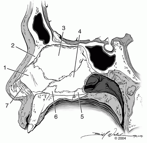 Figure 23.3 Nasal septum. 1, quadrangular cartilage; 2, nasal bone; 3, perpendicular plate of ethmoid bone; 4, vomer; 5, nasal crest of palatine bone; 6, nasal crest of maxilla; 7, membranous septum. Illustration by William E. Walsh, MD, CMI ©2004, used with permission. |
Nasal Septum
The septum separates the two nasal cavities, provides structural support for the nose, and influences airflow in the nasal cavity. The septum is made of a sagittal plate of cartilage and bone covered by respiratory mucosa. The membranous septum connects the columella to the quadrangular cartilage. The quadrangular cartilage comprises the majority of the anterior septum. The perpendicular plate of the ethmoid bone forms the bony upper one-third of the nasal septum and the vomer makes up its bony posteroinferior portion. Finally, the nasal, frontal, maxilla, and palatine bones each contribute nasal crests to the periphery of the septum (Fig. 23.3).
Nasal Valve
The nasal valve is the mobile, airflow-regulating part of the nose that serves as the bridge between the bony skeleton and the nasal tip. This valve is the narrowest part of the nasal airway and poses the greatest resistance to nasal airflow. The nasal valve includes the area between the caudal end of the upper lateral cartilages and the superior septum. These segments usually form an angle of 10 to 15 degrees (Fig. 23.4). A decreased angle can induce airflow turbulence and nasal obstruction. The nasal valve region is bordered superolaterally by the caudal edge of the upper lateral cartilage. The lateral border includes the bony piriform aperture and the fibrofatty tissue of the ala. The nasal valve ends inferiorly at the nasal floor. Finally, the head of the inferior turbinate forms the posterior limit of the nasal valve (Fig. 23.4).
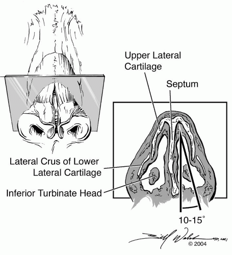 Figure 23.4 Nasal valve. Illustration by William E. Walsh, MD, CMI ©2004, used with permission. |
ANATOMIC CAUSES OF NASAL OBSTRUCTION
Stay updated, free articles. Join our Telegram channel

Full access? Get Clinical Tree


