8 Secondary Glaucoma
CLASSIFICATION OF SECONDARY GLAUCOMA
Secondary glaucomas occur when the intraocular pressure (IOP) is raised as a result of another ocular condition or its treatment. These conditions form a small proportion of all glaucoma but they frequently produce the most difficult problems in diagnosis and management. The diagnosis of secondary glaucoma is often made before the development of glaucomatous cupping and field loss as without treatment these will almost certainly develop with time. Patients presenting with a high IOP secondary to, for example, a traumatic hyphaema are considered to have secondary glaucoma (although strictly speaking they will be suffering from secondary ocular hypertension).
Increased IOP secondary to other conditions is almost always due to a fall in outflow facility. Very rarely raised IOP can be seen with haemodilution from haemodialysis or cardiopulmonary bypass.
Treatment of the secondary glaucomas is directed first at the cause of the condition: inflammation is suppressed, a swollen lens is removed. This may not always control IOP successfully as the trabecular meshwork may have been damaged extensively so that even after removal of the cause long-term glaucoma therapy is still required. Many secondary glaucomas present with an extremely high IOP and require emergency treatment with intravenous or oral carbonic anhydrase inhibitors or hyperosmotic agents. The long-term strategy then depends on the predicted natural history of the condition; the therapeutic approach differs considerably with each individual type of glaucoma. Topical beta-blockers, alpha-agonists and oral or topical carbonic anhydrase inhibitors are very useful and prostaglandin agonists may be effective, even in the presence of angle closure. Pilocarpine may cause vasodilatation and exacerbate blood–aqueous barrier breakdown and is best avoided in patients with these conditions. Many secondary glaucomas respond poorly to maximum tolerated medical treatment in the long term and require trabeculectomy with antiproliferative treatment, aqueous shunt devices or cyclophotocoagulation; fortunately ocular involvement is often asymmetrical.
PRETRABECULAR OUTFLOW OBSTRUCTION
In this group of conditions iris or other tissue obstructs the angle to prevent aqueous from reaching the trabecular meshwork. It is important to ascertain whether pupil block is present as this will affect treatment. On gonioscopy an angle that is closed by apposition indicates pupil block and indentation gonioscopy will help exclude synechial closure. Posterior segment disease must be excluded by fundus examination or, if the media is opaque, by ultrasonography.
SECONDARY ANGLE CLOSURE WITH PUPIL BLOCK
Secondary pupil block occurs when the iris becomes adherent at the pupillary margin to the lens and restricts aqueous flow forwards. Iris bombé shallows the anterior chamber peripherally although it remains relatively deep centrally. Raised IOP may persist after an attack of pupil block from any cause if a large proportion of the angle has been closed by peripheral anterior synechiae (PAS) formed during the attack or if the trabecular meshwork has become damaged.
Possible reasons for pupil block are:
1. Posterior synechiae from inflammation in the anterior segment
2. Occlusion of the pupil in pseudophakic and aphakic eyes by the implant or vitreous gel
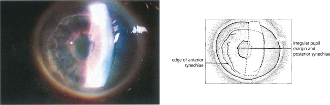
Inflammation in the anterior segment
Inflammation of the anterior segment producing ring posterior synechiae to the lens implant or vitreous face, and occluding the pupillary aperture is an important cause of secondary angle-closure glaucoma with pupil block. Pupil block may develop insidiously in low-grade iritis with gradual seclusion of the pupil or suddenly in acute iritis. Treatment is aimed at preventing seclusion of the pupil with topical steroids and mydriatics but for the established condition laser or surgical iridectomy is necessary to break the block. Before the advent of YAG laser iridotomy peripheral iridectomy was performed routinely during most intraocular surgical procedures to avoid potential pupil block from postoperative inflammation.

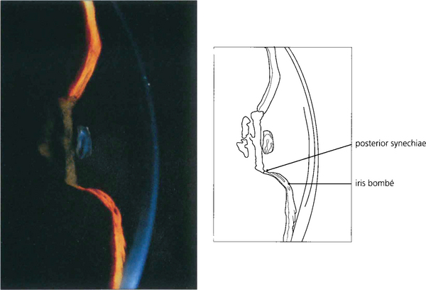
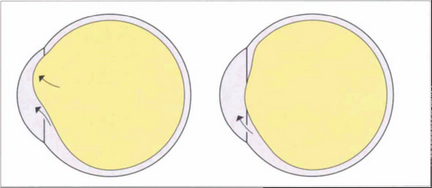
Fig. 8.1 In this patient with uveitis pupil block due to seclusion of the pupil is evident from the forward convexity of the iris resulting in a shallow peripheral anterior chamber, whereas the central chamber remains deep. The deep central anterior chamber distinguishes pupil block from malignant glaucoma with forward movement of the iris–lens diaphragm secondary to posterior segment pathology or pupil block from intumescent cataract.
Pupil block in pseudophakic and aphakic eyes
Modern techniques of phacoemulsification cataract surgery with posterior chamber lens implantation are sufficiently free from postoperative inflammation not to require an iridotomy and pupil block is rarely seen in these eyes. When pupil block does occur it may be due to pupil capture of the implant or from occlusion of the pupil by posterior synechiae to the implant or lens capsule remnants. Intracapsular cataract surgery used to cause pupil block as an acute event although sometimes this did not become apparent until weeks later. Similar findings may be seen with aphakia when posterior synechiae can form between the iris and anterior hyaloid face. Pupil block is suggested by a combination of raised IOP and peripheral shallowing of the anterior chamber. Although extensive prolapse of gel through the pupil sometimes occurs there is usually little vitreous prolapse and the central anterior chamber is of normal depth with peripheral shallowing.
The established condition is treated by laser iridotomy and, when successful, there is immediate deepening of the peripheral anterior chamber. Laser iridotomies, however, are small and may become occluded with fibrin or vitreous gel. A surgical iridectomy can therefore be required or, sometimes, a vitrectomy is necessary performed to re-establish aqueous flow. Eyes containing silicone oil should have a surgical iridectomy performed inferiorly as the oil floats up to occlude a superior iridotomy. Pseudophakic pupil block can occur in eyes with an anterior chamber lens without iridectomy or if an iridotomy is nonfunctioning from blockage by vitreous or capsular remnants. A rigid anterior chamber lens may maintain axial anterior chamber depth with iris ballooning around the edges of the lens making the periphery of the anterior chamber more shallow and occluding the angle.

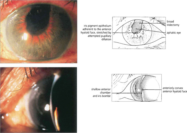
Fig. 8.3 In this patient there is pupil block from posterior synechiae to the vitreous face in an aphakic eye. The slit-lamp image shows peripheral shallowing of the anterior chamber. The extent to which the gel prolapses into the anterior chamber determines the central depth.

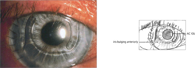
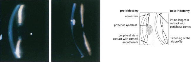
Fig. 8.4 Anterior chamber intraocular lens implantation without peripheral iridectomy is likely to cause pupil block. In this case the iris can be seen bulging forwards around the implant. Although the angle can often be reopened by a laser iridotomy this readily reoccludes with vitreous and so does not reliably prevent recurrence. Surgical iridectomy is definitive but difficult to achieve through a corneal phacoemulsification wound unless it is placed temporally. Temporal iridectomies may cause glare or monocular diplopia, however, and so surgical entry by a separate superior corneal incision is preferable.
Forward movement of the anterior lens surface
Intumescence of the lens, anterior dislocation or age-related growth changes in the lens can all produce pupil block and angle closure by forward movement of the anterior lens surface. These eyes require lens removal or iridotomy. Angle closure may still occur after iridotomy as a result of progressive increase in antero-posterior depth of the lens.

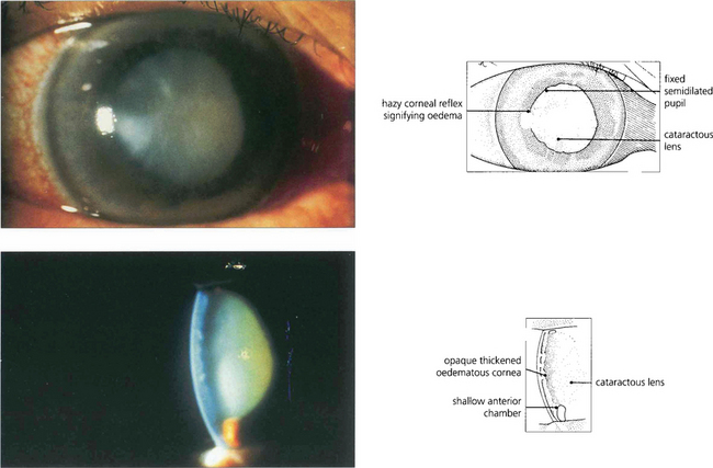
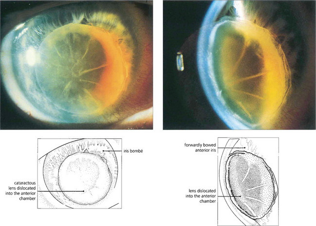
Fig. 8.6 Acute angle closure due to an intumescent cataractous lens. The eye is red with a hazy view of the anterior segment from corneal oedema, with a fixed irregular semidilated pupil from iris infarction. The slit image shows the corneal oedema and a very shallow anterior chamber. Some uveitis may be present because of ischaemia, and this must be differentiated from the larger accumulations of lens material and macrophages seen with phacolytic glaucoma (see Fig. 8.49).

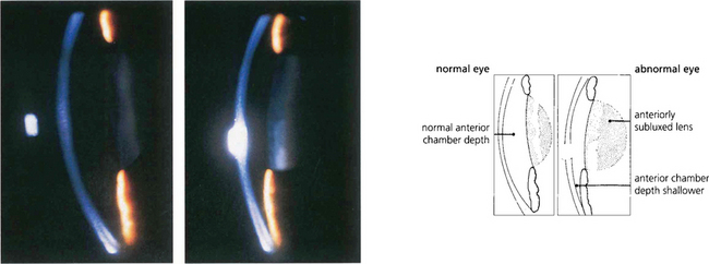
Fig. 8.7 Slit-image photography of the left and right eye of this patient demonstrates a subluxed lens in the left eye. The anterior chamber is shallow and further examination shows that the anterior lens surface is closer to the posterior corneal surface inferiorly than superiorly occluding the pupil and producing pupillary block.

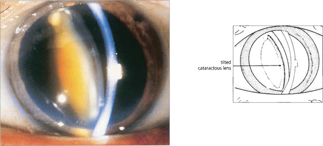
Fig. 8.8 Occasionally pupillary dilatation allows a subluxed lens to swing into the pupillary plane and block communication between the posterior and anterior chamber. Careful positioning of the patient and the use of miotics usually allow the lens to be repositioned safely until definitive treatment can be arranged which in this case is removal of the lens.
SECONDARY ANGLE CLOSURE WITHOUT PUPIL BLOCK
Angle closure may occur without pupil block in four ways:
1. Changes in the posterior segment that push the lens–iris diaphragm forwards
2. Changes in the anterior segment that result in loss of the anterior chamber and iris–trabecular contact with synechial closure
4. Cellular proliferation within the angle of the anterior chamber resulting in iris–trabecular adhesions.
Angle closure from changes in the posterior segment
Tumours form the most important (although the least common) group of conditions in the posterior segment that push the lens–iris diaphragm forwards. Other conditions that may cause an increase in the volume of the posterior segment include choroidal effusions arising either spontaneously or secondary to intraocular surgery, posterior scleritis, encircling bands used in retinal detachment surgery and rarely even ciliary body cysts. An increase in permeability following a breakdown of the blood–retinal barrier may occur after panretinal photocoagulation or central retinal vein occlusion and lead to either a choroidal effusion or a volume increase of the posterior segment pushing the lens–iris diaphragm forwards.

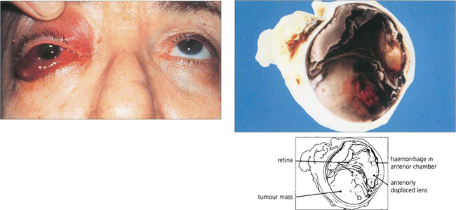
Fig. 8.10 A patient with a long-standing blind eye presented with recent onset of pain, conjunctival oedema and anterior chamber haemorrhage. Posterior segment ultrasonographic examination showed a large choroidal melanoma. Hemisection of the enucleated eye demonstrates a large haemorrhagic choroidal melanoma together with an anteriorly displaced lens and loss of the anterior chamber.

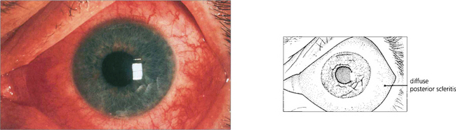
Fig. 8.11 Posterior scleritis may be associated with an annular choroidal effusion that causes the ciliary body to rotate forward about the scleral spur and the iris–lens diaphragm to move forward to produce angle-closure glaucoma. Ciliary body detachment may result in a normal or low IOP, even in the presence of angle closure. This photograph shows a red eye with diffuse anterior and posterior scleritis, although many of these eyes are completely white with inflammation limited to the posterior sclera alone.

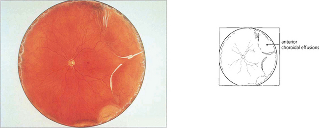
Fig. 8.12 A fundus painting of the same patient reveals an annular choroidal effusion (see also Ch. 5).

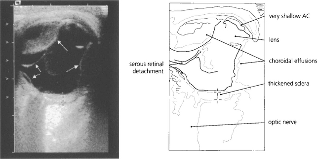
Fig. 8.13 In less obvious cases than the case illustrated, such effusions are easily missed unless the peripheral fundus is inspected carefully under full mydriasis. B-scan ultrasonography is very useful.
By courtesy of Ms M Restori.

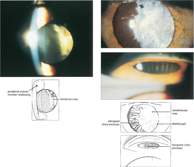
Fig. 8.14 Persistent hyperplastic primary vitreous can produce a contracting fibrotic retrolental mass with forward rotation of the ciliary body and lens–iris diaphragm pushing the iris forwards to occlude the angle. The anterior chamber is often shallow in these eyes making angle occlusion more likely. (Top left) This slit-image photograph shows a shallow anterior chamber and retrolental mass. (Top right) Following mydriasis elongated ciliary processes can be seen being pulled into the mass. (Bottom right) Gonioscopy demonstrates traction and elongation of the ciliary processes.
Malignant glaucoma (ciliolenticular block or aqueous misdirection syndrome)
The term ‘malignant glaucoma’ was originally used to describe this syndrome because the eye did not respond to, and appeared to be made worse by, treatment with pilocarpine. It is used to describe eyes with a very high IOP, absent or shallow anterior chambers and a retrolenticular accumulation of aqueous humour in the absence of pupil block. The most common cause of this uncommon condition is drainage surgery on an eye with a shallow anterior chamber although other forms of intraocular surgery that decompress the anterior chamber can also cause it. Surgery on the fellow eye may be followed by the same result.
The mechanism appears to be an obstruction to forward flow of aqueous humour in the presence of a shallow anterior chamber causing misdirection of aqueous into the posterior segment with pooling in the vitreous gel. Aqueous misdirection appears to occur because of a change in the anatomical relationship between the peripheral vitreous and ciliary processes with the latter rotating forward when the eye is decompressed so that aqueous flows posteriorly. This causes the lens–iris diaphragm to move forward to occlude the angle with central shallowing of the anterior chamber.
The treatment for malignant glaucoma should initially be topical atropine (to relax the ciliary muscle and pull the lens–iris diaphragm posteriorly) together with acetazolamide, beta-blockers and hyperosmotic agents (to lower IOP, dehydrate the vitreous and reduce its volume). Pilocarpine makes the situation worse. Surgical treatment involves decompression of the retrolenticular aqueous pool by pars plana vitrectomy combined with perforation of the anterior hyaloid face and removal of the lens.
For pseudophakic malignant glaucoma the treatment of first choice is YAG laser anterior vitreolysis and posterior capsulotomy. This photodisruption may allow aqueous to percolate from the loculated pools within the anterior vitreous into the posterior chamber and thus relieves the block to aqueous flow. However pars plana vitrectomy is often required as laser vitreolysis may only provide a temporary solution. A process similar to that described above can occur in eyes that are aphakic. Ciliovitreal block can occur if adhesions exist between the anterior hyaloid face and the iris; this may be difficult to distinguish from aphakic pupil block. The diagnosis is confirmed, however, if following YAG laser iridotomy the condition is not resolved and vitreous is seen to be occluding the iridotomy. Surgical or YAG laser rupture of the anterior hyaloid face is curative in this condition.

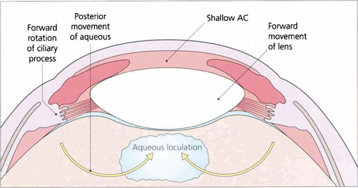
Fig. 8.15 This diagram illustrates how aqueous passes posteriorly and then pushes the lens–iris diaphragm forwards.

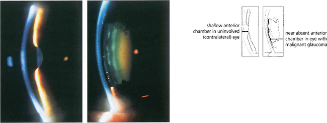
Fig. 8.16 These two slit-lamp photographs of the anterior segments of each eye of a patient demonstrate a shallow anterior chamber in the right eye (left), whereas the left eye (right) has virtually no anterior chamber. The left eye had recently undergone a trabeculectomy followed by loss of the anterior chamber from malignant glaucoma. It is worth using topical atropine in higher-risk eyes at the end of filtration surgery to prevent this.
Synechial closure of the angle
The uveitic eye can develop PAS even if its angle is not narrow. In severe anterior uveitis the angle may become bridged by fibrin which draws the peripheral iris towards the trabecular meshwork.
PAS may also occur when the anterior chamber is shallow enough to allow iris contact with the angle structures. Examples are hypotony from a leaking wound, corneal perforation or cyclodialysis cleft and chronic or intermittent apposition from uncorrected pupil block; these are hastened by coincidental inflammation.

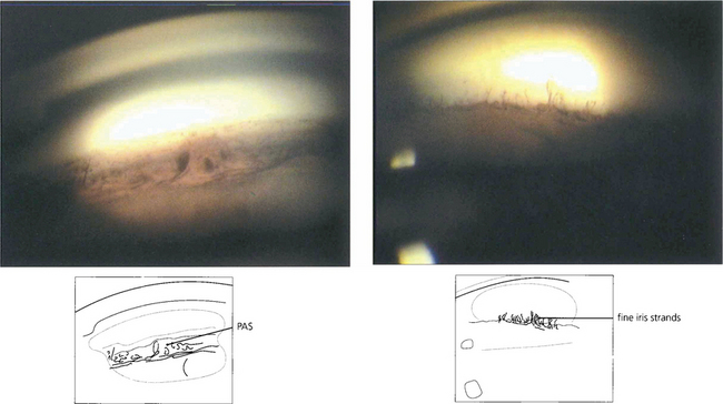
Fig. 8.17 PAS (left) should be distinguished from fine strands from the anterior iris surface to the trabecular meshwork which may be seen in normal eyes (right).

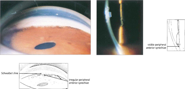
Fig. 8.18 Slit-image and gonioscopic photographs demonstrate the development of PAS in chronic uveitis. Although often confined to the inferior angle such synechiae can extend circumferentially. With sarcoidosis, trabecular granulomas occasionally form as focal lesions around the circumference of the angle and, if untreated, may produce small areas of PAS (see Ch 10).

Full access? Get Clinical Tree

