Salivary Gland Neoplasms


Salivary Gland Neoplasms


Young S. Oh

Matthew S. Russell

David W. Eisele


Neoplasms of the salivary glands represent a diverse group of benign and malignant tumors with varying degrees of behavior. Accurate pathologic diagnosis is key to the management of these lesions because of their varied clinical behaviors. The otolaryngologist-head and neck surgeon must understand the behavior of each tumor type to develop an appropriate treatment plan for a given patient.


ANATOMY AND PHYSIOLOGY

Salivary glands are divided into major and minor glands. The major salivary glands consist of the paired parotid, submandibular, and sublingual glands. The minor salivary glands consist of 600 to 1,000 glands distributed throughout the upper aerodigestive tract. The World Health Organization histologic classification scheme for salivary gland tumors is presented in Table 115.1.

The incidence rate for salivary gland neoplasms is near 1/100,000 with little variance over the last several decades. Approximately 70% of salivary gland tumors originate in the parotid gland. Of parotid gland tumors, approximately 75% to 80% are benign. About 10% of salivary gland tumors arise from the submandibular gland with the distribution of benign and malignant tumors being similar to the parotid gland. Finally, approximately 20% arise from the minor salivary glands, and a higher proportion (50% to 75%) of these neoplasms are malignant (1).

In adults, the vast majority of salivary gland tumors are benign. Malignancies make up 5% of all head and neck cancers and less than 0.5% of all cancers (2). The most common benign tumor for all salivary glands is the pleomorphic adenoma. Tables 115.2, 115.3 and 115.4 list series in the literature detailing the distribution of neoplasms of the parotid, submandibular, and minor salivary glands, respectively (3,4,5).

Salivary gland tumors are far less common in the pediatric population. Only about 5% of all epithelial salivary gland tumors will occur in children and adolescents. Unlike adults, benign vascular and lymphatic tumors are the most common pediatric salivary gland masses (6). Infectious, inflammatory, and congenital conditions should also be considered in the differential diagnosis of pediatric salivary gland lesions. The majority of epithelial neoplasms in children are malignant (Tables 115.5 and 115.6) (7).


BENIGN NEOPLASMS


Pleomorphic Adenoma

Pleomorphic adenoma (benign mixed tumor) accounts for approximately 65% of all salivary gland tumors. These tumors are most often found in the parotid gland, followed by the submandibular gland, and the minor salivary glands. They also represent the most common tumor of each type of salivary gland.

Benign mixed tumor describes the mesenchymal and epithelial components of the tumor. The gross appearance is smooth and lobular with a well-defined capsule. Microscopically, the tumor consists of epithelial and mesenchymal elements. The epithelial component forms a trabecular pattern with a mesenchymal stroma (Fig. 115.1). The mesenchymal portion may be myxoid, chondroid, fibroid, or osteoid. The stroma varies from tumor to tumor and may have a combination of any of these tissue types within it. Histologically, encapsulation may be incomplete with pseudopod extensions of the tumor (8).

Historically, unacceptably high recurrence rates of 20% to 45% have been observed with simple tumor enucleation. Thus, complete surgical resection with an adequate margin of normal tissue is advocated. In the past, for the parotid gland, this constituted a complete superficial parotidectomy. Although this technique is highly effective, there can be some unwanted sequelae such as temporary or permanent facial nerve injury, cosmetic soft tissue defects, or Frey syndrome. Presently, tumor resection by partial parotidectomy
with facial nerve preservation is advocated. This procedure is defined as any parotidectomy in which less than the entire lobe or less than all the branches of the facial nerve are dissected. Lower rates of facial nerve weakness and Frey syndrome are observed with this approach (9).


TABLE 115.1 THE WORLD HEALTH ORGANIZATION HISTOLOGIC CLASSIFICATION OF TUMORS OF THE SALIVARY GLANDS


Malignant epithelial tumors


Benign epithelial tumors


Acinic cell carcinoma


Pleomorphic adenoma


Mucoepidermoid carcinoma


Myoepithelioma


Adenoid cystic carcinoma


Basal cell adenoma


PLGA


Warthin tumor


Epithelial-Myoepithelial carcinoma


Oncocytoma


Clear cell carcinoma, NOS


Canalicular adenoma


Basal cell adenocarcinoma


Sebaceous adenoma


Sebaceous carcinoma


Lymphadenoma


Cystadenocarcinoma


Sebaceous


Low-grade cribriform cystadenocarcinoma


Nonsebaceous


Mucinous adenocarcinoma


Ductal Papillomas


Oncocytic carcinoma


Inverted ductal papilloma


Salivary duct carcinoma


Intraductal papilloma


Adenocarcinoma NOS


Sialadenoma papilliferum


Myoepithelial carcinoma


Cystadenoma


Carcinoma ex pleomorphic adenoma


Soft tissue tumors


Carcinosarcoma


Hemangioma


Metastasizing pleomorphic adenoma


Squamous cell carcinoma


Hematolymphoid tumors


Small cell carcinoma


Hodgkin lymphoma


Large cell carcinoma


Diffuse large B-cell lymphoma


LEC


Extranodal marginal zone B-cell lymphoma


Sialoblastoma


Secondary tumors


LEC, Lymphoepithelial carcinoma; NOS, Not otherwise specified.
From Barnes L, Eveson JW, Reichart P, et al., eds. World Health Organization Classification of Tumors. Pathology and Genetics of Head and Neck Tumours. Lyon, France: IARC Press, 2005, with permission.


TABLE 115.2 DISTRIBUTION OF PAROTID GLAND NEOPLASMS


Histologic Type


Parotid Gland N (%)


Pleomorphic adenoma


256 (66.8)


Warthin tumor


87 (22.8)


Other


40 (10.4)


Total benign


383 (100)


Mucoepidermoid carcinoma


41 (32.5)


Adenoid cystic carcinoma


20 (15.9)


Ca ex pleomorphic adenoma


17 (13.5)


Acinic cell carcinoma


13 (10.3)


Adenocarcinoma NOS


6 (4.7)


Basal cell adenocarcinoma


5 (3.9)


Clear cell adenocarcinoma


4 (3.2)


Myoepithelial carcinoma


3 (2.4)


Salivary duct carcinoma


3 (2.4)


Other malignant


14 (11.2)


Total malignant


126 (100)


From Luksic I, Virag M, Manojlovic S, et al. Salivary gland tumors: 25 years of experience from a single institution in Croatia. J Craniomaxillofac Surg 2011; 40(3):e75-e81. doi:10.1016/j.jcms.2011.05.002.


Rarely, pleomorphic adenoma can metastasize and yet remain benign appearing histologically. Reported sites of metastases include bone, head and neck regions, lungs, and skin. When feasible, surgical resection of the metastatic site is the optimal treatment. Incomplete excision or
rupture of the primary is thought to be a risk factor for the development of distant metastases (10,11).


TABLE 115.3 DISTRIBUTION OF SUBMANDIBULAR GLAND NEOPLASMS


Histologic Type


Submandibular Gland N (%)


Pleomorphic adenoma


479 (97.8)


Other


11 (2.2)


Total benign


490 (100)


Adenoid cystic carcinoma


74 (43)


Mucoepidermoid carcinoma


28 (16)


Ca ex pleomorphic adenoma


27 (16)


Acinic cell carcinoma


7 (4)


Adenocarcinoma NOS


6 (3.5)


LEC


12 (7)


Myoepithelial carcinoma


6 (3.5)


Other malignant


12 (7)


Total malignant


172 (100)


From Zian T, Li L, Wang L, et al. Salivary gland neoplasms in oral and maxillofacial regions: a 23-year retrospective study of 6982 cases in an eastern Chinese population. Int J Oral Maxillofac Surg 2010;39:235-242.


TABLE 115.4 INCIDENCE OF MINOR SALIVARY GLAND NEOPLASMS BY TUMOR TYPE


Tumor Type


Incidence (%)


Pleomorphic adenoma


22.16


Basal cell adenoma


1.08


Cystadenoma


0.54


Myoepithelioma


0.54


Oncocytoma


0.54


Other benign


0.54


Total benign


25.4


Mucoepidermoid carcinoma


34.05


Adenoid cystic carcinoma


14.59


PLGA


9.73


Adenocarcinoma not specified


7.57


Basal cell adenocarcinoma


4.86


Clear cell carcinoma


1.08


Salivary duct carcinoma


1.08


Carcinoma ex pleomorphic adenoma


0.54


Mucinous adenocarcinoma


0.54


Sebaceous carcinoma


0.54


Total malignant


74.6


PLGA, Polymorphous low-grade adenocarcinoma. From Vani NV, Ponniah I. The frequency and distribution pattern of minor salivary gland tumors in a government dental teaching hospital, Chennai, India. Oral Surg Oral Med Oral Pathol Oral Radiol Endod 2011;111:e33.


Warthin Tumor

Warthin tumor (papillary cystadenoma lymphomatosum) is the second most common benign salivary gland neoplasm constituting 5% to 6% of all benign tumors. The majority of Warthin tumors occur in the parotid gland. However, extraparotid sites can be involved as well, typically in the cervical lymph nodes (12). Smoking is a clear etiologic factor for this tumor. Classically, this is a tumor that occurs more commonly in men, but the incidence has increased in women. This is thought to be due to the increased relative rate of smoking in women. Rates of multicentricity for this tumor has been reported from 12% to 50% depending on the series (Fig. 115.2). Bilateral tumor can be seen in up to 12% of patients (13,14).


TABLE 115.5 BENIGN SALIVARY GLAND TUMORS IN CHILDREN


Tumor Type


Number of Patients


Hemangioma


191


Pleomorphic adenoma


182


Lymphangioma


48


Neurogenic


11


Embryoma


5


Lymphoepithelial lesion


3


Cystadenoma


3


Warthin tumor


3


Other


9


Total patients


668


Total benign


68%


From Luna MA, Batsakis JG, El-Naggar AK. Salivary gland tumors in children. Ann Otol Rhinol Laryngol 1991;100:869-871, with permission.


TABLE 115.6 MALIGNANT SALIVARY GLAND TUMORS IN CHILDREN


Histologic Type


N (%)


Mucoepidermoid carcinoma


47 (42)


Acinic cell carcinoma


38 (34)


Rhabdomyosarcoma


8 (7)


Lymphoma/Hodgkin’s


9 (8)


Adenocarcinoma


4 (4)


Adenoid cystic carcinoma


2 (2)


Other malignant


5 (4)


Total malignant


113 (100)


From Shapiro N, Bhattacharyya N. Clinical characteristics and survival for major salivary gland malignancies in children. Otolaryngol Head Neck Surg 2006;134:631-634.


The gross appearance of the tumor is smooth with a well-defined capsule. Cut sections reveal multiple cystic spaces that can be filled with a thick, mucinous exudate. Microscopically, Warthin tumor has a characteristic appearance with a papillary epithelium with a lymphoid stroma projecting into cystic spaces (Fig. 115.3). The epithelium is a double layer of oxyphilic granular cells, with cells of the inner layer having the nuclei oriented toward the basement membrane. The cells in the outer layer have nuclei toward the cystic space.

Complete surgical excision is recommended. Tumor “recurrences” are thought to result from unrecognized multicentric tumors (13).


Figure 115.1 Pleomorphic adenoma. The histologic appearance shows characteristic epithelial and mesenchymal elements.


Figure 115.2 A: Axial contrast-enhanced CT shows a sharply marginated, heterogeneous attenuation lesion in the superficial lobe of the right parotid gland. B: Axial contrast-enhanced CT demonstrates a well-circumscribed enhancing mass, medial to the sternocleidomastoid muscle, which displaces the right thyroid cartilage anteriorly and laterally. This lesion proved to be an additional focus of Warthin tumor.


Oncocytoma

Oncocytoma is an uncommon salivary gland tumor that accounts for about 1% of all salivary gland neoplasms (15). It occurs almost exclusively in the parotid gland. This tumor occurs most frequently in the fifth and sixth decades of life and with equal distribution between men and women (16). Oncocytoma usually presents as a painless mass in the superficial lobe of the parotid gland.


Figure 115.3 Warthin tumor. Lymphoid stroma and double-layered epithelium surround the cystic spaces.

These tumors are noncystic, firm, and rubbery. Microscopically, these neoplasms are composed of brown, plump, granular eosinophilic cells with small indented nuclei (Fig. 115.4). A mitochondria-filled cystoplasm is a characteristic finding on electron microscopy. Malignant degeneration is rare. A possible explanation for these tumors is an acquired genetic defect leading to mitochondrial dysfunction; however, an exact molecular mechanism has not been established (17). Immunohistochemical studies have found that oncocytoma demonstrated positive reactivity to CK5/6, CK8/18, CK19, and EMA. It was negative to S100 and SMA (16).


Figure 115.4 Oncocytoma. The histologic appearance is that of typical plump, granular eosinophilic cells.


Complete tumor resection is the treatment of choice for oncocytoma.


Basal Cell Adenoma

Basal cell adenoma represents about 2% to 3% of all salivary gland tumors. It usually occurs in the sixth to seventh decades of life, and it occurs most often in the parotid gland. The remainder are divided between the submandibular and the minor salivary glands. It affects more women than men (18).

These tumors are well circumscribed and encapsulated. The membranous histologic variant can be multinodular. Microscopically, it can demonstrate four different histologic patterns. The solid pattern consists of sheets of basaloid cells. The tubular pattern consists of small ductules linked by the basaloid cells. The trabecular pattern has plexiform bands of the tumor cells. Finally, the membranous pattern consists of differing sizes of islands of basaloid cells. The membranous pattern can show multicentric tumor patterns and has a higher rate of recurrence after surgery (18).

Basal cell adenoma is considered a benign, nonaggressive tumor. Treatment consists of complete surgical resection with a margin of normal tissue.


MALIGNANT NEOPLASMS


Mucoepidermoid Carcinoma

Mucoepidermoid carcinoma is the most common malignant neoplasm of the salivary glands. It presents most frequently in the parotid gland, for which it is the most common malignant tumor. It is the second most common malignant tumor of the submandibular gland. Mucoepidermoid carcinoma constitutes approximately 30% to 35% of all malignant tumors of the salivary glands (19,20). This is a cancer that usually occurs between the third and sixth decades of life with a slight predominance in women.

Mucoepidermoid carcinomas are usually classified as low-grade or high-grade tumors. However, some pathologists also include an intermediate grade. Low-grade tumors have a higher proportion of mucous cells to epidermoid cells. These lesions behave more like benign neoplasms but in rare cases are capable of local invasion and metastasis. High-grade tumors have a higher proportion of epidermoid cells and may be difficult to differentiate from squamous cell carcinoma. High-grade lesions are aggressive tumors with a high propensity for metastasis. Intermediate-grade lesions have been found to behave like high-grade tumors. Thus, the grade and stage of these neoplasms are important prognostic variables (21).

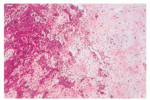
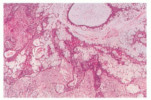
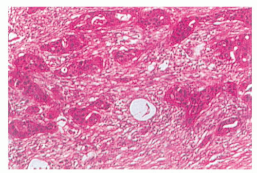
Low-grade tumors are usually small and partially encapsulated. High-grade neoplasms are usually larger and locally invasive. On cut sections, low-grade mucoepidermoid carcinoma may contain mucinous fluid, whereas high-grade tumors are typically solid. Microscopically, low-grade mucoepidermoid carcinoma demonstrates aggregates of mucoid cells separated by strands of epidermal cells (Fig. 115.5). High-grade tumors have few mucoid elements, and the epidermoid cells predominate (Fig. 115.6).


Figure 115.5 Low-grade mucoepidermoid carcinoma. Note the epithelial and glandular elements.

The treatment of this cancer depends on the grade and stage of the tumor. For small, low-grade cancers, complete surgical resection is recommended. Elective treatment of the neck is not recommended for in the clinically N0 neck because of the incidence of occult nodal involvement is very low (21). If the surgical margins are clear and there are no adverse pathologic features, adjuvant radiation therapy is not recommended. Prognosis for this cancer is excellent with locoregional recurrence rates of 10% and 5-year overall survival of 97% (22).

For high-grade tumors, the treatment is tailored to the extent of the disease. As in low-grade disease, complete surgical resection of the primary site is recommended. If the tumor involves the parotid gland, the facial nerve is spared, if possible. Cervical nodal metastases are managed by a comprehensive neck dissection at the time of the resection of the primary. Occult metastases rates have been shown to be approximately 21%; therefore, an elective neck dissection can be considered in a patient with a clinically N0
neck (22). In most cases of high-grade mucoepidermoid carcinoma, adjuvant radiation therapy is recommended to improve locoregional control (23). High-grade mucoepidermoid carcinoma is an aggressive tumor with a poorer prognosis than the low-grade variant. Locoregional recurrence has been shown to occur 43.5% of the time with 3-year disease-free survival reported to be 55% and 5-year disease-free survival at 30% (21,24).


Figure 115.6 High-grade mucoepidermoid carcinoma. Note the relative lack of glandular elements.


Adenoid Cystic Carcinoma

Adenoid cystic carcinoma accounts for approximately 10% of all salivary gland neoplasms. It is the second most common malignancy of the parotid gland but is the most common malignancy of the submandibular and minor salivary glands (25). Adenoid cystic carcinoma occurs with equal frequency in men and women, usually in the fifth decade of life. Facial paralysis and pain occur as initial symptoms in a small proportion of patients.

Adenoid cystic carcinoma has a contradictory clinical course. The tumor is slow growing, but its clinical course is relentless. Multiple local recurrences can occur despite adequate surgical intervention, and although regional metastatic spread is uncommon, distant spread, especially to the lungs, is common.

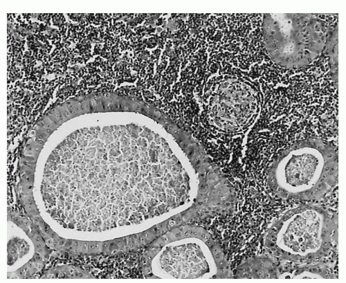
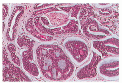
Grossly, the tumor is usually monolobular and either nonencapsulated or partially encapsulated. The mass often demonstrates infiltration of surrounding normal tissue. Microscopically, adenoid cystic carcinoma has a basaloid epithelium arranged in cylindric formations in an eosinophilic hyaline stroma (Fig. 115.7). Different histologic patterns have been identified including cribriform, solid, and tubular. The solid histologic pattern appears to have a worse prognosis in terms of distant metastases and long-term survival (26).

Perineural invasion is a typical feature of adenoid cystic carcinoma. This explains the difficulty in tumor eradication despite the appearance of complete tumor removal. Complete surgical excision and postoperative radiation therapy is recommended for the management of this tumor (27). For select small tumors that are completely excised, however, postoperative radiation therapy may not be needed (28).


Figure 115.7 Adenoid cystic carcinoma, showing the characteristic histologic appearance with eosinophilic hyaline stroma and perineural invasion.

There is evidence that fast neutron radiotherapy may be more effective than conventional photon radiation therapy as primary therapy for select adenoid cystic carcinomas that are not amenable to surgery (29,30).

Long-term follow-up is mandatory for these patients because of the slow, relentless disease progression. Recurrences have been shown to occur at an average of 63 months from the time of initial treatment (31). Thus, 5-year survival is not adequate to detail the outcomes of this malignancy. Ten-year overall survival has been reported to be from 55% to 64%. Recurrence rates can be broken down as 42% with distant metastases only, 39% with locoregional recurrences only, and 19% with both locoregional and distant disease (27,31).

Because of the low incidence of occult metastases, elective neck dissection for the N0 neck is not usually performed. However, the presence of nodal metastases at the time of diagnosis is an important prognostic factor because overall survival is significantly lowered when metastases are present (31).


Acinic Cell Carcinoma

Acinic cell carcinoma constitutes approximately 17% of all salivary gland cancers. Up to 98% of these occur in the parotid gland. It affects women more often than men and occurs in the fourth to sixth decade of life. The tumor can be multicentric in 2% to 5% of cases, and it can involve both parotid glands (32,33).

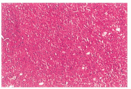
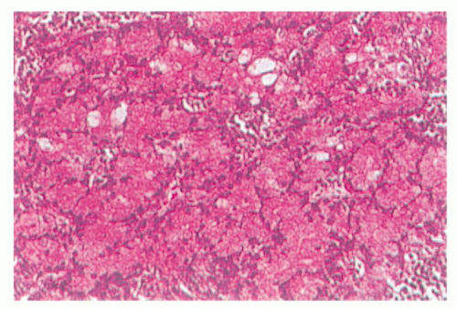
Grossly, this well-circumscribed tumor often has a fibrous capsule. There are two populations of cells: those resembling serous acinar cells of the salivary gland and those with a clear cytoplasm. Tumors occur in several configurations, including cystic, papillary, vacuolated, or follicular. There is often a lymphoid infiltrate, and cells are characteristically positive on periodic acid-Schiff staining (Fig. 115.8).


Figure 115.8 Acinic cell carcinoma. Note cells similar to serous acinar cells and cells with clear cytoplasm.


Treatment consists of complete surgical resection with adjuvant radiotherapy reserved for indicators of a poor prognosis (e.g., facial nerve involvement, neck metastases, and skin involvement). Elective treatment of the neck is indicated for high-grade cases of acinic cell carcinoma (32). Although acinic cell carcinoma is typically categorized as an indolent, slow-growing tumor, the average rate of recurrence has been found to be about 35% (32,33). Long term follow-up is mandatory because late local recurrences have been shown to occur many years after initial treatment. For low-grade lesions, 5- and 10-year disease-free survivals have been reported to be approximately 91% and 88%, respectively (34). Five-year overall survival for high-grade acinic cell carcinoma is much poorer at 33% (33).


Adenocarcinoma

Adenocarcinoma most commonly occurs in the minor salivary glands followed by the parotid gland. This neoplasm represents approximately 12% of malignant parotid neoplasms (35). Adenocarcinomas occur equally in both sexes and usually present as a palpable mass. They behave aggressively with a strong propensity to recur and metastasize.

Grossly, adenocarcinoma is firm or hard and attached to the surrounding tissue. Microscopically, the cylindric cells of variable height form papillae, acini, or solid masses. Most neoplasms produce mucus, which can be detected by mucicarmine stain. Adenocarcinoma can be differentiated from mucoepidermoid carcinoma by the lack of keratin staining. The degree of glandular formation has been used as a means of grading these tumors.

Complete surgical resection with postoperative radiation therapy is the recommended treatment for this highgrade malignancy. Five- and ten-year overall survivals are 52% and 36%, respectively (36).


Polymorphous Low-Grade Adenocarcinoma

Polymorphous low-grade adenocarcinoma (PLGA) occurs almost exclusively in the minor salivary glands. The neoplasm most commonly occurs in the palate, buccal mucosa, and the upper lip. Women are affected more commonly than men, and most of these neoplasms occur in the sixth decade of life. The typical presentation of this tumor is of a long-standing, asymptomatic mass on the palate (37).

Histologically, PLGA demonstrates a variable tumor cell differentiation and organization. Mitotic figures and necrosis are unusual features. Tumors typically have an infiltrative growth pattern with frequent perineural invasion. Typical histologic patterns include solid, trabecular, glandular, cribriform, fascicular, cord-like, and papillary. Because of this, careful pathologic diagnosis is mandatory as this neoplasm can be mistaken for pleomorphic adenoma or adenoid cystic carcinoma. Immunohistochemical staining has not been helpful in differentiating adenoid cystic carcinoma from polymorphous low-grade adenocarcinoma (38).

Treatment is wide local surgical excision. Cervical metastases are very uncommon, so a neck dissection is performed only for clinically positive nodes. In a recent review of the three largest series on PLGA, it was found that local recurrences occur anywhere from 10% to 33% of the time. These recurrences can occur up to 15 years after treatment. Even with recurrences, death from tumor progression is rare (39). In general, adjuvant radiotherapy is not recommended for this cancer (40).


Carcinoma Ex Pleomorphic Adenoma

Carcinoma ex pleomorphic adenoma represents a malignant tumor that has risen from a preexisting or recurrent pleomorphic adenoma. The malignant component and metastases from this tumor are purely epithelial in origin. This malignancy represents 2% to 5% of all salivary gland tumors. Rarely, the malignancy can take the form in which the tumor contains both mesenchymal and epithelial components.

Grossly, the tumors are firm with minimal encapsulation. The lesion is widely infiltrative with regions of necrosis and hemorrhage. Microscopically, the malignant neoplasm arises in a background that is characteristic of a benign mixed tumor (Fig. 115.9

Only gold members can continue reading. Log In or Register to continue

Stay updated, free articles. Join our Telegram channel

May 24, 2016 | Posted by in OTOLARYNGOLOGY | Comments Off on Salivary Gland Neoplasms

Full access? Get Clinical Tree

Get Clinical Tree app for offline access