Pediatric Otolaryngology
Stephen C. Maturo
Michael Cunningham
The management of the otolaryngologic disorders of childhood traditionally has been and remains an integral part of the practice of general otolaryngology. Pediatric otolaryngology has evolved as a surgical subspecialty oriented toward the comprehensive otolaryngologic care specifically of children (1,2). The evolution of this subspecialty can be traced to developments within the fields of pediatrics and anesthesia and within otolaryngology itself (3). The establishment of freestanding children’s hospitals or sections of general hospitals devoted solely to pediatric care reflected a growing appreciation on the part of the medical community that the problems of children differed from those of adults and required a different focus of diagnostic intervention and management (4). The patient population of these children’s hospitals and pediatric wards changed over the years as the development of vaccines and newer generations of antibiotics led to improved control of acute infectious diseases. The focus of pediatric medicine gradually shifted to children with chronic illnesses, progressive noninfectious disorders, malignancies, and disabling or disfiguring handicaps. Patients with these much more complex conditions required the coordinated services of many subspecialty fields, including otolaryngology (5).
Concurrent technologic advances and a better understanding of the pathophysiologic characteristics of cardiorespiratory failure ushered in the modern era of anesthesia and intensive care unit medicine. The establishment of pediatric and neonatal intensive care units, the latter associated with advanced obstetric care hospitals, allowed the treatment and survival of younger and sicker infants and children. These children presented with a new array of medical and surgical problems of interest to the otolaryngologist, particularly regarding airway management.
Scientific advances within the field of otolaryngology-head and neck surgery further contributed to pediatric otolaryngology specialization. The development of fiber optic, illuminated, rigid endoscopes of appropriate size for use in infants and young children allowed safe operative assessment of congenital and acquired airway lesions. Small-diameter flexible endoscopes facilitated the dynamic examination of the upper airway in awake infants and children, and ultrathin versions of these scopes allowed lower airway evaluations even in intubated infants. Newer laryngoscopes and bronchoscopes were developed to allow unrestricted visualization of the pediatric airway for instrumentation and laser applications and to facilitate the delivery of inhalational anesthetic agents through standard endotracheal, Venturi jet, or spontaneous respiration techniques (6). More recently, airway-specific balloon technology and smaller microlaryngeal instruments have made minimally invasive endoscopic airway procedures a viable option.
Radiologic advances in computed tomography (CT), magnetic resonance imaging (MRI), and digital subtraction angiography added to the diagnostic acumen available for evaluating both congenital malformations and mass lesions. The management of extensive benign as well as malignant head and neck neoplasms in this age group has been greatly influenced by the application to children of angiographic embolization and microvascular surgical techniques, more precise radiotherapy protocols including proton beam and gamma knife technologies, and newer chemotherapeutic agents designed to decrease long-term maturation side effects.
Polysomnography studies enhanced the physiologic assessment of children with obstructive lymphoid hypertrophy. Immunologic antibody-antigen detection techniques facilitated the serologic diagnosis of various infections and the confirmation of inhalant atopy in young children. Novel laboratory studies have also helped aid in the diagnosis of immunodeficiency disorders commonly manifested in the upper aerodigestive tract. Electrodiagnostic techniques such as electroneurography, electromyography, brainstem auditory- evoked response testing, and otoacoustic emissions allowed more in-depth assessment of pediatric sensory
impairments. Lasers, cochlear implants, intraoperative radiologic guidance systems, and distraction osteogenesis techniques, to name a few, have all opened new avenues of therapeutic otolaryngologic intervention in children.
impairments. Lasers, cochlear implants, intraoperative radiologic guidance systems, and distraction osteogenesis techniques, to name a few, have all opened new avenues of therapeutic otolaryngologic intervention in children.
The application of these technical innovations to the care of children with acute and chronic otolaryngologic problems stimulated further clinical investigation and bench research activities, leading to the increasing fund of knowledge and expertise that now defines the subspecialty. Pediatric otolaryngology shares with geriatric otolaryngology a focus on a specific age group rather than on an organ system or category of disease (7). This chapter highlights otolaryngologic disorders specific to the pediatric population and relevant issues pertinent to caring for such children.
PATTERNS OF GROWTH AND DEVELOPMENT
Pediatric otolaryngology is the study of the disorders and diseases of the ears, nose, and throat as they relate to the growth and development of the head and neck structures (8). The clinical presentation and sequelae of any disease process are greatly influenced by the age and maturation of the afflicted person. Children differ anatomically and physiologically from adults, and they also change in these respects along a continuum from the neonatal/infancy period through the toddler, preschool, later childhood, and eventually adolescent years. Pediatric otolaryngology requires a working knowledge of the standard patterns of growth and development through this continuum so that deviations from normal can be recognized and age-appropriate diagnostic techniques and methods of management can be applied.
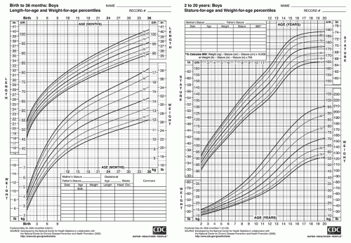 Figure 86.1 Physical growth in boys (Centers for Disease Control and Prevention). |
Measurement of the vital characteristics of somatic growth can provide information about a child’s general state of health or illness regardless of the specific organ system of interest. Height and weight measurements of all children, supplemented by head circumference measurements during the first year of life, provide an early warning system for pathologic processes. Charts documenting serial measurements over months to years construct an accurate record of the child’s general pattern of growth (Figs. 86.1 and 86.2), with deviations from normal being indicative of an intrinsic or extrinsic insult.
Different organ systems mature at different rates and at different times throughout infancy, childhood, and adolescence (Fig. 86.3). The rapid rate of neural tissue growth during fetal life explains the relatively large size of the neurocranium in relation to the face in the newborn. The face of
the infant or young child is not a small image of the eventual adult (Fig. 86.4). The infant’s forehead is prominent and the face comparatively round and diminutive. Elongation of the face occurs secondary to mandibular and maxillary growth in association with the eruption first of the primary and later of the permanent teeth. As this vertical growth continues throughout childhood, the relative proportion of facial to cranial mass gradually changes, and the narrower adolescent/adult facies eventually is achieved (Fig. 86.5). Associated with this progressive increase in facial height is a gradual change in the child’s profile. The child’s cheeks and chin are flat, the nose is diminutive, and the eyes appear comparatively large. With mandibular and maxillary growth, the chin and cheek bones become more prominent, and the growth of the nose and supraorbital rims decreases the relative orbital size.
the infant or young child is not a small image of the eventual adult (Fig. 86.4). The infant’s forehead is prominent and the face comparatively round and diminutive. Elongation of the face occurs secondary to mandibular and maxillary growth in association with the eruption first of the primary and later of the permanent teeth. As this vertical growth continues throughout childhood, the relative proportion of facial to cranial mass gradually changes, and the narrower adolescent/adult facies eventually is achieved (Fig. 86.5). Associated with this progressive increase in facial height is a gradual change in the child’s profile. The child’s cheeks and chin are flat, the nose is diminutive, and the eyes appear comparatively large. With mandibular and maxillary growth, the chin and cheek bones become more prominent, and the growth of the nose and supraorbital rims decreases the relative orbital size.
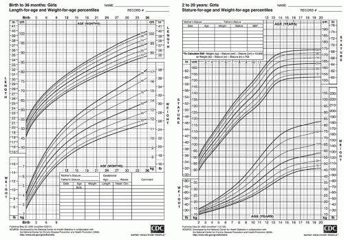 Figure 86.2 Physical growth in girls (Centers for Disease Control and Prevention). |
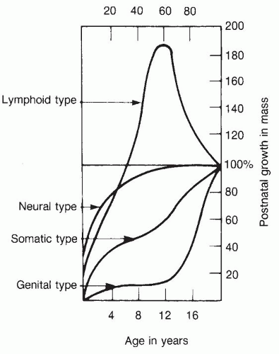 Figure 86.3 Postnatal growth of different anatomic systems with age. |
The nares are small at birth and retain their roughly circular shape until puberty. The oval or oblong nares associated with the adult facies develop in association with the marked increased vertical growth of the nose during adolescence. Major forces in the vertical height and projection of the nose are the osseous and cartilaginous growth centers of the nasal septum and nasomaxillary pyramid.
Elective surgery on the nose usually is deferred until the adolescent years when full facial growth has been achieved. Exceptions to this rule include severe traumatic injuries or nasal deformities associated with congenital anomalies such as cleft lip and palate. Growth may further exaggerate the facial deformity in such cases.
Elective surgery on the nose usually is deferred until the adolescent years when full facial growth has been achieved. Exceptions to this rule include severe traumatic injuries or nasal deformities associated with congenital anomalies such as cleft lip and palate. Growth may further exaggerate the facial deformity in such cases.
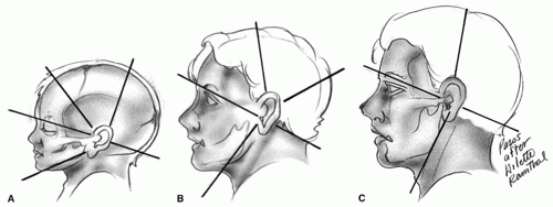 Figure 86.4 Changing facial configuration with age. A: At birth. B: Through 5 years. C: At 15 years. (From Stool SE, Marasovich W. Postnatal craniofacial growth and development. In: Bluestone CD, Stool SE, eds. Pediatric otolaryngology, 2nd ed. Philadelphia, PA: WB Saunders, 1990:18, with permission.) |
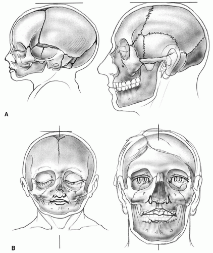 Figure 86.5 Infant and adult skulls for craniofacial skeletal comparison. A: Lateral views. B: Anteroposterior views. |
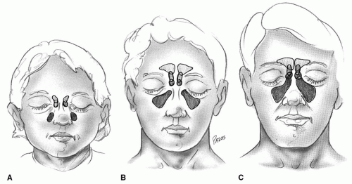 Figure 86.6 Paranasal sinus development. A: At birth. B: At 10 years. C: At 15 years. |
Despite small anatomic nasal dimensions and increased airway resistance, infants are predominantly nasal breathers (the anatomic and physiologic explanations for this preferential nasal breathing pattern are discussed later in this section). Although the degree and duration of this reliance on the nasal airway for respiration vary with each child, complete nasal airway obstruction at birth is usually an airway emergency. Even unilateral obstruction can cause the newborn significant respiratory distress and secondary feeding difficulties. In older children, chronic nasal and nasopharyngeal obstruction, most frequently caused by lymphoid hypertrophy, may be associated with constant mouth breathing, abnormal tongue positioning, dental malocclusion, and suspected craniofacial growth changes. This socalled adenoid facies syndrome remains controversial from a cause and effect standpoint; its description, however, serves to highlight the potential sequelae of a single disease entity on an actively growing child.
TABLE 86.1 PARANASAL SINUS DEVELOPMENT | ||||||||||||||||||||||||||||||||||||||||||||||||
|---|---|---|---|---|---|---|---|---|---|---|---|---|---|---|---|---|---|---|---|---|---|---|---|---|---|---|---|---|---|---|---|---|---|---|---|---|---|---|---|---|---|---|---|---|---|---|---|---|
| ||||||||||||||||||||||||||||||||||||||||||||||||
The development of the paranasal sinuses is connected intimately with nasomaxillary and facial growth (Fig. 86.6). The maxillary and sphenoethmoid sinuses are present at birth, although their small size typically precludes their radiologic appearance (Table 86.1). Conspicuous growth in the maxillary sinuses begins by approximately age 3 years, but inferiorly directed expansion does not occur until eruption of the permanent dentition, when the child is 7 to 8 years of age. The floor of the maxillary sinus approximates the inferior meatus at age 8 years and reaches the level of the floor of the nose by 12 years of age. Adult size is reached by midadolescence. The ethmoid sinuses arise as evaginations of the nasal mucosa from the middle, superior, and supreme nasal meatuses. Although present at birth, significant pneumatization does not occur until the child is between 3 and 7 years of age. Final adult form typically is achieved by age 12 to 14 years. The sphenoid sinuses originate within the nasal cupola; they do not begin to pneumatize the sphenoid
bone and become clinically significant until 4 to 5 years of age. Sphenoid development, although complete by midadolescence, is highly variable in terms of the final extent of sphenoid bone pneumatization. The frontal sinuses originate as outgrowths of the middle meatuses in the frontal recess regions. The frontal sinuses are typically not present at birth; growth begins during the third year of life and continues well into adolescence. Pneumatization is highly variable and is of limited clinical significance until the early adolescent years. The thin posterior table and floor of the frontal sinuses have important anatomic relationships to the anterior cranial fossa and orbital structures, respectively.
bone and become clinically significant until 4 to 5 years of age. Sphenoid development, although complete by midadolescence, is highly variable in terms of the final extent of sphenoid bone pneumatization. The frontal sinuses originate as outgrowths of the middle meatuses in the frontal recess regions. The frontal sinuses are typically not present at birth; growth begins during the third year of life and continues well into adolescence. Pneumatization is highly variable and is of limited clinical significance until the early adolescent years. The thin posterior table and floor of the frontal sinuses have important anatomic relationships to the anterior cranial fossa and orbital structures, respectively.
Discussion of paranasal sinus development is incomplete without mention of the ostiomeatal complex region, which represents the joint location of the maxillary sinus ostium, anterior ethmoid sinus ostia, and frontal recess in the region of the middle meatus. The channels into which these ostia open are bound by the ethmoidal bulla, the uncinate process, and the middle turbinate. Anatomic enlargement or mucosal hypertrophy of these three structures can significantly narrow these channels and obstruct maxillary-ethmoid-frontal sinus drainage. The relatively small size of the child’s nose has traditionally made intranasal sinus surgery comparatively risky because of limited surgical exposure. Although still challenging, this situation has improved with the development of instruments and telescopes of appropriate size for the application of functional endoscopic sinus surgery directed at the ostiomeatal region in the pediatric population.
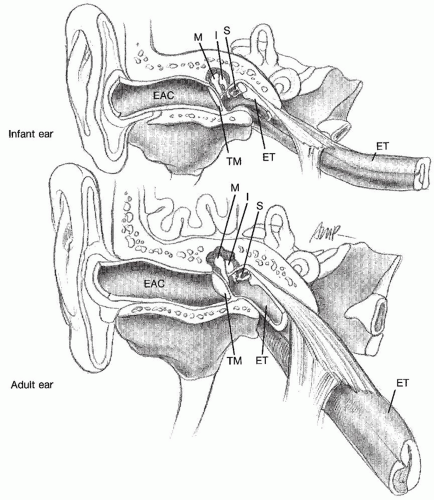 Figure 86.7 Comparison of infant and adult middle ear and eustachian tube development. M, malleus; I, incus; S, stapes; ET, eustachian tube; TM, tympanic membrane; EAC, external auditory canal. |
At birth, the pinna has an adult configuration and location, although the ears appear to rise in position as a result of the vertical growth of the lower third of the face. The pinna reaches near adult size at 4 to 5 years of age and obtains full adult size by age 9 years. The soft and pliable nature of the young child’s ear cartilage also matures during this same period, which influences the timing of auricular reconstruction, specifically those procedures requiring cartilage manipulation.
The tympanic membrane is adult sized at birth, but due in part to incomplete ossification of the external auditory canal, lies in a nearly horizontal position, impairing its visualization on neonatal ear examination. The final vertical orientation of the eardrum is achieved with completion of canal ossification by approximately 2 years of age. Middle ear ossicular formation is complete at birth. Middle ear pneumatization is likewise near complete. The mastoid antrum, in contrast, enlarges significantly over the first years of life with generalized mastoid pneumatization and development continuing well into early childhood. Most postnatal mastoid growth occurs in a lateral and posterior direction, with a fully developed mastoid and styloid process not appearing until the child is about age 3 (Fig. 86.7).
The extratemporal portion of the facial nerve is relatively unprotected during this period of development, predisposing it to obstetric injury and potential iatrogenic injury during tympanomastoid and parotid surgery. The thin mastoid cortex at this young age also accounts for the potential subperiosteal postauricular spread of mastoid infection.
The extratemporal portion of the facial nerve is relatively unprotected during this period of development, predisposing it to obstetric injury and potential iatrogenic injury during tympanomastoid and parotid surgery. The thin mastoid cortex at this young age also accounts for the potential subperiosteal postauricular spread of mastoid infection.
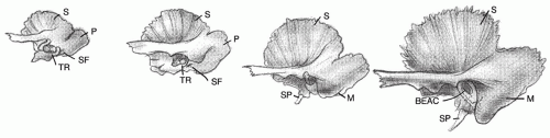 Figure 86.8 Postnatal temporal bone development. Note mastoid bony external ear canal and styloid process growth. BEAC, bony external auditory canal; M, mastoid; P, petrosa; S, squamosa; SF, stylomastoid foramen; SP, styloid process; TR, tympanic ring. (Adapted from Kenna M, Hirose K. Embryology and developmental anatomy of the ear. In: Bluestone CD, et al., eds. Pediatric otolaryngology, 4th ed. Philadelphia, PA: WB Saunders, 2003:136.) |
Eustachian tube development plays a prominent role in the predisposition of infants and young children to middle ear infection (Fig. 86.8). At birth, the eustachian tube is about 50% of its adult length and lies in a fairly horizontal position, entering the nasopharynx at the level of the hard palate. With growth, the eustachian tube lengthens, widens, and angles inferiorly, achieving its final nasopharyngeal position by the time the child is 5 to 7 years old. The petrous portion of the temporal bone, including the bony and membranous labyrinths, is formed completely at birth. The neonate should be fully functional from both a hearing and a vestibular standpoint.
The neonate’s oral cavity is small and the comparatively large tongue fills it entirely, contributing significantly to the infant’s status as a preferential nasal breather. The fully formed palatal structures provide the infant with velopharyngeal competence and the more superior cervical position of the larynx allows potential overlap of the epiglottis and the velum, establishing a nasopharyngeal airway during suckle feeding. The flow of milk or formula is channeled around the dorsum of the tongue and laterally around the epiglottis, protecting the airway. With mandibular growth, the oral cavity enlarges and the base of the tongue descends to its final hypopharyngeal position. The infant’s suckle gradually changes to a more mature swallow pattern, which is functionally quite complex, consisting of an extremely well-synchronized series of oral, pharyngeal, and esophageal neuromuscular movements.
The larynx serves the infant immediately as a conduit for breathing. No other head and neck structure is initially so essential to life. The larynx additionally protects the lower airway by means of two mechanisms: glottic and supraglottic closure during swallowing and the cough reflex. The phonatory function of the larynx provides the infant with a means of expressing basic needs; this communicative function obviously increases in complexity and importance later in childhood.
The pediatric larynx has considerable anatomic differences from that of the adult. These differences involve the specific breathing and airway protection demands of suckle feeding in the newborn infant. The newborn neck is relatively short, and the infant larynx is positioned high, approximating the third or fourth cervical vertebra at rest and rising to the height of the first or second cervical vertebra with swallowing (Fig. 86.9). As discussed previously, this high position allows overlap of the epiglottis with the soft palate. With growth of the neck, the larynx gradually descends to its adult position opposite the fifth cervical vertebra. The still relatively high childhood position of the larynx is highlighted by the ease with which the epiglottis can be visualized on oropharyngeal examination in many children.
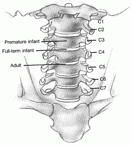 Figure 86.9 Comparative neck positions of the infant and adult larynx (glottic level). |
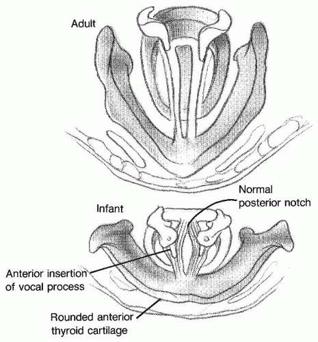 Figure 86.10 Endoscopic view of infant and adult demonstrating the comparatively large arytenoids and rounded thyroid cartilage configuration in the infant compared with those of the adult larynx. |
The predisposition of the infant airway to obstruction is related to its absolute small size, the pliability of its constituent connective tissues, and some intrinsic anatomic features. The infantile epiglottis is furled or omegashaped and the arytenoids are relatively large, covering a significant percentage of the posterior glottis (Fig. 86.10). This anatomic configuration contributes to the entity of laryngomalacia. In the infant, the cricoid cartilage is smaller in diameter than the length of the true vocal cords, making the subglottic region the narrowest portion of the pediatric airway. The resultant funnel-shaped internal dimension (Fig. 86.11) has important consequences for the young child in terms of both the sequelae of inflammatory airway edema and the effects of endotracheal intubation.
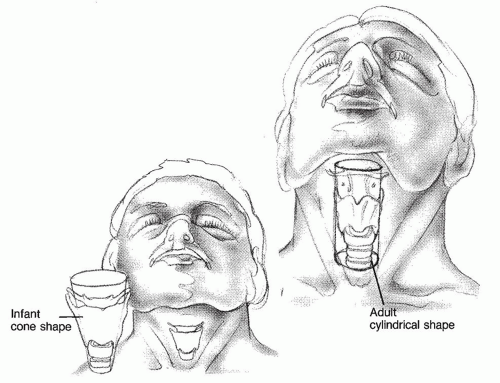 Figure 86.11 Narrowing cone-shaped internal dimension of the infant larynx is due to the smaller diameter of the cricoid cartilage compared with that of the glottis. These glottic-subglottic dimensions approximate one another in the adult larynx. |
The infant larynx grows rapidly in terms of both width and length in the first 3 years of life, which may obviate the need for airway intervention in certain congenital anomalies. Laryngeal growth then slows until adolescence, when there is a rapid increase in all airway dimensions. The adolescent growth spurt of the cricoid and thyroid cartilages also changes the angulation of the true vocal cords as they insert into the anterior commissure region. This contributes, in part, to the voice changes associated with puberty.
Of additional anatomic importance is the comparative underdevelopment of the thyroid cartilage in the infant. The thyroid cartilage is relatively flat without a vertical midline prominence (Fig. 86.12) and tends to be overlapped by the hyoid bone because of the high laryngeal position. The cricoid cartilage is also small, and the cricothyroid membrane is more of a slit than a true palpable space. The standard landmarks for tracheotomy and cricothyroidotomy are not very demonstrable, making the emergent performance of either of these procedures difficult in the newborn infant. Endotracheal intubation is a far preferable choice of airway maintenance in acute emergencies in young children.
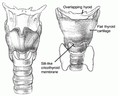 Figure 86.12 Anterior view demonstration of the flat infantile thyroid cartilage and overlap by the hyoid cartilage above and over the cricoid cartilage below. These cartilage elements separate with increasing age. |
The neck of the infant and young child also differs from that of the adult in the prominence of the cervical lymphoid tissue. The cervical lymph nodes increase in size proportionally to the growth curve for the body’s lymphoid tissue in general (Fig. 86.3). The variability of cervical lymphadenopathy palpable on routine pediatric neck examination can make the decision regarding when to perform a nodal biopsy a diagnostic challenge. Children at risk for significant pathology include those with supraclavicular adenopathy, worrisome clinical symptoms such as persistent fever or weight loss, and local fixation of the node(s) to the overlying skin or underlying deep tissues. The retropharyngeal and parapharyngeal lymph nodes are of additional importance in young children. Suppurative lymphadenitis in these regions can rapidly progress to abscess formation with aerodigestive tract compromise and sepsis risk.
CONGENITAL MALFORMATIONS
Diagnosis and management of congenital malformations of the head and neck structures are an integral part of the practice of pediatric otolaryngology. Children with these conditions require a thorough otolaryngologic and general pediatric assessment to ensure that their head and neck anomaly is not a manifestation of an underlying craniofacial or systemic syndrome. The discovery of additional syndromic manifestations can be of prognostic importance, altering surgical or long-term care plans, and also be of potential use from a genetics and family planning standpoint. A comprehensive list of all congenital malformations of the head and neck is beyond the scope of this chapter. A brief review of several of the more common lesions highlights the diversity of a pediatric otolaryngology practice.
Auricular Malformations
The complex origin of the middle and external ear from the first two branchial arches lends itself to varying degrees of sporadic malformation. Minor malformations include preauricular pits and tags. Although typically an isolated finding in normal-hearing children, preauricular tags may be associated with ossicular malformations and secondary conductive hearing loss. Preauricular pits can occur in the presence of renal disease and mixed conductive/sensorineural hearing loss in children with branchial-oto-renal syndrome. Not all children with preauricular pits or tags require formal audiologic or renal ultrasound examinations; such evaluations, however, should definitely be performed in those with additional congenital anomalies and in those with a family history of hearing loss or renal disease (9,10).
Major auricular malformations are microtia and atresia. These may be isolated entities, or they may occur in the setting of a generalized craniofacial disorder, such as Goldenhar syndrome. Intervention in these children is typically dictated by the unilaterality or bilaterality of the condition, the child’s hearing status, and the feasibility or desirability of cosmetic or hearing restoration. Such children may develop acute otitis media (AOM) in the rudimentary ear space of the microtic ear, and they have an increased risk of developing cholesteatoma in the atretic ear canal. The normal ear in unilateral cases must be monitored closely for otitis media with effusion (OME) and secondary hearing compromise. Comprehensive care of children with these conditions includes parental counseling, detailed audiologic assessment with amplification if needed, and surgical intervention including auricular reconstruction, atresia repair, or bone-anchored hearing aides.
Nares Malformations
Choanal atresia is representative of a major malformation in nares development that, when bilateral, usually causes immediate respiratory compromise in the newborn. Modified oral airways or endotracheal intubation provide acute airway relief until definitive surgical management is possible. High-resolution CT scanning is a requisite part of the preoperative evaluation of these patients. Their operative management has been enhanced greatly by the application of endoscopic surgical techniques. Osseous as well as membranous atresias may be approached in transnasal or combined transoral-transnasal fashion, rendering the classic transpalatal approach near obsolete. Choanal atresia needs to be additionally recognized as one potential manifestation of the CHARGE syndrome. Such children have a propensity for additional airway and systemic anomalies that may dictate neonatal tracheotomy to be the most appropriate initial treatment measure, delaying choanal atresia repair until the child is older. CHARGE syndrome also highlights the importance of a thorough
cardiology evaluation in most children with syndromic abnormalities prior to committing to a surgical procedure. The long-term effect of successful choanal atresia repair on midfacial growth and future velopharyngeal function is still unknown. Debate also continues on the necessity of nasal stents and the application of mitomycin-C for improved surgical outcomes (11).
cardiology evaluation in most children with syndromic abnormalities prior to committing to a surgical procedure. The long-term effect of successful choanal atresia repair on midfacial growth and future velopharyngeal function is still unknown. Debate also continues on the necessity of nasal stents and the application of mitomycin-C for improved surgical outcomes (11).
Less severe malformations of nares development include pyriform aperture stenosis and choanal stenosis. These cause varying degrees of nasal obstruction. Most affected neonates can be treated conservatively with artificial airways or topical steroid drop preparations to decrease nasal mucosal hypertrophy. Such measures often must be continued through the infant’s preferential nasal breathing period. Surgical intervention to enlarge the nasal passages is required in a small percentage of pyriform aperture stenosis patients typically via a sublabial approach. Children with pyriform aperture stenosis are also at risk for associated holoprosencephaly (central mega-incisor syndrome) and their overall evaluation should include central nervous system imaging (12).
Midline Malformations
Encephaloceles, gliomas, and dermoids are congenital midline nasal masses whose typical manifestations are signs and symptoms of nasal obstruction. Their greatest risk is that of potential central nervous system infection, either spontaneously or from inappropriate surgical intervention. The complementary use of both CT and MRI is recommended as the preoperative evaluation of such lesions. Surgical resection via a transnasal endoscopic approach, an external lateral rhinotomy or open rhinoplasty, or an anterior craniofacial resection using the combined skills of both the otolaryngologist and the neurosurgeon provides definitive treatment.
Malformations of the Oral Cavity
Stay updated, free articles. Join our Telegram channel

Full access? Get Clinical Tree


