Retinal Artery Occlusion
Occlusion of retinal arterioles in the form of a branch retinal artery occlusion (BRAO) or central retinal artery occlusion (CRAO) may be a precursor to further visual loss, stroke, or death.
EPIDEMIOLOGY AND ETIOLOGY
The causes of retinal artery occlusion may be embolic or thrombotic. Different etiologies are more prevalent in different age groups. For example, in the vasculopathic age group (over 50 years of age), carotid atheroma is a more likely cause than it would be in a younger population. In the younger age group, hyperviscosity syndromes, vasculitis, or cardiac abnormalities tend to be more frequent causes.
There are several types of retinal emboli:
• Platelet-fibrin emboli: These appear as nonrefractile white or gray emboli usually seen in the distal arterioles. The source of the emboli is thrombotic (carotid or aortic arch atheroma), cardiac, or cardiac prosthesis.
• Cholesterol: These are often multiple yellow, refractile emboli often located at the arterial bifurcation (Fig. 4-1). They suggest an atheroma in the ipsilateral carotid artery or aortic arch.
• Calcium: These are usually isolated, large white emboli located in the proximal segment of the central retinal artery or its branches. They suggest calcified atheromatous plaque or cardiac valve (Fig. 4-2).
• Other
Fat: It appears as multiple whitish spots associated with hemorrhages and/or cotton wool infarcts. They occur most frequently with damage/injury to long bones.
Talc: These multiple yellow refractile emboli are associated with intravenous drug use.
Infectious: Multiple white spots (Roth spots) suggest underlying infectious endocarditis.
Neoplasm: Cardiac myxoma may produce multiple white gray emboli.
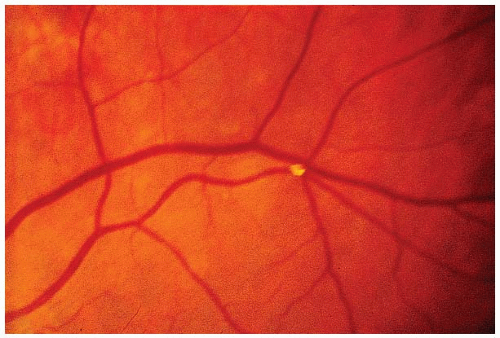 FIGURE 4-1. A bright plaque that appears larger than the artery in which it resides is seen at a retinal arteriole bifurcation. This glistening appearance suggests a cholesterol embolus of carotid artery origin. |
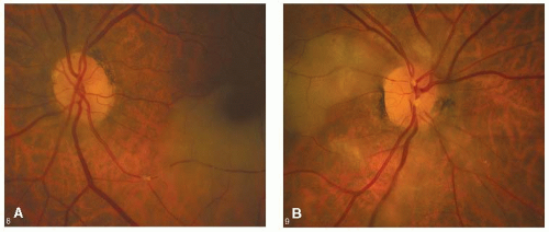 FIGURE 4-2. A. Hour-glass-shaped embolus lodged in the inferior temporal artery OS with adjacent retinal whitening. B.The following day the patient presented with decreased vision OD with multiple large white emboli (not present the previous day) at the optic disc with retinal whitening. |
CLINICAL CHARACTERISTICS
Symptoms
• The primary symptom is acute, usually unilateral painless visual loss, which is sudden, but may be stepwise and stuttering in onset. The visual loss may be partial, with a scotoma that may involve or spare fixation, or may be nearly total, with the preservation of a small island of peripheral vision.
• The presence of a cilioretinal artery will often determine the configuration of the scotoma and, at times, the level of visual acuity.
Signs
• Decreased acuity if the macula area is involved.
• Visual field defects that correspond to the area of retinal infarction (Fig. 4-3).
• Relative afferent pupillary defect (RAPD) is usually detected.
• Areas of retinal whitening that correspond to the scotoma develop within hours but fades over days or weeks. The fundus may appear almost normal initially (Fig. 4-4).
• Intravascular embolic or thrombotic material, at times characteristic of its site of origin, is sometimes identified on ophthalmoscopy.
• Diabetic patients with carotid occlusions will have less diabetic retinopathy on the side of the occluded carotid artery (Fig. 4-5).
• Occlusion at different locations of the retinal vasculature may produce different clinical features:
Stay updated, free articles. Join our Telegram channel

Full access? Get Clinical Tree


