General Concepts
The latissimus dorsi flap can be used as a myofascial flap or as a myocutaneous flap for reconstruction of scalp defects (Fig. 38.1). It is critically important to consider the nature of the scalp defect when planning the design of a latissimus dorsi flap for scalp reconstruction, as the nature of the defects of the scalp skin, calvarium, and dura all have a significant impact on flap design. For soft tissue defects of the scalp where there is not a fullthickness defect of the calvarium, a myofascial flap combined with a skin graft often results in the best possible aesthetic reconstruction (Fig. 38.2). As they are harvested from a sun-protected donor site, myocutaneous latissimus dorsi flap often are pale in color, so they result in a poor color match for native scalp skin, especially when used in the common situation of patients with scalp skin cancers, who invariably have significant chronic actinic damage to their scalp skin. In addition, the flank skin is very thick, and the skin paddle of a latissimus dorsi myocutaneous flap is often too bulky to restore an appropriate scalp contour. This often results in a pincushioned appearing scalp reconstruction (Fig. 38.3). As a broad, thin muscle that will undergo postoperative denervation atrophy when inset over the rigid infrastructure of an intact calvarium, latissimus dorsi myofascial flaps often produce superior aesthetic results with regard to the thickness and contour of the resulting scalp reconstruction, and the color match of a skin-grafted myofascial flap is often superior to that achieved using a latissimus dorsi myocutaneous flap.
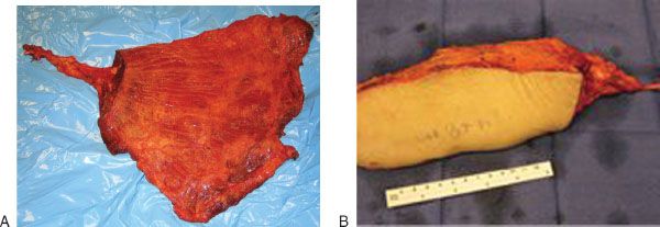
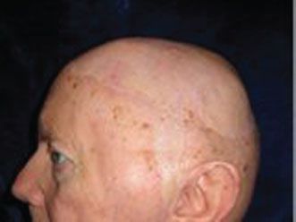
FIGURE 38.2 Two-year postoperative appearance of scalp reconstruction using latissimus dorsi myofascial flap with meshed split-thickness skin graft.
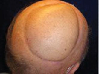
FIGURE 38.3 Five-year postoperative appearance of scalp reconstruction using latissimus dorsi myocutaneous flap with duraplasty and titanium mesh cranioplasty.
In situations where there is a scalp skin defect occurring in conjunction with a full-thickness defect of the calvarium, a latissimus dorsi myocutaneous flap may be the best option. In this situation, the increased thickness of the flap helps to camouflage contour defects of the skull in patients who have small calvarial defects. Many patients with larger defects of the calvarium will undergo cranioplasty reconstruction using nonvascularized bone grafts or alloplastic materials. In this situation, scalp reconstruction using a myofascial flap is ill-advised, as there will be a risk of cranioplasty extrusion after a myofascial flap becomes very thin due to postoperative denervation atrophy. The risk of cranioplasty extrusion may be reduced in patients with thick myocutaneous flap reconstructions, although the aesthetics of the resulting reconstruction are usually compromised.
Myocutaneous flaps may be preferred for reconstruction of scalp defects that occur in conjunction with defects of the dura. This is the case because it is difficult to achieve a watertight closure of the scalp skin defect when using a myofascial latissimus dorsi flap that is resurfaced with a skin graft, thereby increasing the risk of a postoperative cerebrospinal fluid leak that can lead to meningitis. In this situation, the use of a myocutaneous latissimus dorsi flap may lessen the risk of cerebrospinal fluid leak by allowing for a watertight scalp wound closure, thereby increasing the safety of the reconstruction at the expense of the aesthetic results.
Multiple other flap components that are perfused by the subscapular vascular pedicle are available for transfer with the latissimus dorsi muscle in patients with large or complex defects of the scalp. When the soft tissue defect of the scalp is larger than what can reliably be resurfaced using a latissimus dorsi flap, additional soft tissue components can be incorporated, including the serratus anterior muscle that is perfused by the serratus anterior branch of the thoracodorsal artery, and the parascapular and scapular fasciocutaneous flaps that are perfused by the circumflex scapular artery. Using these additional soft tissue components, even near-total defects of the scalp can be reconstructed using a single subscapular system “megaflap” (Fig. 38.4). Osseous reconstruction of the calvarium can be accomplished using vascularized ribs, which are perfused either by the serratus anterior branch of the thoracodorsal artery or by the perforating branches from the intercostal blood vessels. In addition, a vascularized graft of the lateral border of the scapula and scapular tip can be transferred based upon the circumflex scapular vessels or the angular branch of the thoracodorsal vessels. Vascularized thoracolumbar or serratus anterior fascia can be used for reconstruction of associated defects of the dura.
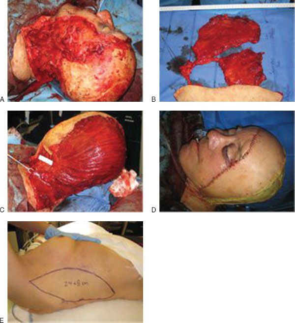
FIGURE 38.4 A. Defect of the left cheek, forehead, and subtotal resection of scalp sparing only the right temporal scalp after excision of angiosarcoma. B. A latissimus dorsi myofascial—serratus anterior myofascial—parascapular fasciocutaneous flap allows for single-flap reconstruction of this large defect. C. The latissimus dorsi myofascial—serratus anterior myofascial flap components are used to resurface temporal, parietal, and occipital calvarium. D. The parascapular fasciocutaneous flap component is used to resurface the cheek and forehead defect. E. The myofascial flaps have been covered with a meshed split-thickness skin graft.
Identification of the borders of the latissimus dorsi muscle is usually a straightforward endeavor using visible and palpable landmarks. The latissimus dorsi muscle is a broad thin muscle that covers most of the lower back from the inferior tip of the scapula to the iliac crest (Fig. 38.5). The anterior border of the latissimus dorsi muscle is an important landmark that is usually identified early during the flap harvest procedure and is a key landmark with regard to identifying the flap’s vascular pedicle. The anterior border of the latissimus dorsi muscle can be palpated in thin patients by asking patients to press their hands against their hips. The location of the anterior border of the latissimus dorsi muscle can also be estimated drawing a line from the humerus in an inferior direction toward the ipsilateral iliac crest at approximately the midaxillary line.
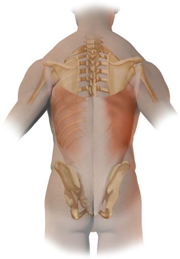
FIGURE 38.5 The latissimus dorsi muscle. The latissimus dorsi muscle is a broad thin muscle that covers most of the lower back from the inferior tip of the scapula to the iliac crest.
HISTORY
With regard to the patient’s history, because free tissue transfer provides the advantage of transferring well-vascularized tissue to a recipient site, the condition of the recipient site is important. Any history of prior radiation may impede healing. This should be elicited in the history to prepare the patient for the potential for wound healing problems. A history of prior surgery or trauma to the donor site(s) may eliminate the use of this flap.
PHYSICAL EXAMINATION
The physical examination should include an examination of the recipient site and the donor site. In many cases, the patient will have sustained the scalp defect as a result of excision of skin cancer. It is important to assess both sites for skin cancers that may have been missed or incompletely excised. The condition of the recipient site should also be evaluated to anticipate the potential for wound healing complications. The donor site must be examined to rule out previous trauma or surgery in this tissue.
INDICATIONS
Latissimus dorsi free flaps are indicated for reconstruction of scalp defects that cannot reliably be reconstructed using local tissues, skin grafts, or regional flaps. As previously discussed, this indication varies according to the size of the defect in different regions of the scalp as determined by laxity of the scalp tissue, the location of the defect with regard to the utility of regional flaps as determined by their arc of rotation, prior therapies that affect the reliability of local and regional flaps, and the nature of associated defects of the calvarium and dura.
CONTRAINDICATIONS
Prior surgeries that might have adversely affected the integrity of the thoracodorsal vascular pedicle need to be recognized. In this regard, the most common prior surgeries to consider include prior axillary lymph node dissection and prior thoracotomy. In these patients, preoperative flap angiography or an alternative flap donor site should be considered. In addition, use of a large skin paddle can produce a restrictive pulmonary defect caused by tight primary closure of the flank skin after latissimus dorsi flap harvest, so large myocutaneous flaps should be used with caution in patients with limited pulmonary reserve. In this situation, the flap donor site can be repaired with a skin graft, but skin graft healing is notoriously slow due to a shearing effect on the skin graft caused by respiratory motion of the flap donor site.
PREOPERATIVE PLANNING
Stay updated, free articles. Join our Telegram channel

Full access? Get Clinical Tree


