R
radiant flux See flux, radiant.
radiation 1. Emission or transfer of energy in the form of electromagnetic waves or particles. 2. A group of nerve fibres that diverge in all directions from a point of origin. Example: the optic radiations.
See spectrum, electromagnetic.
radiation, black body The radiation emitted by a heated black body.
See colour temperature.
radiations, optic That part of the visual pathway which consists of axons arising in the lateral geniculate body and terminating in a fan-shaped manner in the visual area of the occipital lobe. As they emerge from the lateral geniculate body the inferior fibres loop forward in the temporal lobe before swinging back toward the occipital cortex. These fibres form what is called Meyer’s loop (or Archambault’s loop). They receive impulses from the inferior retinal quadrants (corresponding to the superior aspect of the contralateral visual field) and terminate on the inferior lip of the calcarine fissure. Syn. optic radiations of Gratiolet; geniculocalcarine pathway; geniculostriate pathway.
radiology A science dealing with techniques that use radiant energy (e.g. X-rays) for diagnosis and therapy.
See angiography, fluorescein; magnetic resonance imaging; tomography, computed.
radiuscope Instrument used for measuring the radius of curvature of the surfaces of a contact lens. It is based on the Drysdale method. Syn. optical microspherometer.
See method, Drysdale’s; Toposcope.
Raman effect See effect, Raman.
Ramsden eyepiece See eyepiece, Ramsden.
ramus communicans See ganglion, ciliary; nerve, ophthalmic.
random-dot stereogram; E test See stereogram, random-dot.
randomized controlled trial See trial, randomized controlled.
range of accommodation See accommodation, range of.
ranibizumab See anti-VEGF drugs; macular degeneration, age-related; retinopathy, diabetic.
raphe, retinal See retinal raphe.
ratio, Arden See electrooculogram.
ratio, AV The ratio of the diameter of the retinal arteries to that of the retinal veins. It is usually around two-thirds. Deviations from this value may indicate a vascular disease (e.g. hypertension).
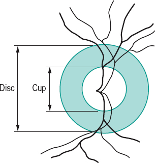
ratio, cup-disc (C/D) The ratio of the horizontal diameter of the physiological cup to that of the horizontal diameter of the optic disc. It should be less than 0.5. If it exceeds that value, or if there is a difference in ratio between the two eyes, or if there is a progressive enlargement of the cup, glaucoma may be suspected (Fig. R1).
See cup, glaucomatous.
Raubitschek chart See chart, Raubitschek.
ray In geometrical optics, a straight line representing the direction of propagation of light.
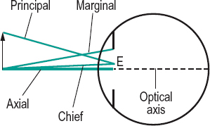
axial r. A ray that is coincident with the axis of an optical system.
chief r. A ray joining an object point to the centre of the entrance pupil of an optical system (Fig. R2).
emergent r. A ray of light in image space either after reflection (reflected ray) or after refraction (refracted ray).
extraordinary r. See birefringence.
incident r. A ray of light in object space that strikes a reflecting or refracting surface.
marginal r. A ray joining the axial point of an object to the edge or margin of an aperture or pupil (Fig. R2).
ordinary r. See birefringence.
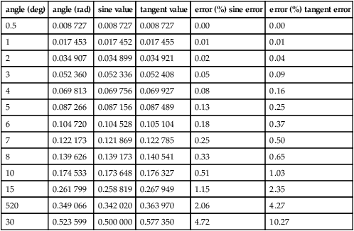
paraxial r. A light ray that forms an angle of incidence so small that its value in radians is almost equal to its sine or its tangent. (i.e. sin θ = θ or tan θ = θ. These are approximate expressions referred to as the paraxial approximation (or the gaussian approximation).
See optics, paraxial; paraxial region; theory, gaussian.
principal r. A ray joining the extreme off-axis object point to the centre of the entrance pupil or aperture (Fig. R2).
r. tracing Technique used in optical computation consisting of tracing the paths of light rays through an optical system by graphical methods or by using formulae. Nowadays, computer methods are used.
See sign convention.
Rayleigh criterion See criterion, Rayleigh.
Rayleigh equation A colour equation representing a match of yellow (usually 589 nm) with a mixture of red (usually 670 nm) and green (usually 535 nm). It is used to differentiate certain types of colour deficiencies. The anomaloscope is built on this principle.
See colour vision, defective.
Rayleigh scattering See scattering, Rayleigh.
reaction time The time interval between the onset of a stimulus and the response of a subject. Visual stimulations with a flash of light give rise to reaction times varying between 130 and 180 ms. This figure increases significantly with age.
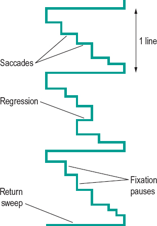
reading The act of viewing and interpreting letters, words, sentences, etc. It consists of a pattern of eye movements. The eyes proceed along a line in a series of step-like saccades, separated by fixation pauses during which information from the text is acquired. The amount of reading matter correctly identified during the fixation pause is called the span of recognition or the perceptual span. Most saccades are made from left to right, but some occur in the opposite direction (called regression) to return to text recently read but not yet fully perceived. At the end of the line the eyes make a return sweep to the next line of text (Fig. R3).
See movement, saccadic eye; test, developmental eye movement.
reading addition See addition, near.
reading distance; lens See under the nouns.
reading portion See segment of a bifocal lens.
Table R1
Differences between the sine and the tangent values of various angles (in degrees and radians). The error is calculated between the sine value and the value in radians and between the value in radians and the tangent value
| angle (deg) | angle (rad) | sine value | tangent value | error (%) sine error | error (%) tangent error |
| 0.5 | 0.008 727 | 0.008 727 | 0.008 727 | 0.00 | 0.00 |
| 1 | 0.017 453 | 0.017 452 | 0.017 455 | 0.01 | 0.01 |
| 2 | 0.034 907 | 0.034 899 | 0.034 921 | 0.02 | 0.04 |
| 3 | 0.052 360 | 0.052 336 | 0.052 408 | 0.05 | 0.09 |
| 4 | 0.069 813 | 0.069 756 | 0.069 927 | 0.08 | 0.16 |
| 5 | 0.087 266 | 0.087 156 | 0.087 489 | 0.13 | 0.25 |
| 6 | 0.104 720 | 0.104 528 | 0.105 104 | 0.18 | 0.37 |
| 7 | 0.122 173 | 0.121 869 | 0.122 785 | 0.25 | 0.50 |
| 8 | 0.139 626 | 0.139 173 | 0.140 541 | 0.33 | 0.65 |
| 10 | 0.174 533 | 0.173 648 | 0.176 327 | 0.51 | 1.03 |
| 15 | 0.261 799 | 0.258 819 | 0.267 949 | 1.15 | 2.35 |
| 520 | 0.349 066 | 0.342 020 | 0.363 970 | 2.06 | 4.27 |
| 30 | 0.523 599 | 0.500 000 | 0.577 350 | 4.72 | 10.27 |
reading slit See typoscope.
receptive field See field, receptive.
receptor See photoreceptor.
recession A surgical procedure used in strabismus in which an extraocular muscle is removed from its insertion and repositioned elsewhere on the globe, posteriorly to weaken it and anteriorly to strengthen it (called advancement procedure).
See resection; strabismus surgery.
recessive inheritance See inheritance.
reciprocal innervation See law of reciprocal innervation, Sherrington’s.
reciprocal metre See curvature of a surface.
reciprocity, law of See law, Bunsen–Roscoe.
von Recklinghausen’s disease See disease, von Recklinghausen’s.
recovery point See point, recovery.
rectus muscles See muscles.
recumbent spectacles See spectacles, recumbent.
recurrent corneal erosion See corneal erosion, recurrent.
red One of the hues of the visible spectrum evoked by stimulation of the retina by wavelengths beyond 630 nm. The complementary colours to red are blue-green (between 490.4 and 492.4 nm).
red blindness See protanopia.
red glass test See test, red glass.
red reflex See reflex, fundus.
red-green colour deficiency A general term indicating a colour vision deficiency, which is either of the deutan (green colour vision defect) or of the protan (red colour vision defect) type. These defects are mostly hereditary and affect both eyes equally. Most cases are inherited in X-linked recessive manner.
See colour vision, defective; rule, Kollner’s.
reduced eye; vergence See under the nouns.
refixation reflex See reflex, refixation.
reflectance A measure of reflection equal to the ratio of reflected luminous flux φ’ to the incident luminous flux φ, i.e. ρ = φ’/φ. Syn. reflection factor.
See Fresnel’s formula.
reflecting prism; telescope See under the nouns.
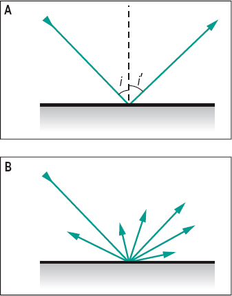
reflection Return or bending of light by a surface such that it continues to travel in the same medium.
angle of r. See angle of reflection.
diffuse r. Reflection from a surface that is not polished and light is reflected in many or all directions (Fig. R4). Syn. irregular reflection.
See diffusion; glossmeter; matt surface.
direct r. See reflection, specular.
r. factor See reflectance.
irregular r. See reflection, diffuse.
law of r. See law of reflection.
mixed r. The simultaneous occurrence of diffuse and specular reflection.
regular r. See reflection, specular.
specular r. Reflection from a polished surface in which there is no scattering and light travels back in a definite direction (Fig. R4). Syn. direct reflection; regular reflection.
See microscope, specular.
surface r . Light reflected at a surface according to Fresnel’s formula.
total r. Reflection occurring when light is incident at an angle greater than the critical angle. Syn. total internal reflection.
See prism, reflecting.
total internal r. See reflection, total.
reflector Any device that reflects light (e.g. glass-plate, prism, mirror).
reflex 1. Involuntary response to a stimulus. 2. Reflection or an image formed by reflection (e.g. corneal reflex).
accommodative r. See accommodation, reflex; reflex, near.
r. arc See reflex, pupil light.
blinking r. Blinking in response to various stimulations such as a light source or a mechanical threat.
See pathway, retinotectal.
cat’s eye r. A whitish, bright reflection observed in the normally black pupil in several conditions, such as leukocoria, retinoblastoma, Coats’ disease or persistent hyperplastic primary vitreous. It resembles the reflection from the tapetum lucidum of a cat when a light is shined at night.
consensual light r. See reflex, pupil light.
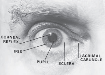
corneal r. 1. Blinking in response to a threat, or to tactile stimulation of the cornea, as for example when measuring objectively the corneal touch threshold. Associated responses include lacrimation and miosis. 2. Image formed by reflection of light from the cornea (Fig. R5).
See aesthesiometer; method, Hirschberg’s; method, Krimsky’s; pupillometer; strabismus, apparent.
direct light r. See reflex, pupil light.
eyeball compression r. See reflex, oculocardiac.
fixation r. Psycho-optical reflex consisting of an involuntary movement of the eye (or eyes) aimed at placing on the foveola the retinal image of an object that was formed in the retinal periphery.
See reflex, psycho-optical; reflex, re-fixation.
foveal r. Tiny reflection from the concave surface of the foveal depression of the retina seen in ophthalmoscopy. It is not usually visible in old eyes.
fundus r. Light reflected by the fundus of the eye, as seen in retinoscopy and ophthalmoscopy. It appears as a red glow in the plane of the pupil in retinoscopy. It is absent when the eye has a dense cataract. Syn. red reflex.
hemianopic pupillary r. In hemianopia, a loss of pupillary constriction when light falls on the blind side of the retina while pupillary constriction is maintained when light stimulates the unaffected side of the retina. Syn. Wernicke’s hemianopic pupil; Wernicke’s pupillary reaction; Wernicke’s pupillary reflex; Wernicke’s sign.
indirect light r . See reflex, pupil light.
lacrimal r. Secretion of tears in response to irritation of the cornea or conjunctiva as, for example, when first wearing contact lenses (hard in particular), but it may also be induced by eyestrain, glare, laughing, etc. Syn. lacrimation reflex; tearing reflex; weeping reflex.
See lacrimal apparatus; tear secretion; test, Schirmer’s.
lacrimation r. See reflex, lacrimal.
light r. 1. That light which appears in the pupil in retinoscopy. It is light reflected by the retina. Syn. retinoscopic light. 2. Any reflected light.
See reflex, pupil light.
near r. Reflex evoked by a blurred retinal image, as when fixating from far to near. It consists of three responses: (1) increased convexity of the crystalline lens; (2) constriction of the pupils; and (3) convergence of the eyes. This reflex is not a pure reflex since each of the three components can act independently of the other two; convergence by means of prisms, accommodation by means of lenses and miosis by light stimulation. Syn. accommodative reflex; near-triad reflex; synkinetic near reflex.
See accommodation, mechanism of; accommodation, reflex; accommodative response.
near-triad r. See reflex, near.
oculocardiac r. A decrease in pulse rate following compression of the eyeball or traction on the extraocular muscles during ocular surgery. It may produce a systolic cardiac arrest. Syn. Ascher’s phenomenon; Ascher’s reflex; eyeball compression reflex.
optokinetic r. See reflex, vestibulo-ocular.
postural r. A reflex which helps to maintain static or dynamic posture of the body, for example, the righting reflex, in which visual stimuli help to maintain a correct position of the head in space by activating the muscles of the neck and limbs.
See reflex, static eye.
psycho-optical r. Reflexes involving the eye which are mediated by the occipital cortex such as the accommodative, fixation, fusion, version and vergence reflexes.
pupil r. Any alteration of the pupil size in response to stimuli other than light (e.g. a sudden noise).
See reflex, pupil light.
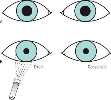
pupil light r. 1. Constriction of the pupil in response to light stimulation of the retina. The response of an eye to light stimulation can occur either with a light shining on it directly (the direct light reflex) or when the other eye is stimulated (the consensual or indirect light reflex). The reflex arc consists of four neurons beyond the ganglion cells. The first afferent neuron transmits nervous impulses from the retina to the two pretectal olivary nuclei, located on the lateral and anterior side of the superior colliculi, in response to light stimulation of the photoreceptors. The second neurons, called the internuncial neurons, connect each pretectal olivary nucleus to both Edinger–Westphal nuclei which form part of the oculomotor nuclei. The third efferent neurons connect the latter nuclei, via the third nerve (oculomotor nerve) to the ciliary ganglion where there is a synapse. The fourth efferent neurons connect the latter, via the short ciliary nerves, to the sphincter pupillae muscle of each iris and constrict the pupil. Light stimulation of the central region of the retina produces a greater pupillary response than peripheral stimulation. The efferent path for pupil constriction represents the parasympathetic innervation (Fig. R6). Note: recent research points to a different pathway for the second afferent neuron: almost all of the fibres from each pretectal olivary nucleus project to the contralateral Edinger–Westphal nucleus. 2. Dilatation of the pupil in response to a reduction of the light stimulation of the retina. It is effected by sympathetic innervation, which originates in the hypothalamus and descends down the brainstem to the ciliospinal centre (of Bulge), located between C8 and T2. From there fibres pass to the superior cervical ganglion in the neck, then efferent fibres ascend along the internal carotid artery until they join the ophthalmic division of the trigeminal nerve. The fibres reach the dilator pupillae muscle of the iris via the nasociliary and long ciliary nerves, which enter the eyeball behind the equator. Syn. light reflex.
See fibres, pupillary; pathway, retinotectal; pretectum; pupillary defect, efferent.
re-fixation r. This reflex occurs while fixating one object and another in the visual field attracts the attention. The eye then turns to fixate on the new object. This is a special case of the fixation reflex.
retinoscopic r. See reflex, light; retinoscope.
righting r. See reflex, postural.
static eye r . A higher order postural reflex which helps to maintain the eye static with respect to the visual environment by action on the extraocular muscles (possibly via the utricular receptors of the vestibular system) during head or body movements. Syn. compensatory eye movements.
r. tearing See reflex, lacrimal.
tonic neck r. Orientation of the head, eyes and body in response to proprioceptive information provided by the activity of the muscles of the neck.
See proprioception.
vergence r. A disjunctive fixation reflex in response to an object that moves closer or further than the original position of the fixation point.
See movements, disjunctive eye.
version r . A conjugate fixation reflex in response to an object moving in the same frontal plane.
See version.
vestibulo-ocular r. A conjugate movement of the eyes in the direction opposite to a head movement. This reflex is triggered by stimulation of the semicircular canals. It is aimed at maintaining a stable image on the retina during head movement. This reflex responds best at high velocities and frequencies of the visual stimulus. At low velocities and frequencies the stabilization of the retinal image is attempted by the optokinetic reflex, which is triggered only by retinal stimulation: this latter reflex complements the vestibulo-ocular reflex.
See nystagmus; optokinetic.
weeping r. See reflex, lacrimal.
Wernicke’s pupillary r. See reflex, hemianopic pupillary.
white pupillary r . See leukocoria.
refract 1. To bend a ray of light when it passes through a surface separating media of different refractive indices. 2. To measure the refractive state of the eye.
refracting angle See angle, prism.
refracting unit See phoropter.
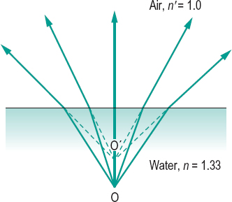
refraction 1. The change in direction of the path of light as it passes obliquely from one medium to another having a different index of refraction (Fig. R7). 2. The process of measuring and correcting the refractive error of the eyes. Syn. refraction of the eye; sight testing (obsolete term). 3.
See refractive error.
See law of refraction.
angle of r . See angle of refraction.
binocular r. A clinical procedure in which the subjective measurement of refraction of each eye is performed while both eyes are viewing a test. The visual examination is thus carried out under more natural conditions than when one eye is closed; the sizes of the pupils are similar and the accommodationconvergence relationship is maintained.
There are various such methods; using polarized targets (e.g. Vectograph slides; Parallel-testing Infinity Balance test), using a septum (e.g. Turville Infinity Balance test), or fogging (e.g. Humphriss Immediate Contrast test). These methods give better results than refracting monocularly, especially in latent hyperopia, hyperopic anisometropia, pseudomyopia, cyclophoria, etc. and no additional step for binocular balancing is necessary.
See method, Humphriss; test, balancing; test, Turville, infinity balance; testing in parallel.
cycloplegic r. Assessment of the refractive state of the eye when accommodation has been totally or partially paralysed by a cycloplegic (e.g. cyclopentolate 1% eyedrops, or atropine 0.5 or 1% ointment). This may be carried out in children to reveal the full extent of a hyperopia or in the initial assessment of accommodative esotropia, but only occasionally in adults as fogging methods usually suffice for them.
See cycloplegia; strabismus, accommodative.
double r. The splitting of an incident ray into two (ordinary and extraordinary) by a birefringent medium.
See anisotropic; birefringence; prism, Nicol; prism, Wollaston.
dynamic r. Determination of the refractive state of the eye when accommodation is stimulated, as distinguished from static refraction which is the determination of the refractive state of the eye when accommodation is at rest or paralysed.
See refractive error; retinoscopy, dynamic.
error of r. See ametropia; refractive error.
r. of the eye 1. See refraction. 2. Refraction of light by the optical media of the eye. 3. Syn. for ametropia.
See ametropia; refractive error.
index of r. See index of refraction.
laser r. A method of subjective refraction in which the patient observes a slowly rotating drum, on the surface of which is perceived a speckle pattern resulting from illumination by a laser. The speckle pattern appears to move only when the eye is not focused for the fixation distance. If the perceived movement of the pattern is opposite to that of the drum, the eye is myopic and if the perceived movement of the pattern is in the same direction as the drum, the eye is hyperopic. Correction can be determined by placing a lens in front of the eye, which will neutralize the movement; at that point the eye is focused for the fixation distance. Astigmatism can be measured by rotating the drum in various meridians. The drum can be placed at infinity or at near (an allowance for the radius of curvature of the drum and the distance must then be made). This method can be useful for mass screening, especially children, as accommodation is not stimulated as much as with Snellen letters. It has been very useful as a research tool for accommodation studies where it is arranged as part of a Badal optometer.
manifest r. The refractive error or the process of determining it, when accommodation is at rest (but not paralysed).
See refraction, cycloplegic.
objective r. Measurement of the refraction of the eye that is not based on the patient’s judgements, as when using an objective optometer or a retinoscope.
See photorefraction.
static r. See refraction, dynamic; refractive error.
subjective r. Measurement of the refraction of the eye based on the patient’s judgements.
See method, fogging; optometer; test, duochrome; test, fan and block; test, plus 1.00 D blur.
refractionist One who measures and corrects the refractive state of the eye.
See optometrist.
refractive amblyopia See amblyopia.
refractive correction See correction.
refractive error The dioptric power (K) of the ametropia of the eye. It is equal to 1/k in dioptres, where k is the distance between the far point and either the spectacle plane (spectacle refraction), or the principal point of the eye, or the refracting surface of the reduced eye (ocular refraction), in metres. Thus

when the eye is situated in air. Syn. ametropia (although this is not strictly so as ametropia is the anomaly); refraction of the eye; refractive status; static refraction.
See accommodation, far point of; experiment, Scheiner’s.
refractive index See index of refraction.
refractive surgery
Stay updated, free articles. Join our Telegram channel

Full access? Get Clinical Tree


