FIGURE 27.1 A,B. The high-resolution CT scan (1-mm cuts) provides a sensitive method to evaluate the infraglottic airway. These images demonstrate invasion of the trachea that may not be appreciated on office endoscopy.
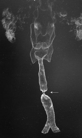
FIGURE 27.2 Three-dimensional reconstruction can provide important information related to the site of the stenosis.
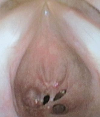
The intraoperative endoscopy typically provides the best examination. A rigid fiberoptic endoscope provides a high-resolution evaluation of the airway that can be exceptionally helpful in determining the nature of the disease and predict the best approach to reconstruction (Fig. 27.3).
INDICATIONS
The indications for primary (end-to-end) tracheal reconstruction are defects that are less than 4 cm in length. In select patients, defects that are less than 6 cm can be managed with primary end-to-end reconstruction but may require infrahyoid muscle release and/or a suprahyoid muscle release. However, these techniques impede elevation of the larynx during swallowing and may result in aspiration.
CONTRAINDICATIONS
The contraindications to primary tracheal reconstruction are patients with defects greater than 6 cm in length and patients with defects greater than 4 cm in length if there is a history of a previously failed reconstruction, external beam radiation therapy, or compromised healing. It is critical to understand that every patient is an individual and therefore each patient should be evaluated as such. Most contraindications are “relative” contraindications. The patient’s anatomy, body habitus, and personal disposition all play a role in the decision-making process concerning reconstruction.
PREOPERATIVE PLANNING
Prior to surgery, I evaluate airway resistance in all subjects. A flow-volume loop is generated by having the patient inhale deeply to total lung capacity (TLC), forcefully exhale until the lungs have been emptied to residual volume, and rapidly inhale to reach TLC. A maximal expiratory flow 50%: Maximal inspiratory flow 50% ratio is, therefore, usually less than 1. In variable extrathoracic lesions, the ratio is increased (usually >1), while in variable intrathoracic lesions, the ratio is diminished (0.2 or less). In fixed obstructions, the ratio is expected to be close to 1. This study provides an excellent method to determine diagnosis and eligibility for surgery.
SURGICAL TECHNIQUE
Stay updated, free articles. Join our Telegram channel

Full access? Get Clinical Tree


