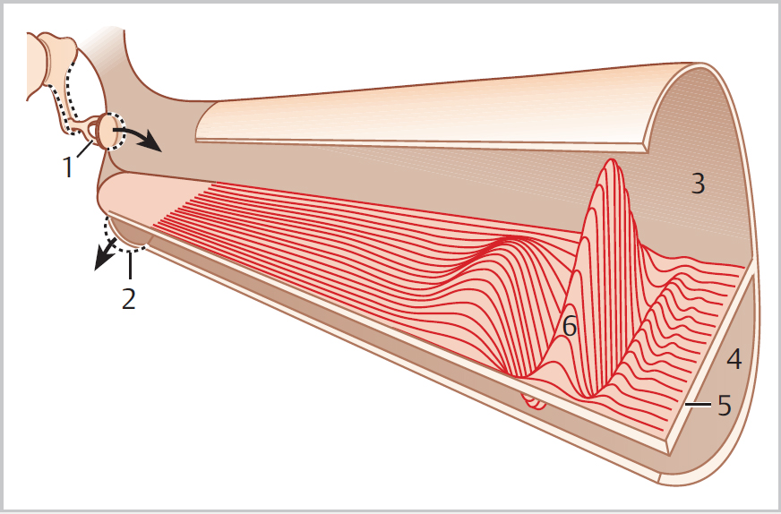
2 Physiology of the Ear • The tympanic membrane (TM) outer layer is keratinizing squamous epithelium • Superficial cells are shed and cleared from the centre of the TM by an “escalator” effect; retraction pockets may affect this process leading to keratin accumulation and potential cholesteatoma formation • The middle ear (ME) is a sound–pressure transformer • The resistance to the passage of sound through a medium is its acoustic impedance—if small, most sound is transmitted; from middle to inner ear, sound passes from air to fluid, resulting in impedance mismatch • The acoustic role of the TM is to transform sound pressure over its outer surface to malleus vibration (the umbo moves most) • The lever effects of the ossicles (mechanical advantage of incudomalleolar joint is 1:1.3) and the area ratio of TM to stapes footplate (1:17) allows for sound–pressure transformation (and ~25 dB gain of energy to cochlea) • The total pressure on the stapes footplate is therefore enhanced by a factor of 22 (17 × 1.3) • Sound is transmitted from ear canal to cochlea via tympano-ossicular system and direct acoustic stimulation of oval and round windows (acoustic coupling) • The ME loses gases to the tissues constantly; this is compensated for by influx from the nasopharynx via the eustachian tube; when gas input < output, negative ME pressure is created, which is a factor in TM retraction • Vibration at oval window results in wave that travels up basilar membrane, increases to a maximum amplitude, then dies away; point of maximum amplitude depends on frequency introduced (high frequency = apical, low frequency = basal) • Each hair cell has an optimal frequency that it responds to, but responds to other frequencies to lesser extent • Humans can discriminate between sounds differing by 0.75 Hz frequency, and we can hear over the range 20 to 20,000 Hz • Vibration of basilar membrane boosted by active mechanical amplifier mechanism; as basilar membrane moves, get shearing forces between it and overlying tectorial membrane, resulting in deflection of stereocilia of hair cells • Ion channels open in cell membrane, and neurotransmitter is released at base of cell • Most afferent auditory nerve fibres are stimulated in this way by inner hair cells (95% afferents); they convert the mechanical energy to bioelectrical energy • Deflection of hair cell cilia towards the cells’ longest cilia leads to depolarization, whereas deflection away leads to hyperpolarization • Outer hair cells (OHCs) have role in active mechanical amplifier mechanism; they may amplify the effect of the sound stimulus and increase the sensitivity and frequency selectivity of the cochlear output (in presbyacusis, lose OHCs and therefore lack of amplification so thresholds rise and discrimination falls) • Utricle has receptor cells orientated horizontally, saccule vertically • The maculae of the saccule and utricle have hair cells with stereocilia embedded in overlying fibrogelatinous mass (otoconial membrane); on its surface are otoconia (calcium carbonate) that make it denser than the endolymph • Linear acceleration (and gravity) moves the otolithic membrane relative to the hair cells, bends stereocilia, thus stimulating the sensory cell by alteration of the resting potential • If accelerated, the inertia of the otoconia causes them to lag behind the maculae (which move with skull), hence stereocilia deflected; when acceleration is over, elastic recoil restores steady state
2.1 External Ear
2.2 Middle Ear
2.3 Inner Ear
2.3.1 Cochlear Mechanism (Figs. 2.1 and 2.2)
2.3.2 Vestibular Mechanism (Fig. 2.3)
< div class='tao-gold-member'>
![]()
Stay updated, free articles. Join our Telegram channel

Full access? Get Clinical Tree


