Pharyngitis


Pharyngitis


Stephen R. Hoff

Kay W. Chang


Pharyngitis refers to inflammation of the pharyngeal mucosa, lymphoid tissue, musculature, and surrounding fatty and fascial tissues. This can be infectious or noninfectious, and can herald more extensive systemic diseases such as autoimmune disease or immunocompromise with human immunodeficiency virus (HIV). Acute pharyngitis is one of the frequent common causes of visits to a physician or health care provider, with over 15 million office visits per year for streptococcal pharyngitis alone (1). Approximately one-third of patients with upper respiratory infection (URI) have sore throat as the chief complaint. The abundance of lymphoid tissue in the pharynx, and the constant communication with the outside world during respiration, leads to the frequency of pharyngitis, and accounts for many of the symptoms.

Pharyngitis typically presents with sore throat, with odynophagia and dysphagia. This may be accompanied by fevers, hoarseness, nasal congestion, cough, halitosis, and malaise, depending on the etiology. Although there is a wide range of possible diagnoses, an accurate assessment of the time course, symptoms, and associated physical exam findings can rapidly and efficiently narrow the differential diagnosis (Tables 52.1 and 52.2). Viral infections are the most common cause of pharyngitis in both children and adults, but pediatric patients have a much higher rate of bacterial infection than adults (30% to 40% and 5% to 15%, respectively) (2,3).

Generally, most patients are treated supportively for the symptoms of pain and dysphagia, but antibiotics may be used when bacterial infection is suspected, or with opportunistic infections in the immune-compromised host. Of paramount importance is distinguishing pharyngitis from an acute infection with abscess formation, including peritonsillar, retropharyngeal, and parapharyngeal abscesses, which require more aggressive management. These are discussed further in Chapter 55. The symptoms of a pharyngeal malignancy may also mimic the clinical presentation of pharyngitis, and a high index of suspicion on the part of the physician is necessary, with biopsy and histologic examination as warranted.

Although there is overlap, the presentation and etiology of pharyngitis is different in the child and the adult. Therefore, this chapter discusses them separately, with a focus on the more common clinically encountered pathologies. Because adolescents tend to have pharyngitis profiles similar to adults, the pediatric section is specific to children less than 14 years of age. Adenotonsillar-specific disease, which obviously overlaps with pharyngitis, is discussed in Chapter 95.


ANATOMY

Anatomically, the pharynx has three divisions: the nasopharynx, oropharynx, and hypopharynx. These divisions are contiguous with each other, but contain different amounts of lymphoid tissue and unique specialized structures, and so are a helpful clinical distinction. The mucosal lining of the pharynx transitions from respiratory epithelium (pseudostratified ciliated) to stratified squamous from superior to inferior.

The nasopharynx is bounded by the nasal cavity and choanae anteriorly, soft palate inferiorly, sphenoid rostrum superiorly, and clivus posteriorly. The adenoidal lymphoid tissue sits on the roof of the nasopharynx, and is typically largest in children 5 to 7 years old. This may lead to obstruction between the nasal cavity and nasopharynx, with rhinorrhea, sleep apnea, and “adenoid facies” as a result. Typically, the adenoids begin to regress in late childhood, and are nearly absent in the adult. Presence of prominent adenoids in the adult should lead the physician to suspect pathologies such as HIV, lymphoma, or malignancy, and biopsy or testing may be warranted. Posterolaterally, the nasopharynx contains the eustachian tube orifices and associated muscles (salpingopharyngeus, levator veli palatini) and torus tubarius cartilage. Rosenmüller fossa (pharyngeal recess) is
located adjacent to the medial part of the eustachian tube orifice in the nasopharynx, and contains a small amount of lymphoid tissue known as Gerlach tonsil.


TABLE 52.1 INFECTIOUS CAUSES OF PHARYNGITIS


Viral


Rhinovirus


Coronavirus


Adenovirus


Parainfluenza virus


Influenza virus types A and B


Measles virus


Respiratory syncytial virus


Coxsackie virus


EBV


CMV


HSV


HIV


Bacterial


GABHS (S. pyogenes)


Groups C and G Beta-Hemolytic Streptococcus


C. diphtheriae


N. gonorrhea


A. haemolyticum


Staphylococcus aureus


T. pallidum (Syphilis)


Fusobacterium


Francisella tularensis


Yersinia enterocolitica


M. tuberculosis


Mycobacterium leprae


C. trachomatis


Mycoplasma pneumonia


Fungal


C. albicans


Protozoal


Toxoplasma gondii


The oropharynx is bounded by the circumvallate papillae anteriorly, and contains the lingual tonsil of the posterior one-third of the tongue. Laterally, it is bounded by the pharyngeal tonsils, which are supported by the palatoglossus and palatopharyngeus muscles. The tonsillar capsule sits on the superior pharyngeal muscle, with the peritonsillar space between them. Together, the pharyngeal, lingual, and adenoidal tonsils form Waldeyer’s ring. Just deep to the superior pharyngeal muscle sits the glossopharyngeal nerve, which can transmit the pain and odynophagia of inflammation during pharyngitis and tonsillitis.

The hypopharynx extends from the level of the hyoid bone to the inferior border of the cricoid cartilage, where it meets the cervical esophageal introitus, and contains the pyriform sinuses, postcricoid region, and posterior pharyngeal wall. The superior, middle, and inferior constrictor muscles form the open-tubed structure of the pharynx, and fuse in the posterior midline at the pharyngeal raphe. Due to this raphe, retropharyngeal abscesses form posterior to the constrictor muscles and tend to be bounded to one side of the retropharynx without crossing the midline. There are several important muscular/fascial layers and spaces between the pharyngeal wall and vertebrae. From anterior to posterior these are: the buccopharyngeal fascia (covering of pharyngeal constrictor muscles), retropharyngeal space, alar fascia, “danger space,” prevertebral fascia, prevertebral space and musculature, and bony vertebral body. The deep spaces of the neck are covered in more detail elsewhere in this text.


TABLE 52.2 NONINFECTIOUS CAUSES OF PHARYNGITIS


Autoimmune/Inflammatory


KD


Pemphigus


Pemphigoid


EB


Erythema Multiforme/Stevens-Johnson syndrome


Crohn disease


Sarcoidosis


Behcet syndrome


Reactive arthritis (Reiter syndrome)


Other


Foreign body


Trauma


PND


LPR


Neoplasm (squamous cell carcinoma, lymphoma, leukemia)


Caustic ingestion/thermal injury


Smoking/environmental exposures


Innervation of the pharyngeal musculature is via the vagus nerve through the pharyngeal plexus, except the stylopharyngeus muscle is innervated by the glossopharyngeal nerve. The arterial supply of the pharynx is mainly from the external carotid branches, including the ascending pharyngeal arteries, dorsal lingual arteries, tonsillar branches of the facial arteries, and palatine branches of the maxillary arteries. In the posterior pharyngeal submucosa, there is an extensive venous plexus, which drain inferiorly into the internal jugular veins. Lymphatic drainage from the pharynx is via the retropharyngeal, lateral pharyngeal, and deep jugular nodes.


INFECTIOUS CAUSES OF PHARYNGITIS IN THE CHILD


BACTERIAL


Streptococcal Pharyngitis

The most important distinction to make in a child with acute pharyngitis is to determine whether the etiologic agent is Group A Beta-Hemolytic Streptococcus (GABHS) a.k.a
Streptococcus pyogenes. This is the only common pathogen that necessitates the use of antibiotics (4). The overuse of antibiotics in pharyngitis from other pathogens, usually viral, should be avoided, with the exception of the rare bacterial infections. Treatment with antibiotics for GABHS will shorten the patient’s symptoms by 24 to 48 hours and reduce infectivity as well as suppurative complications, but the most important reason to give antibiotics is to prevent late sequelae (rheumatic fever and rheumatic heart disease) (2,3,4,5,6,7,8). Because the symptoms of viral and bacterial pharyngitis are similar, and physicians using clinical judgment have been shown to overestimate the occurrence of GABHS, laboratory testing is required in pediatric patients (1,4,5,7).

GABHS infection is common in the 5- to 15-year-old age group, with a peak incidence during the early school years, and a decline in incidence in late adolescence and adulthood. In temperate climates, it is most common in the winter and early spring months (4,7). GABHS accounts for 20% to 30% of cases of pharyngitis in children, compared to 5% to 15% in adults, and is rare in patients less than 2 to 3 years (2). According to a 2008 study, the economic burden from pediatric streptococcal pharyngitis in the United States is estimated between $224 and $539 million per year, with most of the cost attributed to parent’s time off from work (9). In addition to GABHS, infection with other serogroups of streptococcus is possible but much less common. These include Streptococcus C and G, which have been responsible for foodborne and waterborne outbreaks of pharyngitis, but with generally less severe symptoms than GABHS (8).

The incubation period for streptococcal pharyngitis is 1 to 4 days, after which patients will develop rapid onset of severe sore throat and odynophagia, with fevers, malaise, and possibly headache and gastrointestinal (GI) symptoms, such as abdominal pain and vomiting. In general, viral symptoms, such as cough, coryza, and nasal congestion are absent, and the presence of these symptoms makes the diagnosis of GABHS less likely. Tender, enlarged cervical lymph nodes are common. Pharyngeal examination shows erythematous oropharyngeal mucosa, including a beefy red uvula and possibly soft palate petechiae. Tonsils are also erythematous and inflamed, and may have a whitish, creamy exudate. Infants often have red, excoriated nares, with minimal rhinorrhea. Occasionally, patients will have a characteristic “scarlet fever” rash, consisting of fine, blanching, erythematous papules beginning on the trunk and spreading to the extremities, with sparing of the palms and soles (3). This rash has a texture like sandpaper, and will last 6 to 9 days. In addition to the rash, enlarged tongue papillae with erythema (strawberry tongue) may be present, with circumoral pallor. Infants and children less than three with GABHS tend to present predominately with irritability, fevers, and abdominal symptoms, such as vomiting.

Diagnosis of GABHS depends on clinical judgment and laboratory testing, and there are several different clinical guidelines available. A scoring system has been developed and validated, which assigns points for symptoms consistent with GABHS, and may help determine the risk of GABHS in each patient (10). All guidelines recommend against further testing if the patient does not have symptoms typical of GABHS. However, physicians have been shown to overestimate the incidence of GABHS, and overtreat with antibiotics (10). Therefore, in patients less than 15 years of age, with typical symptoms of sore throat, fever, adenopathy, and inflamed pharynx, laboratory testing should always be performed. The gold standard laboratory test is a pharyngeal swab, with culture on a blood-agar plate, which has a sensitivity of 90% to 95% (7,10). Throat swabs should be taken from bilateral tonsils and the posterior oropharyngeal wall, and not the oral cavity. However, culture results typically take greater than 24 hours to obtain, and a “rapid-strep” (rapid antigen detection test, RADT) has been developed. RADT results are available in 5 to 10 minutes, which allows for immediate antibiotic treatment of a positive result. Unfortunately, traditional RADT tests only have a sensitivity of 80% to 90%, making a false-negative result more likely (5,6,
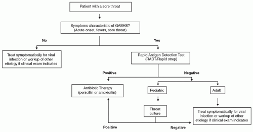
11). Newer RADT, which use optical immunoassays and chemiluminescent DNA probes, are 90% to 99% sensitive, but these are more expensive and may not be available. Therefore, a reasonable approach to the pediatric patient with sore throat is as follows: If the patient has symptoms typical of GABHS (fever, sore throat, lymphadenopathy), RADT should be performed in the office. If positive, the patient is started on antibiotic therapy immediately. If negative, a throat culture is obtained, and the patient is discharged home with a prescription for oral antibiotics. If the culture is positive, the patient is informed to fill the prescription and start therapy; if negative, they do not fill the prescription and are treated symptomatically. Practitioners should rely solely on the RADT if they have independently verified that their RADT has sensitivity greater than 90% (Fig. 52.1).

Penicillin or amoxicillin, which is more palatable for children, is an effective treatment for GABHS. First-generation cephalosporins, azithromycin, or clindamycin may be used in patients with penicillin allergy (1,6). Corticosteroids have not been proven to have benefit in acute streptococcal pharyngitis (12). Patients are not considered contagious after 24 hours of antibiotics, and should be allowed to return to school and regular activities at that time. If untreated, the GABHS pharyngitis is typically self-limited, with symptom resolution at 8 to 10 days, although throat cultures may be positive for up to 6 weeks in 50% of patients (8). Patients are considered contagious during the acute illness and 1 week thereafter if not treated (3). Testing or treatment of close contacts is generally not warranted, except in cases with recurrent pharyngitis between family members, or severely invasive strains of GABHS (e.g., necrotizing fasciitis). In children with recurrent strep throat and tonsillitis, tonsillectomy with or without adenoidectomy may be of benefit.

Approximately 10% of patients are considered carriers of GABHS, and will have positive strep testing without
acute illness or immune response. Treatment of carrier patients, or their family members (and pets) has not been shown to be useful and should be avoided (13). However, antibiotic therapy may be appropriate with a personal history or close contact with someone who has had acute rheumatic fever or poststreptococcal glomerulonephritis, invasive strains of GABHS, or workers in health care facilities or nursing homes. Carrier patients can still develop pharyngitis from another serotype of GABHS or viral infection, and treatment should be started as appropriate.


Figure 52.1 Algorithm for the diagnosis and management of sore throat and GABHS pharyngitis.

Complications from streptococcal pharyngitis are rare but well described. These include rheumatic fever; rheumatic heart disease; poststreptococcal glomerulonephritis; suppurative complications such as peritonsillar, retropharyngeal, and parapharyngeal abscess; suppurative lymphadenopathy; mastoiditis; bacteremia; and sepsis. Rheumatic fever is diagnosed based on the Jones Criteria, which includes major and minor criteria such as carditis, polyarthritis, and chorea, and carries a mortality risk. Traditionally, the major role of antibiotic therapy is prevention of rheumatic fever, and rheumatic heart disease. It is estimated that 3% of patients with untreated GABHS infection will develop rheumatic fever, compared to 0.3% of patients treated with antibiotics. In the United States, rheumatic heart disease has largely been eliminated as a complication, but rates remain relatively high in undeveloped and underdeveloped countries, such as India. Antibiotics also decrease the rate of suppurative complications and associated abscess development.

Of special note is a relatively recently described but controversial entity, known as Pediatric Autoimmune Neuropsychiatric Disorders Associated with Streptococcal (PANDAS) infections. After streptococcal infection, including pharyngitis, these patients rapidly develop neuropsychiatric symptoms of obsessive-compulsive disorder (OCD) and tic disorders (14,15,16,17). The OCD and tics will last for several weeks before remitting, then abruptly return after the next streptococcal infection. In general, neuropsychiatric symptoms are from minimal to absent between episodes. It has been hypothesized that GABHS antineuronal antibodies cross-react with cells in the basal ganglia, giving rise to behavioral and motor disturbances (16). In several case series, tonsillectomy has been of benefit, with resolution of neuropsychiatric symptoms (17). The etiology of PANDAS is yet unknown, and its association with streptococcal infection has not been absolutely proven.


Corynebacterium Diphtheriae

Although now extremely rare, Corynebacterium diphtheriae historically claimed the lives of millions of children and adults. Death usually occurred from cardio- or neurotoxicity from the exotoxin, or from asphyxiation from membranous pharyngitis. Vaccine introduction began in the 1920s, and there has not been a confirmed case in the United States since 2003, although diphtheria remains endemic in developing countries with low vaccination coverage (18). C. diphtheriae is a gram-positive filamentous rod, which is transmitted via nasal, eye, or oral secretions, or from skin lesions.

In addition to the typical symptoms of sore throat and malaise, diphtheria is characteristically associated with a gray pharyngeal/tonsillar membrane, which is produced by the local tissue response and necrosis from the bacterial exotoxin
(present in approximately one-third of patients). This can extend to cover the oropharynx and larynx, and cause rapid death by asphyxiation, particularly in the young child. The pseudomembrane is firmly attached to the mucosa, and removal characteristically leads to bleeding of the underlying site with an edematous submucosa. Patients may also have a prominent reactive cervical adenopathy leading to a “bull-neck” appearance (1). Exotoxin-induced systemic cardiotoxicity and neurotoxicity are also possible and account for many of the adverse consequences of infection (19).

When strongly suspected based on clinical grounds, the diagnosis is confirmed with culture of the pseudomembrane on Loeffler or tellurite medium. Treatment consists of antibiotics and antitoxin, typically with penicillin or erythromycin, and the equine hyperimmune diphtheria antitoxin. Current recommendations for immunization include combined vaccination during childhood and booster injection every 10 years in adults (19).


Viral

In both children and adults, viral infection is the most common cause of pharyngitis, and is typically self-limited. Viruses that produce the common cold predominate, particularly the rhinovirus family followed by coronavirus and adenovirus (Table 52.1). The onset of viral pharyngitis may be more gradual than of bacterial pharyngitis, and symptoms typically include rhinorrhea, cough, and diarrhea. A viral etiology is suggested by the presence of conjunctivitis, coryza, hoarseness, and cough, and presence of these symptoms makes GABHS infection much less likely (1,7).

Preschool-aged children have an average of five to seven episodes of viral URI per year. This rate steadily decreases into adulthood, and plateaus at two to three episodes per year. Spread of viral pharyngitis is by direct contact and inhalation/ingestion of respiratory secretions, and is greatly increased by contact with other children at home or in daycare and school environments. Especially in the northern states, there is a seasonal incidence during the fall, winter, and early spring months, from September to April. Parainfluenza virus, the main etiologic agent of acute croup attacks, peaks in the late fall and late spring, whereas respiratory syncytial virus (RSV) and influenza viruses are highest between December and April (20).

Symptoms begin 1 to 3 days after transmission, and a sore, scratchy throat and inflamed pharynx is often the first sign. This is followed by the typical symptoms of URI, including nasal congestion and rhinorrhea. Influenza and RSV infections are more likely to produce fevers. The symptoms of URI last for approximately 1 week, then spontaneously resolve. Traditionally, patients were treated with over-the-counter medications such as saline rinses, decongestants, antipyretics, and antitussives, if necessary. However, recent evidence has shown that there is a high risk for overdose and serious adverse consequences in infants under 2 years of age; in addition, there is no conclusive evidence that these medications are effective for symptom resolution in pediatric patients. Therefore, over-the-counter cough and cold medications are no longer recommended for infants under 2 years of age, and caution should be used in recommending these medications to patients 2 to 11 years of age (21).

Treatment is symptomatic, and reassurance should be given. Parents may bring their child to the pediatrician or emergency room requesting antibiotics, but antibiotics should be avoided for viral pharyngitis. Aside from the cost of medication, development of antibiotic resistance is a concern when overused.


Coxsackie Virus

Herpangina, characterized by palatal vesicular enanthem and throat pain, is caused by the Coxsackie virus, and is often mistaken for acute bacterial tonsillitis. Coxsackie virus is in the enterovirus family, and was traditionally divided into Group A or Group B Coxsackie virus types, each of which has multiple strains. New molecular techniques have reclassified enteroviruses into five species, with the Coxsackie viruses coming under Human Enterovirus B and C groups (22). Herpangina usually occurs in children 3 to 10 years of age. Spread is by direct contact with infected saliva, fluids, or feces, commonly on unwashed hands in children, with a predilection for the summer months. Outbreaks in military barracks and other close quarter situations have been reported in older populations.

Only gold members can continue reading. Log In or Register to continue

Stay updated, free articles. Join our Telegram channel

May 24, 2016 | Posted by in OTOLARYNGOLOGY | Comments Off on Pharyngitis

Full access? Get Clinical Tree

Get Clinical Tree app for offline access