P
P cell See cell, ganglion; cell, P.
p value See significance.
pachometer A device, mounted on a slit-lamp, that is used for measuring corneal thickness (or the depth of the anterior chamber). It consists of an optical system that provides two half-fields by means of two glass plates with parallel sides placed in front of one objective of the microscope, the other being occluded. These plates rest one on top of the other with the junction between them situated so as to horizontally bisect the objective. The top plate can be rotated while the bottom one is fixed. The observer viewing through the microscope sees two corneal optical sections and adjusts the top plate until the outer surface of the epithelium appears aligned with the inner surface of the endothelium (Fig. P1). The corneal thickness is then read directly from a scale attached to the pachometer and calibrated in millimetres. To increase the accuracy of the measurement a special eyepiece is used with the microscope. It has a magnification of × 10 and has two additional components: a horizontal slit and a bi-prism. The role of the eyepiece is to remove from the field of view half of the two optical sections. The measurement of the depth of the anterior chamber is made with a similar device but with a different scale. Note: also spelt pachymeter. Pachometry can also be carried out using slit-scanning topography in which a computerized system integrates a series of slit-beam images to produce a map of the curvature and elevation of the anterior and posterior surfaces as well as corneal thickness. An instrument with greater magnification (called a micropachometer) has been devised, principally for research purposes, using a projection system which incorporates variable doubling plates and forms two slit images on the cornea in conjunction with the viewing system of a slit-lamp and a magnification of up to × 100, mounted on another arm. This instrument allows the measurement of the thickness of the corneal epithelium alone with a precision that can reach ±1µm. The above pachometers are referred to as optical pachometers to differentiate them from ultrasonic pachometers, which use high-frequency ultrasound waves, reflected from the anterior and posterior corneal surfaces and a transducer probe placed against the cornea. Ultrasound and slit-scanning pachometers have higher reproducibility and less interobserver variation than subjective optical pachometers. Usage of pachometers (pachymeters) includes evaluation of contact lens wear, pre- and post-refractive surgery (e.g. PRK, LASEK, LASIK), glaucoma detection and monitoring corneal oedema.

See ultrasonography.
pad arm An extension of a spectacle frame either integral with the bridge or as a separate attachment to which a pad is fitted.
pad, nose One of a pair of protuberances attached to the bridge of a spectacle frame or mounting that rests against the side of the nose. Syn. nose pad.
See bridge, pad; pince-nez; spectacle frame, metal.
Paget’s disease See disease, Paget’s.
palinopsia Visual persistence of the image of an object in the absence of its original stimulus. There is usually a latent period, which may amount to several minutes between the visual stimulation and the corresponding mental image. The latter typically disappears within seconds, although it may persist in some cases for several minutes. The subsequent mental image is quite faithful to the original stimulus. It is usually associated with a lesion in the parieto-occipital or temporal-occipital areas as a result of a cerebral infarction, epilepsy, tumour, or brain injury. Syn. visual perseveration.
palisades of Vogt The crests of epithelium folds that run radially towards the cornea, at the limbus, from the bulbar conjunctiva. They are often seen in slit-lamp examination, especially in pigmented individuals, and clearly in fluorescein angiography. They may contain stem cells, which play a role in the regeneration of corneal epithelium cells.
palpebrae See eyelids.
palpebrae muscle, levator See muscle, levator palpebrae.
palpebral aperture; conjunctiva See under the nouns.
palpebral, elephantiasis See elephantiasis oculi.
palpebral fissure See aperture, palpebral.
palpebral ligament See ligament, palpebral.
palsy Synonym for paralysis, although it often implies partial paralysis.
abducens nerve p. See paralysis of the sixth nerve.
Bell’s p. A paralysis of the upper and lower muscles of the face on one side, due to an inflammation of the facial nerve. It results in a wider palpebral aperture and inability to close the eye on the affected size and drying of the cornea.
See sign, Bell’s; tears, artificial; tears, crocodile.
double elevator p. A condition characterized by limited or complete inhibition of the upward rotation of an eye, due either to paresis of its superior rectus and inferior oblique muscles, or to entrapment of the inferior orbital tissues. It may be congenital or acquired (e.g. a lesion in the pretectum). Treatment is principally surgical.
gaze p . Inability of the eyes to make conjugate movements due to a lesion in the cortical or subcortical oculomotor centres.
See paralysis of the fourth nerve; paralysis of the sixth nerve; paralysis of the third nerve.
supranuclear gaze p. A disturbance of the conjugate movements of the eye. If the lesion is in the frontal lobe, the patient is unable to direct the eyes to the contralateral side of the lesion (frontal gaze palsy). In bilateral lesion the patient is unable to turn the eyes voluntarily in any direction but is able to maintain fixation and perform pursuit movements. If the lesion is in the midbrain it produces Parinaud’s syndrome, in which there is an inability to elevate (and sometimes depress) the eyes on command and the pupils are large and may not react to light. If the lesion is in the paramedian pontine reticular formation there is ipsilateral horizontal gaze palsy, while lesions in the medial longitudinal fasciculus produce internuclear ophthalmoplegia.
See ophthalmoplegia, internuclear.
pannus Abnormal superficial vascularization of the cornea covering the upper half, or sometimes the entire cornea. It is characterized by a thick plexus of vessels. It is found in some cases of contact lens wear, mainly soft lenses. Pannus following contact lens wear is referred to as corneal vascularization. If induced by soft lenses, it can be reduced by changing to lenses of high oxygen transmissibility or ceasing contact lens wear. Deep corneal vascularization involving the stroma is usually the result of a disease process (e.g. interstitial keratitis, phlyctenular keratitis, severe long-standing trichiasis, trachoma).
panophthalmitis Acute inflammation of the eyeball involving all its structures and extending into the orbit. The disease develops very rapidly. The eyelids are red and swollen and there is severe chemosis of the conjunctiva. The cornea is often a whitish mass of necrotic tissue and there may be severe ocular pain.
See endophthalmitis.
pantoscopic angle See angle, pantoscopic.
Panum’s area; fusional space See under the nouns.
papilla Any small elevation shaped like a nipple.
Plural : papillae.
See follicle, conjunctival.
Bergmeister’s p. A cone-shaped, sheath of glial cells (astrocytes) and connective tissue covering the hyaloid artery formed during embryonic development over the optic disc and projecting into the vitreous humour. It usually atrophies before term but in some individuals it persists and a proliferation of glial cells form a glial veil. It may obscure the full view, or usually only part, of the optic disc and may sometimes suggest a disc tumour but it is in fact benign, stable and does not interfere with vision.
See hyaloid remnant.
lacrimal p. See lacrimal papilla.
p. lacrimalis See lacrimal tubercle.
papillary conjunctivitis, giant See conjunctivitis, giant papillary.
papillitis See neuritis, optic; pseudopapilloedema.
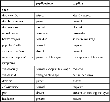
papilloedema A non-inflammatory oedema of the optic nerve head produced by raised intracranial pressure, and due most commonly to a cerebral tumour. It can also result from cerebral abscesses, meningitis, encephalitis, subarachnoid haemorrhages, head injury, hydrocephalus, etc. The optic disc appears raised above the level of the retina and its margins are blurred, the central vessels on the surface of the disc are displaced forward, the retinal veins are dilated and there is nearly always a loss of induced venous pulsation. The swollen disc displaces the retina and this causes an enlargement of the blind spot on visual field measurement. In the early stages visual acuity is not affected (unlike in papillitis), although if the condition persists there will be some loss. In advanced stages, there may be haemorrhages around the disc, secondary optic atrophy, exudates, as well as headaches and vomiting. The condition is usually bilateral. Note: also spelt papilledema. Syn. choked disc.
Table P1
Differential diagnosis between papilloedema and papillitis
| papilloedema | papillitis | |
| signs | ||
| disc elevation | raised | slightly raised |
| disc hyperaemia | present | present |
| disc margins | blurred | blurred |
| retinal veins | congested | congested |
| haemorrhages | near disc | some in late stage |
| pupil light reflex | normal | impaired |
| venous pulsation | absent | present |
| secondary optic atrophy | present in late stage | may appear in late stage |
| symptoms | ||
| visual acuity | normal, except in late stage | reduced |
| visual field | enlarged blind spot | central scotoma |
| diplopia | present | absent |
| colour vision | normal | impaired |
| pain | absent | present on moving the eyes |
| headache | present | absent |
See atrophy, optic; neuritis, optic; pseudopapilloedema; syndrome, Foster Kennedy.
papilloma A tumour most commonly found on the conjunctiva, the limbus or the lid margins. It is usually benign. It should be excised but it is likely to recur.
papillomacular bundle; fibres See fibres, papillomacular.
paracontrast See metacontrast.
paradoxical ARC See retinal correspondence, abnormal.
paradoxical diplopia See diplopia, incongruous.
paraffin See tears, artificial.
parallax Apparent displacement of an object viewed from two different points not on a straight line with the object.
binocular p . The difference in angle subtended at each eye by an object that is viewed first with one eye and then with the other.
chromatic p. Apparent lateral displacement of two monochromatic sources (e.g. a blue object and a red object) when observed through a disc with a pinhole placed near the edge of the pupil. When the pupil is centred on the achromatic axis (in some people the pinhole may have to be placed away from the centre of the pupil), the two images appear superimposed. The relative displacement of the two images becomes reversed when the pinhole is on the other side of that axis. This phenomenon is attributed to the chromatic aberration of the eye.
See chromostereopsis; aberration, longitudinal chromatic.
monocular p. The apparent change in the relative position of an object when the eye is moved from one position to another.
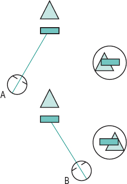
motion p . Apparent difference in the direction of movement or speed produced when the subject moves relative to his environment (Fig. P2). Example: when viewing the landscape through the window of a moving train near objects appear to move much more quickly than distant objects. See perception, depth; stereopsis.
relative binocular p. See acuity, stereoscopic visual.
paralysis Loss of action of a muscle due to injury or disease of that muscle or its nerve supply.
See palsy.
abducens p. See paralysis of the sixth nerve.
p. of accommodation See accommodation, paralysis of.
p. of convergence A condition characterized by an inability of the eyes to converge while all other monocular eye movements are unaffected. The patient notices diplopia in near vision, which usually occurs suddenly. It is presumably due to some lesion in the nuclei responsible for convergence, as may happen in tabes dorsalis or Parkinson’s disease.
divergence p. A condition characterized by an inability of the eyes to diverge while all other monocular eye movements are unaffected. It is characterized by a sudden development of diplopia with marked esotropia at distance and sometimes headaches. The key difference with divergence insufficiency is the sudden onset of symptoms. Its association includes encephalitis, multiple sclerosis, head trauma, cerebral haemorrhage, brain tumour and vascular lesions of the brainstem.
p. of the fourth nerve A condition characterized by a hypertropia of the eye with the affected superior oblique muscle. It may be due to a lesion of the fourth cranial nerve or its nucleus as a result of injury (the most common cause), vascular lesions, aneurysm or tumour. The patient usually presents with an abnormal head posture to avoid diplopia. If the condition does not recover by itself following therapy of the underlying cause, surgery is usually the only alternative treatment. Syn. trochlear paralysis.
See head posture, abnormal; nerve, trochlear; strabismus, paralytic.
oculomotor p . See paralysis of the third nerve.
p. of the sixth nerve A condition characterized by an esotropia of the eye with the affected lateral rectus muscle. It may be due to a lesion of the sixth cranial nerve or its nucleus as a result of a vascular disease (e.g. diabetes, hypertension), injury, or tumour. The patient presents with an abnormal head turn to avoid diplopia. If the condition does not recover by itself following therapy of the underlying cause, surgery is usually the only alternative treatment. Syn. abducens paralysis; lateral rectus palsy.
See head posture, abnormal; nerve, abducens; strabismus, paralytic; syndrome, Gradenigo’s; transposition.
p. of the third nerve A condition that leads to a wide impairment of motor function, as this nerve innervates most of the muscles of the eye. It may be due to a vascular disease (e.g. diabetes, hypertension), aneurysm (especially of the internal carotid artery), injury or tumour. In total paralysis only the lateral rectus and the superior oblique muscles will be spared and the eye will be in a position of abduction, slight depression and intorsion. Ptosis will also be present and the pupil will be dilated and non-reactive, and there will also be paralysis of accommodation. If the condition does not recover by itself following therapy of the underlying cause, surgery is usually the only alternative. Syn. oculomotor paralysis.
See circle of Willis; nerve, oculomotor; ophthalmoplegia; strabismus, paralytic; syndrome, Benedikt’s; syndrome, Weber’s; test, forced duction; transposition.
trochlear p . See paralysis of the fourth nerve.
parastriate area See areas, visual association.
parasympathetic nervous system See system, autonomic nervous.
parasympatholytic Pertains to a drug that blocks the effects of the parasympathetic nervous system. Examples: atropine, cyclopentolate. Syn. antimuscarinic, anticholinergic, atropine-like.
See acetylcholine; cycloplegia; mydriatic.
parasympathomimetic drug A drug with an action resembling that caused by stimulation of the parasympathetic nervous system. Example: a miotic of which there are two types: a direct-acting cholinergic, such as pilocarpine or carbachol; and the other, indirect-acting anticholinesterase, such as physostigmine, neostigmine, echothiophate iodide, demecarium bromide. Syn. cholinergic drug.
See miotics.
paraxial Pertains to light rays situated near enough to the axis of an optical system for the gaussian theory to apply.
paraxial approximation See ray, paraxial.
paraxial equation, fundamental Equation based on gaussian theory and dealing with refraction at a spherical surface:
where n and n’ are the refractive indices of the media on each side of the spherical surface, r is the radius of curvature of the surface and l and l’ the distances of the object and the image from the surface, respectively. n/l and n’ /l’ are the vergences (or reduced vergences) of the incident and refracted light rays respectively. L’ – L corresponds to the change produced by the surface in the vergence of the light and is called the focal power (or vergence power, or refractive power) F of the surface. Thus
Focal power is usually expressed in dioptres and can be either positive or negative.
At a reflecting surface or a mirror the equation becomes
where r is the radius of curvature of the surface or mirror. Syn. general refraction formula. See distance, image; distance, object; sign convention; theory, gaussian; vergence.
paraxial optics; ray See under the nouns.
paraxial region The hypothetical cylindrical narrow space surrounding the optical axis within which rays of light are still considered paraxial. Syn. gaussian space.
See ray, paraxial; theory, gaussian.
paraxial theory See theory, gaussian.
paresis Slight or partial paralysis.
Parinaud’s syndrome See syndrome, Parinaud’s.
Parks three-step test See test, three-step.
pars An anatomical part.
pars ciliaris muscle See muscle of Riolan.
pars plana; plicata See ciliary body.
pars planitis See uveitis, intermediate.
partial sight See vision, low.
parvo cells See cell, ganglion; geniculate bodies, lateral.
parvocellular layer See geniculate bodies, lateral.
parvocellular visual system See system, parvocellular visual.
past-pointing See pointing, past-.
patellar fossa See fossa, hyaloid.
pathological myopia See myopia, pathological.
pathology, ocular The discipline which deals with the nature of diseases of the eye and its surrounding structures, their effect on the ocular tissues and on ocular functions, as well as the causes and management.
pathway 1. A collection of nerve fibres (axons) along which nervous impulses travel. 2. A path or a route.
geniculocalcarine p. See radiations, optic.
geniculostriate p. 1. See radiations, optic. 2. Some authors consider this term to be a synonym of visual pathway.
magnocellular p . See system, magnocellular visual.
motor p. Pathway from the cortex to the muscles that control the movements of the eyes enabling them to act as a unit.
parvocellular p . See system, parvocellular visual.
retinotectal p. 1. The nervous pathway connecting the retina to the pretectal region (anterior to the superior colliculi) and from there to the Edinger–Westphal nucleus. It is involved in the pupillary light reflexes. 2. The nervous pathway between the retina and the superior colliculus. It is involved in the involuntary blink reflex to a dazzling light and in the eye movements occurring in response to the sudden appearance of a novel or a threatening stimulus.
See blind sight; fibres, pupillary; reflex, pupil light; pretectum.
uveoscleral p . An unconventional route through which the aqueous humour drains out of the eye. The aqueous passes from the anterior chamber across the iris root through small spaces between the ciliary muscle fibres into the supraciliary space and suprachoroid space. The fluid is believed to escape the eye via veins in the ciliary muscle and anterior choroid. The amount of aqueous outflow through this route amounts to between 10% and 15%, the rest flows out through the conventional pathway via the trabecular meshwork and into Schlemm’s canal. Prostaglandin drugs reduce the intraocular pressure by increasing the outflow through the uveoscleral pathway. This pathway is occasionally made use of in cyclodialysis.
visual p . Neural path starting in the photoreceptors of the retina and travelling through the following structures: the optic nerve, the optic chiasma, the optic tract, the lateral geniculate bodies, the optic radiations and the visual cortex where the pathway ends. The fibres of the optic nerve of one eye meet with the fibres from the other eye at the optic chiasma, where approximately half of them (the nasal half of the retina) cross over to the other side. Thus, there is semidecussation in the visual pathway (Fig. P3).
See area, visual; decussation; magnification, cortical; retinotopic map.
patient Term originating from the Latin patior meaning to suffer; one who suffers or is ill and requires treatment.
pattern A combination of acts or parts, forming a consistent or characteristic arrangement or behaviour.
A p. Neuromuscular anomaly of the eyes characterized by an increase in exotropia when the eyes fixate downward, or increase in esotropia when the eyes fixate upward. Upgaze and downgaze are usually measured at 25 degrees from the horizontal. Syn. A syndrome.
See strabismus, convergent; strabismus, divergent.
checkerboard p.
Stay updated, free articles. Join our Telegram channel

Full access? Get Clinical Tree


