 Orbital Myogenic Tumors
Orbital Rhabdomyosarcoma
General Considerations
Tumors of skeletal muscle derivation that can develop in the orbit include rhabdomyoma, rhabdomyosarcoma (RMS), and malignant rhabdoid tumor (1,2,3,4,5,6,7,8,9,10,11,12,13,14,15,16,17,18,19,20,21,22,23,24,25,26,27,28,29,30). Tumors of smooth muscle derivation include leiomyoma and leiomyosarcoma. Orbital rhabdomyoma is extremely rare with only a few cases reported in the literature (28,29,30). It is generally a circumscribed soft tissue tumor of infancy that is composed of well-differentiated striated muscle cells with a mixture of collagen fibers. We have no examples of orbital rhabdomyoma to illustrate. RMS is the most important myogenic tumor of the orbit.
Rhabdomyosarcoma is the most common primary orbital malignancy of childhood (1,2,3,4,5,6,7,8,9,10,11,12,13,14,15,16,17,18,19,20,21,22,23,24,25,26,27). In the author’s clinicopathologic series, it accounted for only 1% of all biopsied orbital masses (2) and for 4% of all biopsied orbital masses in children (3). In the clinical series of 1,264 patients, the 35 cases of orbital RMS accounted for 97% of myogenic tumors and for 3% of all orbital lesions (4). There have been many articles on orbital RMS and most of the articles are quoted in a large review on the subject that was published in 2003 (6). Orbital RMS generally occurs in the first two decades of life with a mean age at diagnosis of 8 years (6). The tumor can originate primarily in the orbit or it can develop in the sinuses or nasal cavity and secondarily extend to involve the orbit. Orbital RMS has been observed many years after orbital irradiation for retinoblastoma (11).
Clinical Features
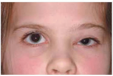
The clinical features of orbital RMS can vary considerably from case to case. The patient generally presents with proptosis (80%-100%), globe displacement (80%), blepharoptosis (30%-50%), conjunctival and eyelid swelling (60%), palpable mass (25%), and pain (10%). Most patients have proptosis and downward and lateral displacement of the globe owing to the superior or superonasal location of the mass in 70% (6,7). It can occasionally occur as an epibulbar mass without deep orbital involvement (6,7,22).
Diagnostic Approaches
The differential diagnosis of orbital RMS includes orbital cellulitis, nonspecific orbital inflammation (“pseudotumor”), ruptured dermoid cyst, capillary hemangioma, lymphangioma, Langerhans cell histiocytosis, myeloid sarcoma, lymphoma, and most other childhood orbital tumors. The clinical features that serve to differentiate them are described elsewhere in this book and in the literature (6).
Imaging studies are usually helpful in the differentiation. Computed tomography (CT) demonstrates a moderately well-circumscribed but irregular orbital mass that is generally confined to soft tissue and usually spares extraocular muscles. Less often it can extend to involve the adjacent orbital bones or sinuses. The tumor shows enhancement with contrast material. It usually has a hypointense signal with respect to orbital fat, but is isointense with respect to extraocular muscles. It generally shows moderate to marked enhancement
with gadolinium and is best delineated with fat suppression techniques. On T2-weighted images, the lesion is hyperintense to extraocular muscles and orbital fat. The mass is usually solid but can occasionally show cavitary changes, suggesting the diagnosis of lymphangioma (21).
with gadolinium and is best delineated with fat suppression techniques. On T2-weighted images, the lesion is hyperintense to extraocular muscles and orbital fat. The mass is usually solid but can occasionally show cavitary changes, suggesting the diagnosis of lymphangioma (21).
Pathology
Orbital RMS probably arises from primitive pluripotent mesenchymal cells with a propensity to differentiate toward skeletal muscle (25). Several histologic variations of RMS occur in the orbit. The embryonal type is most common, whereas the alveolar type appears to be the most malignant (6,7,25). Embryonal RMS is characterized histopathologically by spindle to round cells that show features characteristic of skeletal muscle in various stages of embryogenesis. The predominant cell is an elongated spindle cell that can assume a variety of arrangements and degrees of differentiation. The cytoplasm is generally highly eosinophilic and cross-striations can sometimes be identified on routine histopathologic sections or with special histochemical stains. The alveolar type appears as loosely arranged, malignant cells with septae that are reminiscent of the pulmonary alveoli. The botryoid type may be a variant of the embryonal type that assumes a papillary configuration.
Management
Current management of orbital RMS is reviewed in considerable detail in the recent literature (6,7). Suspected orbital RMS should be managed by a systemic evaluation to exclude metastatic disease including lung, lymph nodes, and other sites. This should be followed by prompt biopsy with histopathologic confirmation of the diagnosis (6,7). When possible, the entire tumor should be removed intact. When that cannot be easily accomplished without damage to extraocular muscles or optic nerve, a generous incisional biopsy is sufficient. Once the diagnosis is established histopathologically, most patients are treated with irradiation and chemotherapy, according to guidelines established by the Intergroup Rhabdomyosarcoma Study (6,13).
Prognosis
Orbital RMS can be highly aggressive locally and can invade the brain and adjacent tissues and metastasize to lung, lymph nodes, and other distant sites. Using the modern therapeutic regimen, however, the survival has improved dramatically in recent years. Reports from the 1970s indicated that only 30% were alive after 5 years (12). Today, the survival is .95% for orbital RMS (6,7). Factors that appear responsible for the better prognosis for RMS in the orbital region include the more favorable anatomic location, the earlier stage of the disease at the time of diagnosis, more favorable tumor morphology, and perhaps patient age.
Selected References
1. Shields JA. Myogenic tumors. In: Shields JA, ed. Diagnosis and Management of Orbital Tumors. Philadelphia: Saunders; 1989:244-252.
2. Shields JA, Bakewell B, Augsburger JJ, et al. Classification and incidence of space-occupying lesions of the orbit. A survey of 645 biopsies. Arch Ophthalmol 1984;102:1606-1611.
3. Shields JA, Bakewell B, Augsburger JJ, et al. Space-occupying orbital masses in children: A review of 250 consecutive biopsies. Ophthalmology 1986;93:379-384.
4. Shields JA, Shields CL, Scartozzi R. Survey of 1264 patients with orbital tumors and simulating lesions: The 2002 Montgomery Lecture, part 1. Ophthalmology 2004;111:997-1008.
5. Seregard S. Management of alveolar rhabdomyosarcoma of the orbit. Acta Ophthalmol Scand 2002;80:660-664.
6. Shields JA, Shields CL. Rhabdomyosarcoma: review for the ophthalmologist. The 2001 Henry Dubins Lecture. Surv Ophthalmol 2003;48:39-57.
7. Shields CL, Shields JA, Honavar SG, et al. Clinical spectrum of primary ophthalmic rhabdomyosarcoma. Ophthalmology 2001;108:2284-2292.
8. Shields CL, Shields JA, Honavar SG, et al. Primary ophthalmic rhabdomyosarcoma in 33 patients. Trans Am Ophthalmol Soc 2001;99:133-142.
Stay updated, free articles. Join our Telegram channel

Full access? Get Clinical Tree


