(1)
Department of Ophthalmology and Visual Sciences, University of Iowa Hospitals and Clinics, Iowa City, IA, USA
The association of carotid artery disease with various serious ophthalmic disorders is well known. That has been discussed in previous chapters dealing with central retinal artery occlusion (Chap. 13), branch retinal arteriolar occlusion (Chap. 15), posterior ciliary artery occlusion (Chap. 18), and ocular ischemic syndrome (Chap. 21). Following is a brief composite account.
Atherosclerosis is the major factor in producing carotid artery stenosis or occlusion. However, other uncommon conditions can also produce carotid artery disease, and those include aneurysm of the internal carotid artery [1], dissection of the common carotid artery/spontaneous internal carotid artery dissection [2–7], internal carotid artery thrombosis [8], carotid endarterectomy [9], after carotid angiography [10], after manipulation of the neck by a chiropractor [11], and rarely arterial fibromuscular dysplasia [12, 13] and pseudoxanthoma elasticum [14]. Embolization from the external carotid artery system can also cause CRAO [15, 16].
Carotid artery disease can cause various serious ophthalmic disorders by the following four types of lesions: (1) occlusion, (2) stenosis, (3) ulcerative lesions, and (4) atheromatous plaques.
Pathophysiological Mechanisms
There are two mechanisms by which carotid artery disease can produce ophthalmic complications: (1) by embolism and (2) by hemodynamic disturbance. These two produce ophthalmic disorders independent of each other.
Embolic Disorders
These usually originate from ulcerative lesions or atheromatous plaques and less commonly from stenosis of the internal or common carotid arteries. The emboli may go to the retina, choroid, or optic nerve head (Fig. 22.1). Therefore, embolism is by far the most common cause of retinal ischemic lesions and may also sometimes cause choroidal and optic nerve head ischemic lesions.
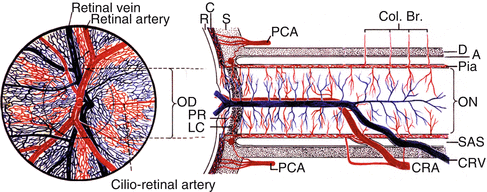
Fig. 22.1
Schematic representation of the course and branches of the central artery and vein of the retina (Modified from Hayreh SS [17].) Abbreviations: A arachnoid, C choroid, CRA central retinal artery, Col. Br. collateral branches, CRV central retinal vein, CZ circle of Haller and Zinn, D dura, LC lamina cribrosa, OD optic disc, ON optic nerve, PCA posterior ciliary artery, PR prelaminar region, R retina, S sclera, SAS subarachnoid space
Retinal Emboli
In the retina, these produce either central retinal artery occlusion (CRAO), branch retinal arteriolar occlusion (BRAO), or cilioretinal artery occlusion. Arruga and Sanders [18], in 70 patients with evidence of retinal embolism, found cholesterol emboli (Hollenhorst plaque) in 74 %, calcific emboli in 10.5 %, and platelet-fibrin emboli in 15.5 %. They stated that the carotid artery and the heart are the most common sources of embolism to the retinal arteries. My study [19] of 289 eyes with CRAO and 210 eyes with BRAO showed the same. Sheng et al. [20] in a review of a series of 62 patients with CRAO found ipsilateral extracranial carotid occlusive disease in 56 %; these patients did not generally have carotid bruits and had normal noninvasive carotid tests. They concluded that over half of patients with CRAO who undergo carotid angiography will have a carotid lesion on the ipsilateral side. This suggests that CRAO is a significant marker for extracranial carotid disease and should be an indication for complete carotid evaluation. Figure 15.11 in Chap. 15 shows the various types of retinal emboli.
In my study [19] of 234 eyes with CRAO, which had carotid Doppler/angiography, plaque(s) was seen in 71 % and stenosis of 0–15 % in 55 %, 50–79 % in 16 %, and 80–99 % in 7 %. In my study [19] of 128 cases with BRAO, who had carotid Doppler/angiography, plaque(s) was seen in 66 % and carotid artery stenosis of 0–15 % in 54 %, 50–79 % in 16 %, and 80–90 % in 9 %. Thus, plaque(s) in the carotid arteries is the most common source of retinal emboli. In this study 67 % of CRAO and 70 % of BRAO cases had 0–45 % stenosis, i.e., normal or minimal narrowing of the lumen of the carotid artery.
Visual Loss Caused by Retinal Emboli in CRAO
This is of two types:
1.
Amaurosis fugax: This is usually produced by platelet emboli or cholesterol emboli because they can migrate easily. Amaurosis fugax is an important manifestation of carotid artery disease, although it can occur from cardiac emboli also. It can last from a few seconds to minutes, with normal recovery of vision. The usual description given is “a blind over the eye from above.” These episodes may recur.
In a study [21] of 70 patients with amaurosis fugax, CT scan or MRI showed evidence of silent cerebral infarcts in 43 % and cerebral atrophy in 25 %. This is because small emboli in the retina can cause symptoms but produce no detectable, immediate symptoms in the brain. In my study [22] of 271 eyes with CRAO, there was a history of amaurosis fugax in 12.18 %; of those carotid Doppler or angiography revealed plaque in 58 % and carotid artery stenosis of 50–79 % in 7 % and 80–90 % in 3 %.
2.
Permanent occlusion: This results when the embolus gets impacted and that produces permanent visual loss. Usually calcific emboli are rough in texture and they get stuck permanently, but other types of emboli also do, less commonly. Various aspects of CRAO are discussed at length in Chap. 13.
Visual Loss Caused by Retinal Emboli in BRAO
In these eyes, the embolus almost invariably gets stuck at the site of bifurcation of a retinal arteriole. Visual loss, as in CRAO, is also of two types:
1.
Amaurosis fugax: This is usually produced by platelet emboli or cholesterol emboli, because they can migrate easily. In my study [22] of 169 eyes with BRAO, 14.20 % of patients gave a history of amaurosis fugax. These episodes involve a segment of the visual field, depending upon the location of the involved branch retinal arteriole, and usually they tend to recur and have a similar pattern.
Dr. William M. Haining, of Dundee in Scotland, more than 30 years ago, provided me with fluorescein cineangiographies of cases showing moving recurrent emboli in the retinal arterioles, a phenomenon not previously described or published. Recurrent moving emboli went through the same branch retinal arteriole again and again, repeatedly, as if the hemodynamic factors caused them to follow the same course every time. That would explain why the transient visual loss in BRAO often occurs repeatedly in the same region.
2.
Permanent occlusion: This results when the embolus gets impacted and it produces permanent visual loss in the involved visual field. Usually calcific emboli are rough in texture and they get stuck permanently, but other types can also get impacted sometimes.
Symptomless retinal emboli are also seen from time to time. Those are not benign because they indicate carotid artery or cardiac disease and require thorough evaluation.
Various aspects of BRAO are discussed at length in Chap. 15.
Visual Loss Caused by Emboli in the Optic Nerve Head
Since the optic nerve head is supplied by the posterior ciliary artery circulation, if an embolus gets into that circulation, it results in transient or permanent optic nerve head ischemia. I have seen some such cases, and Fig. 22.2 is an example where it produced anterior ischemic optic neuropathy.
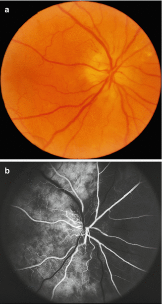
Fig. 22.2
Fundus photograph (a) and fluorescein fundus angiogram (b) of the right eye with embolic non-arteritic anterior ischemic optic neuropathy. Temporal artery biopsy for giant cell arteritis was negative. Angiogram shows normal filling of the area supplied by the lateral posterior ciliary artery (including the temporal half of optic disc) but no filling of the area supplied by the medial PCA (including the nasal half of optic disc)
Choroidal Ischemia Following Embolism into the Choroidal Vascular Bed
An atheromatous lesion in the carotid artery can cause embolic acute choroidal ischemic lesions. Acute choroidal ischemic lesions may present as a variety of distinct clinical entities, depending upon the size and number of the involved vessels and the severity of ischemia [23, 24]. The various lesions are discussed in Chap. 18.
There are some prevalent misconceptions about using the findings from carotid Doppler to evaluate the source of emboli [25]. There is a widespread belief that the absence of any abnormality on Doppler evaluation of the carotid artery always rules that out as the source of embolism or cause of ocular ischemia. This is not always true, for the following reasons:
(i)
I have found that when evaluating the carotid artery by Doppler, vascular surgeons, neurologists, and other physicians are almost invariably interested in the degree of stenosis of the carotid artery, because their primary interest is hemodynamic insufficiency and carotid endarterectomy. As discussed above, arterial occlusions in the eye are due to microembolism in the vast majority of cases, and the major source of microemboli is plaque – which may be present with or without any significant carotid artery stenosis. Thus, the absence of significant stenosis or of plaque in the carotid artery does not necessarily rule out the carotid artery as the source of microembolism. Vascular insufficiency in the eye caused by a significant carotid artery stenosis is seen only rarely and occurs primarily in ocular ischemic syndrome, but not every patient with ocular ischemia shows severe carotid artery stenosis or occlusion (see Chap. 21) [26, 27].
(ii)
Carotid Doppler evaluates the artery only in the neck and not above and below that (in the skull and the thorax, respectively) – the source of microembolism may be at those locations. Moreover, the resolution of Doppler is not always sensitive enough to detect plaques which, though very small, may be enough to produce microembolism in the eye. I have observed patients with visible retinal emboli who showed no abnormality on routine carotid Doppler or echocardiography. Similarly, I have previously reported that 26 % of patients with ocular ischemia had no significant carotid Doppler stenosis on the side of the ocular ischemia [27]. Likewise, Nicolaides et al. [21] reported the presence of cerebral infarcts with no evidence of internal carotid artery stenosis on the side of the infarct. Most retinal occlusions are due to relatively small emboli (“microemboli”). Microemboli may be derived from a non-hemodynamically significant obstruction, as it requires only a small intraluminal plaque which may be present with or without any significant carotid artery stenosis. Thus, contrary to the prevalent belief, an inability to find any abnormality on carotid Doppler does not rule out the carotid artery as the source of embolism.
Therefore, from the ophthalmic point of view, when evaluating the results of a carotid Doppler study for ocular microembolism, the presence of plaques is usually of much greater importance than the degree of stenosis; and the absence of marked carotid artery stenosis on Doppler does not rule out ocular ischemia.
Hemodynamic Disturbance
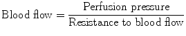
In carotid artery disease, this plays an independent role, unrelated to embolism, in producing ocular ischemic lesions. To understand this, one has to determine the factors which control the ocular blood flow. Ocular blood flow is calculated by using the following formula:
 Perfusion pressure can be calculated in two ways:
1.
Perfusion pressure = mean arterial blood pressure (BP) minus venous BP in the retinal/choroidal vessels.
2.
Perfusion pressure = mean arterial BP in the retinal/choroidal vessels minus intraocular pressure (IOP). Normally the pressure in the central retinal vein at the optic disc is slightly higher than the IOP, so that for all practical purposes, IOP is usually a good index of the ocular venous pressure.
Mean BP = diastolic BP + 1/3 (systolic minus diastolic BP).
Thus, the following three factors decrease the blood flow in the ocular vascular bed: (a) an increase in vascular resistance to blood flow, (b) a fall of BP, and (c) an increase of IOP. I have discussed the pathophysiology of these three factors at length in Chap. 10.
Stay updated, free articles. Join our Telegram channel

Full access? Get Clinical Tree


