Operative Technologies
Paul G. van der Sloot
Runhua Hou
INTRODUCTION
The nature of surgery is such that operative technologies provide impetus for change and progress with improved operative outcomes. Operative technologies are numerous and diverse, maybe more so in otolaryngology than any other surgical specialty. This chapter represents an overview of operative technologies in otolaryngology. It is by no means comprehensive. An array of technologies are presented that include new technologies that have come into widespread practice, developing technologies that are at the cusp of general use and old technologies for which new applications have been recently developed.
DISSECTION/HEMOSTASIS TECHNOLOGIES
A number of recent tools have been developed that provide the ability to dissect and achieve hemostasis virtually simultaneously. These instruments tend to be most useful in operative cases where space is limited and a clean operating field is particularly important. These include cases like parotidectomy, thyroidectomy, parathyroidectomy, tonsillectomy, and microvascular free flap harvest.
Tonsillectomy
In the case of tonsillectomy, technologies have been adopted that attempt to minimize pain associated with conventional cautery without increasing the risk of post-tonsillectomy bleeding. Coblation involves the dissociation of isotonic saline into sodium ions between the electrodes of the coblator, which breaks molecular bonds between cells. The temperatures generated by the coblator are between 45°C and 85°C as compared to 400°C and 600°C with electrocautery. This, along with the fact that coblation tonsillectomy is usually performed using an intracapsular technique, has been shown to decrease the duration of postoperative pain and speed the return to a normal diet (1). The disposable coblator components do create an added cost for the procedure. Coblation has also been used for a variety of nasal surgeries like turbinate reduction, hemostatic debulking of tumors to allow endoscopic visualization, and the removal of encephaloceles.
Other techniques that have been used successfully include argon plasma scalpels where argon gas is ionized by an electrode creating a beam of argon plasma through which current flows allowing for tissue dissection using molecular resonance technology where alternate current high-frequency electron waves at well-defined and varying wavelengths create electron energy quanta, which, when in resonance with cell molecular bonds, causes them to break (2, 3). The main thrust of these technologies is to decrease pain without increasing the risk of hemorrhage in the post-operative period.
Thyroidectomy and Parotidectomy
Ultrasonic shears (or the harmonic scalpel) involve the use of ultrasonic blade vibrations at 55,000 Hz that denature proteins and create a coagulum that seals vessels. Vessels of up to 2-mm diameter can be sealed using this technique. Because no electrical energy is transferred to the patient, it may be ideally suited for areas of dissection near nerves. Some studies have suggested decreased blood loss and decreased frequency of facial nerve injury are seen with the use of ultrasonic shears for superficial parotidectomy (4). Similar results have been obtained for thyroidectomy. Despite the common use of this instrument for these procedures, basic science researchers have shown that activation of the shears near the nerve, particularly closer than 2 mm or when it is activated in the vicinity for greater than or equal to 3 seconds, does result in a change in peak latency for the recurrent laryngeal nerve in a rabbit model (5).
Microvascular Free Tissue Harvest
Several donor sites for free tissue harvest involve the harvest or division of large muscle bellies. The fibular osteocutaneous free flap and the anterolateral thigh myofasciocutaneous flap with or without significant vastus lateralis muscle are commonly used donor sites that require substantial muscle division. Vascular perforators in the muscle at irregular intervals make the muscle division tedious, and conventional harvest can be prolonged with blood loss hampering field visualization. Increasingly, ultrasonic shears are used to limit time required to harvest, decrease blood loss, and improve field visualization.
TECHNOLOGIES TO IMPROVE VISUALIZATION OF THE OPERATIVE FIELD
Sinus Surgery
Endoscopes with variably angled lenses have been in conventional use for sinus surgery for many years. They have paved the way for less morbid surgery by allowing the sinus surgeon to visualize the tissues more precisely. The surgeon is thus able to limit trauma to those areas of the nose and sinuses that have no bearing on the desired outcome of surgery. In this way, it has decreased the morbidity of sinus surgery while improving outcomes.
Skull Base Surgery
The combination of improved endoscopes and cameras coupled with image-guided navigation and even real-time imaging has expanded the bounds of endoscopic skull base surgery (6). Although the basic instruments are similar to sinus surgery instruments, more varied angles for cutting and grasping instruments, extended guards for drills, and other modifications like endoscopic bipolar cautery have all contributed to the advancement of endoscopic skull base surgery. Self-irrigating endoscope technology has also allowed for more efficient surgery by limiting the need for scope removal and cleaning during the procedure. Although robotic surgery has not become a standard for skull base surgery at this point, increasing miniaturization of instruments and cameras as well as work on multiply articulated instruments may make robot-assisted surgery of the skull base a standard in the future (7).
Thyroid and Parathyroid Surgery
Minimally invasive procedures of the thyroid and parathyroid glands have evolved as a result of a desire for improved cosmesis, particularly in certain cultural groups, and a desire to limit pain and hospital stay. Video-assisted thyroidectomy involves a reduced central neck incision, subsequent blunt dissection to divide the muscular raphe in the midline, blunt dissection over the capsule of the thyroid, and subsequently the use of a 5-mm 30-degree laparoscope combined with the harmonic scalpel to take down the superior pole vessels and subsequently identify the recurrent laryngeal nerve. The remainder of the lobe is then separated from the parathyroids, Berry’s ligament is divided, and the lobe is dissected off the trachea. Nodules greater than 3 cm and a history of thyroiditis may limit this technique, but with appropriately selected patients, complication rates that are comparable to conventional surgery are quoted in the literature (8, 9).
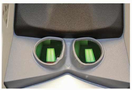 Figure 4.1 Robot stereo viewer. |
An extension of the desire to limit any visible scar is the transaxillary thyroidectomy, often done with robotic assistance. The da Vinci Surgical System (Intuitive Surgical, Sunnyvale, CA) provides binocular (Fig. 4.1), three-dimensional (3D) magnification, tremor filtration, and greater degrees of freedom in the mobility of instruments. The surgeon, after preparing the surgical field, sits in a console with 3D visualization of the field and ergonomic hand and foot controls. Scaling of movement up to 5 to 1 can be set, and any tremor is filtered as the surgeon’s movements in the console are transferred in real time to the instruments in the patients (Fig. 4.2). Clutching of the wrist


Stay updated, free articles. Join our Telegram channel
Full access? Get Clinical Tree
