Ocular Tumors of Childhood


Ocular Tumors of Childhood


Carol L. Shields

Jerry A. Shields


OVERVIEW

There are several benign and malignant ocular tumors that can occur in childhood. Tumors in the ocular region can lead to loss of vision, loss of the eye and, in the case of malignant neoplasms, loss of life. Therefore, it is important for the clinician to recognize childhood ocular tumors and to refer affected patients for further diagnostic studies and appropriate management. Based on our extensive clinical experience with ocular tumors over the past 50 years, we review general concepts of childhood eye tumors and discuss the clinical manifestations of specific tumors of the eyelid, conjunctiva, intraocular structures, and orbit in children (1,2,3,4,5).


Clinical Signs of Childhood Ocular Tumors

The clinical characteristics of childhood ocular tumors vary with its location: whether the tumor is located in the eyelids, conjunctiva, intraocular tissues, or the orbit. Each location imparts different signs and symptoms.


Eyelids and Conjunctiva

Eyelid and conjunctival tumors are generally more evident than intraocular or orbital tumors, prompting an early visit to a physician. Most tumors in the eyelid or conjunctival region have characteristic features, so an accurate diagnosis can generally be made with inspection. However, additional diagnostic studies of optical coherence tomography (OCT) or magnetic resonance imaging (MRI) can be helpful.


Intraocular Tumors

Intraocular tumors are often hidden until the patient is visually symptomatic. Young children often do not complain of visual loss and visual acuity can be difficult to assess due to cooperation. However, several features should alert the pediatrician to consider the possibility of an intraocular tumor and prompt a timely referral, particularly the presence of leukocoria (white pupil), strabismus (crossed eye), or lack of visual response.

Leukocoria There are several causes of leukocoria in children (2,4,5,6,7) (Fig. 19.1). The more common causes include congenital cataract, retinal detachment from retinopathy of prematurity, persistent fetal vasculature (persistent hyperplastic primary vitreous), and Coats’s disease. Retinoblastoma is likely the most serious condition to cause leukocoria in children. Any child with leukocoria should be referred promptly to an ocular oncologist for diagnostic evaluation and management.

Strabismus Most children with strabismus do not have an intraocular tumor. However, about 30% of patients with retinoblastoma present initially with either esotropia or exotropia, due to the tumor location in the macular area which disrupts central fixation. It is important that a complete retinal examination with indirect ophthalmoscopy be performed to exclude an underlying tumor in these cases.

Visual Impairment An older child with an intraocular tumor might complain of visual impairment or could be found to have decreased vision on visual screening in school. This can occur from reduction of central retinal function by a tumor or by the presence of vitreous hemorrhage, hyphema, or secondary cataract formation.


Orbital Tumors

Unlike tumors of the eyelid and conjunctiva, orbital tumors are not directly visualized by the physician. Therefore, these tumors can attain a large size before becoming clinically evident. These patients generally present with proptosis or displacement of the eye (3). Pain, diplopia, and conjunctival edema can also be early clinical features of an orbital tumor. Computed tomography (CT) and MRI have revolutionized the diagnosis and treatment of orbital tumors (8). Caution is issued for use of CT in children to reduce radiation exposure (9).


Diagnostic Approaches

Although some atypical tumors can defy clinical diagnosis, most ophthalmic tumors in children can be accurately diagnosed by a competent ophthalmologist or ocular oncologist.


FIGURE 19.1. Leukocoria secondary to retinoblastoma.


Eyelid and Conjunctiva

Most eyelid and conjunctival tumors are recognized by their classic clinical features. Diagnostic studies can provide additional help. Smaller suspicious tumors can be removed by excisional biopsy and larger tumors are best diagnosed by incisional biopsy and definitive treatment withheld until a definite diagnosis is established.


Intraocular Tumors

Concerning intraocular tumors, lesions of the iris can often be recognized with external ocular examination or slit-lamp biomicroscopy. Tumors of the retina and choroid can be visualized with ophthalmoscopy, which can reveal typical features depending on the type of tumor. Many small tumors are difficult to visualize and may only be detected by an experienced ophthalmologist using binocular indirect ophthalmoscopy. Ancillary studies such as fundus photography, autofluorescence, OCT, fluorescein angiography, indocyanine green angiography, ocular ultrasonography, and occasionally CT or MRI are of supplemental value in establishing the diagnosis. OCT is a relatively newer fundus scanning method using a rapid, noncontact, comfortable technique providing cross-sectional imaging of the retina to 4 to 5 µm resolution in a few minutes. Children comfortably tolerate this technique (10). OCT can provide in vivo, high-resolution information of the retina to the 10 µm level. Fine needle aspiration biopsy under general anesthesia has recently been employed in selected intraocular tumors of children (11).


Orbital Tumors

Some orbital tumors occur in an anterior location and can be recognized by their extension into the conjunctiva and eyelid area. This is particularly true of childhood vascular tumors such as capillary hemangioma and lymphangioma. Other tumors reside in the deeper orbital tissues and are less accessible. Orbital ultrasonography can be performed in the office, but often more formal imaging with CT or MRI is necessary.


Therapeutic Approaches

The treatment of an ocular tumor in a child depends on the type of tumor as well as the location and size of the lesion, and the general health of the patient.


Eyelid and Conjunctiva

Neoplasms of the eyelid and conjunctiva can be removed surgically by a qualified ophthalmologist or ocular oncologist. Inflammatory lesions that simulate neoplasia can be managed by antibiotics or corticosteroids, depending on the diagnosis. Some malignant neoplasms such as leukemias and lymphomas are best managed with a limited diagnostic biopsy followed by irradiation and/or chemotherapy.


Intraocular Tumors

The management of intraocular tumors is more complex. Certain benign intraocular tumors that are asymptomatic are usually managed by serial observation. Some symptomatic benign tumors can be treated with laser or cryotherapy, depending on the mechanism of visual impairment. Malignant tumors, such as retinoblastoma, are managed with chemotherapy (intravenous chemotherapy, intra-arterial chemotherapy [IAC], sub-Tenon chemotherapy, or intravitreal chemotherapy approaches), radiotherapy (external beam or plaque radiotherapy), laser photocoagulation, thermotherapy, cryotherapy, or enucleation of the eye (2,4,12,13). More recently, there is a strong trend toward using methods of chemotherapy for retinoblastoma control (13,14).


Orbital Tumors

The treatment of an orbital tumor varies greatly with the clinical signs of histopathologic diagnosis. Benign vascular tumors, such as capillary hemangioma and lymphangioma, can be managed by serial observation or amblyopia patching of the opposite eye to decrease the severity of associated amblyopia. Circumscribed tumors in the anterior orbit may be managed by excisional biopsy. Many malignant tumors, such as rhabdomyosarcoma and orbital leukemia, require limited biopsy to establish the diagnosis, followed by irradiation or chemotherapy (3,5).


EYELID TUMORS

There are several pediatric cutaneous tumors that affect the skin of the eyelids (3,14).


Capillary Hemangioma

The capillary hemangioma or strawberry hemangioma can occur on skin in 10% of infants and is recognized to be
more common in premature infants and twins. Capillary hemangioma of the eyelids can be a reddish, diffuse, or circumscribed mass (15) (Fig. 19.2). It usually has clinical onset at birth, or shortly thereafter, tends to enlarge for a few months, and then slowly regress. The main complications of this benign tumor are strabismus and amblyopia. In the recent years, the most frequently used treatment has been refraction, glasses for refractive error, patching of the opposite eye, and close follow-up. More recently, there has been a trend toward oral propranolol or complete surgical excision of those lesions that are relatively small and localized (16). Propranolol can hasten tumor regression with little side effect. Corticosteroids or radiotherapy are reserved for lesions that do not respond to standard measures.


FIGURE 19.2. Capillary hemangioma of the eyelid. A: Eyelid hemangioma in an infant twin #1 managed with observation as it did not obstruct visual acuity. B: Cutaneous hemangioma in twin #2 on the hand.


Facial Nevus Flammeus

Facial nevus flammeus is a congenital cutaneous vascular lesion that occurs in the distribution of the fifth cranial nerve (Fig. 19.3). It may be an isolated entity or it may occur with variations of the Sturge-Weber syndrome. Infants with this lesion have a higher incidence of ipsilateral glaucoma, diffuse choroidal hemangioma, and secondary retinal detachment. Affected infants should be referred to an ophthalmologist as early as possible in order to diagnose and treat these serious ocular conditions. Management of the cutaneous lesion includes observation, cosmetic make-up, or tunable-dye laser treatment delivered at infancy.


Kaposi Sarcoma

Opportunistic neoplasms such as Kaposi sarcoma can be found in immunosuppressed children, particularly those with acquired immunodeficiency syndrome. Although the affected patient can display red cutaneous lesions elsewhere, the eyelid can occasionally be the initial site of involvement. The lesion appears as a reddish-blue subcutaneous mass near the eyelid margin. This tumor is managed by improvement of immunosuppression. In children with human immunodeficiency virus (HIV) infection, treatment with highly active anti-retroviral therapy is employed.


Basal Cell Carcinoma

Although basal cell carcinoma is primarily a disease of adults, it is occasionally seen in younger patients, particularly if there is a family history of the basal cell carcinoma (nevus) syndrome. It generally occurs on the lower eyelid as a slowly progressive mass that can develop a central ulcer (rodent ulcer) and loss of eyelashes. Treatment generally involves local excision, frozen section margin control, and eyelid reconstruction.

Taylor and coauthors reviewed 39 patients with basal cell carcinoma (nevus) syndrome (Gorlin-Goltz syndrome) and found the age of presentation between 5 and 72 years (17). The presenting clinical features included odontogenic
keratocyst (n = 17 patients), basal cell carcinoma (n = 13), and congenital malformations (n = 2). Family history of the syndrome was present in 17 of 39 patients. Basal cell carcinoma developed in 18 of 28 (64%) patients before the age of 30 years. Newer treatment with systemic hedgehog inhibition has been a breakthrough in the control of this disease (18).


FIGURE 19.3. Nevus flammeus of the face in a child with Sturge-Weber syndrome.


Melanocytic Nevus

Melanocytic nevus can occur on the eyelid as a variably pigmented well-circumscribed lesion, similar to those that occur elsewhere on the skin. Nevus does not usually cause loss of cilia. In some instances, the nevus is congenital and large and involves both the upper and lower eyelids and is termed “kissing nevus” or “divided nevus” (Fig. 19.4). There is some evidence that early intervention with curettage of the lesion within the first 3 to 4 weeks after birth can successfully remove the lesion without the need for extensive grafting. At infancy, the nevus is superficial and can be scraped off the skin whereas later it deepens into the subcutaneous tissue, making surgical removal difficult. Following curettage, topical antibiotic ointment is applied and the skin heals by granulation.

The blue nevus is often apparent at birth, whereas the junctional or compound nevus may not become clinically apparent until puberty. Transformation into malignant melanoma is rare and usually occurs later in life. Although most eyelid nevi in children can be safely observed, they are occasionally excised because of cosmetic considerations or because of fear of malignant transformation.


Neurofibroma

Neurofibroma can occur on the eyelid as a diffuse or plexiform lesion that is often associated with von Recklinghausen’s neurofibromatosis. In the earliest stages, the lesion produces a characteristic S-shaped curve to the upper eyelid. Larger lesions produce thickening of the eyelid with secondary blepharoptosis (Fig. 19.5). Since these diffuse tumors are often difficult or impossible to completely excise, they should be managed by periodic observation or surgical debulking if they cause a major cosmetic problem.


FIGURE 19.4. Eyelid margin kissing nevus.


FIGURE 19.5. Neurofibroma of the eyelid and orbit in an infant.


Neurilemoma (Schwannoma)

Neurilemoma is a benign peripheral nerve sheath tumor that is composed purely of Schwann cells of peripheral nerves. It can appear in the orbit or on the eyelid and is managed by surgical resection. This tumor may or may not be associated with neurofibromatosis.


CONJUNCTIVAL TUMORS


Introduction

Tumors of the conjunctiva and epibulbar tissues involve a large spectrum of conditions ranging from benign lesions such as limbal dermoid, myxoma, and scleral melanocytosis to aggressive, life-threatening malignancies such as melanoma, Kaposi sarcoma, and sebaceous carcinoma (1,3,19,20,21,22). The clinical differentiation of the various tumors is based primarily on the clinical features of the tumor as well as the patient history. The clinical features as well as the management of each tumor are discussed, based on the authors’ personal experience with approximately 2,000 patients with conjunctival tumors over a 40-year period (22). Herein, we review and illustrate the features of conjunctival tumors in children.


Spectrum of Tumors in Children

Several previously published surveys (20,21,22) have reported on the incidence of conjunctival lesions in adults. However, the epidemiologic features, anatomic characteristics, and
malignant potential of such lesions differ in the pediatric age group. There have been only three large series of conjunctival tumors in children (1,23,24). Elsas and Green, and Cunha and coworkers evaluated the incidence of these lesions from a pathology laboratory standpoint, whereas Shields and associates presented their data from a clinical standpoint (1,23,24) (Tables 19.1 and 19.2).

In a clinical series of 262 children referred to an Oncology Service with a conjunctival tumor, Shields and coworkers found that the most common lesions were of melanocytic (67%), choristomatous (10%), vascular (9%), and benign epithelial (2%) origin (1) (Table 19.1). They noted that 10% of cases were non-neoplastic lesions simulating a tumor such as epithelial inclusion cyst, nonspecific inflammation/infection, episcleritis, scleritis, and foreign body.


Specific Tumors

The following tumors are classified based on tissues of origin, including choristomatous, epithelial, melanocytic, vascular, fibrous, xanthomatous, and lymphoid/leukemic origin.


TABLE 19.1 CLINICAL DIAGNOSTIC CATEGORIES OF CONJUNCTIVAL TUMORS IN 262 CHILDREN (1)


Classification of Tumors


Number of Patients (%)


Choristomatous


26 (10%)


Benign epithelial


5 (2%)


Premalignant and malignant epithelial


1 (<1%)


Melanocytic


175 (67%)


Vascular


23 (9%)


Fibrous


2 (<1%)


Neural


0 (0%)


Xanthomatous


1 (<1%)


Myxomatous


0 (0%)


Lipomatous


0 (0%)


Lacrimal gland


0 (0%)


Lymphoid


4 (1.5%)


Leukemic


0 (0%)


Metastatic


0 (0%)


Secondary


0 (0%)


Non-neoplastic lesions simulating a tumor


25 (9.5%)


Data from the Oncology Service at Wills Eye Institute.


Choristomatous Conjunctival Tumors

A variety of tumors can be present at birth or become clinically apparent shortly after birth. Most of the lesions are choristomas, consisting of displaced tissue elements normally not found in these areas. A simple choristoma is comprised of one tissue element such as epithelium, whereas a complex choristoma represents variable combinations of ectopic tissues like bone, cartilage, and lacrimal gland.

Dermoid Conjunctival dermoid is a congenital wellcircumscribed yellow-white solid mass that involves the bulbar or limbal conjunctiva (25,26). It characteristically occurs inferotemporally and often this tumor has fine white hairs (
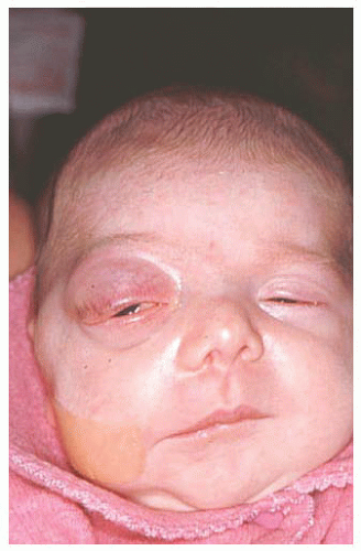
Fig. 19.6). In rare cases, it can extend to the central cornea or be located in other quadrants on the bulbar surface. Most often dermoid straddles the limbus, but in rare cases it can be extensive and involves the full-thickness cornea, anterior chamber, and iris stroma. The more severe dermoids occur earlier in embryogenesis.

The conjunctival dermoid can occur as a solitary lesion or can be associated with Goldenhar’s syndrome. The patient should be evaluated for ipsilateral or bilateral preauricular skin appendages, hearing loss, eyelid coloboma, orbitoconjunctival dermolipoma, and cervical vertebral anomalies.

Histopathologically, the conjunctival dermoid is a simple choristomatous malformation that consists of dense fibrous tissue lined by conjunctival epithelium with deeper dermal elements including hair follicles and sebaceous glands. The management of an epibulbar dermoid includes observation if the lesion is small and visually asymptomatic. Anterior segment OCT can assist in judging depth of involvement. It is possible to excise the lesion for cosmetic reasons, but the remaining corneal scar can be cosmetically unacceptable. Larger or symptomatic dermoids can produce visual loss from astigmatism. These can be approached by lamellar keratosclerectomy with primary closure of overlying tissue if the defect is superficial, or closure using corneal graft if the defect is deep or of full thickness. The cosmetic appearance might improve, but the refractive and astigmatic error and visual acuity might not change. When the lesion involves the central cornea, a lamellar or penetrating keratoplasty is necessary and long-term amblyopia should be anticipated.

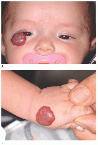
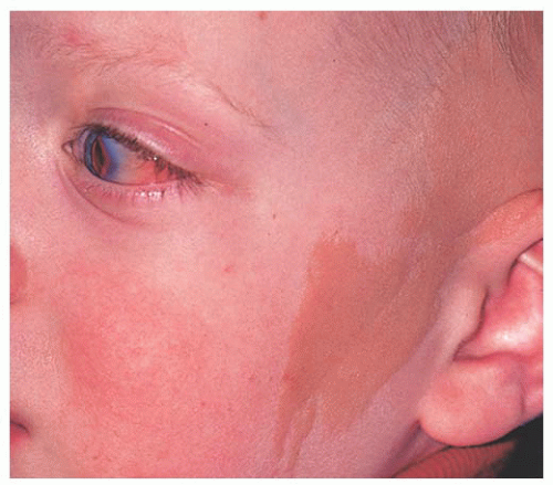
Dermolipoma Dermolipoma is believed to be congenital, but it classically remains asymptomatic for years and might not be detected until adulthood. This tumor tends to occur in the conjunctival fornix superotemporally and appears as a yellow, soft, fluctuant mass with fine white hairs on its surface (Fig. 19.7). It can extend into the orbital fat and onto the bulbar conjunctiva, sometimes reaching the limbus.

Dermolipoma has features similar to orbital fat on CT and MRI scans. Histopathologically, it is lined by conjunctival epithelium on its surface and the subepithelial tissue has variable quantities of collagenous connective tissue and adipose tissue. Pilosebaceous units and lacrimal gland tissue might be
present. The majority of dermolipomas require no treatment, but larger ones or those that are cosmetically unappealing can be managed by excision of the entire orbitoconjunctival lesion through a conjunctival forniceal approach or by removing the anterior portion of the lesion in a manner similar to that used to remove prolapsed orbital fat. Amniotic membrane graft might be necessary to repair the defect.


TABLE 19.2 COMPARISON OF DATA FROM THREE SERIES OF CONJUNCTIVAL LESIONS IN YOUNG PATIENTS


Classification of Tumors


% Tumors


% Tumors


% Tumors


Data source


Clinical series (1)


Pathology series (24)


Pathology series (23)


(n = 262)


(n = 282)


(n = 302)


Choristomatous


10


22


33


Benign epithelial (papilloma)


2


10


7


Premalignant and malignant epithelial


< 1


0


1


Melanocytic


67


23


29


Vascular


9


6


2


Fibrous


< 1


na


< 1


Neural


0


1


na


Xanthomatous


< 1


na


na


Myxomatous


0


na


na


Lipomatous


0


4


2


Lacrimal gland


0


na


na


Lymphoid


1.5


3


na


Leukemic


0


na


na


Metastatic


0


na


na


Secondary


0


na


na


Non-neoplastic lesions simulating a tumora


9.5


30


23


a Includes epithelial inclusion cyst, inflammatory lesions, vernal conjunctivitis, pyogenic granuloma, nonspecific granuloma, foreign body, scar tissue, keloid, and others.


FIGURE 19.6. Conjunctival dermoid.


FIGURE 19.7. Conjunctival dermolipoma.


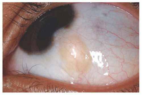
Epibulbar Osseous Choristoma Epibulbar osseous choristoma is a tumor comprised of mature bone, usually located in the bulbar conjunctiva superotemporally (3,27) (Fig. 19.8). It is believed to be congenital and typically remains undetected until palpated by the older patient. On ultrasonography or CT scanning, the mass demonstrates calcium. This tumor is usually managed by observation. Occasionally, a foreign body sensation necessitates excision of the mass using a conjunctival forniceal incision followed by dissection of the tumor to bare sclera.

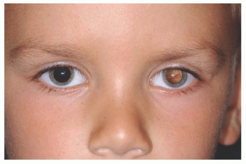
Lacrimal Gland Choristoma Lacrimal gland choristoma is a congenital lesion, discovered in young children as an asymptomatic pink stromal mass, typically in the superotemporal or temporal portion of the conjunctiva. It is speculated that this lesion represents small sequestrations of the embryonic evagination of the lacrimal gland from the conjunctiva. The lacrimal gland choristoma can masquerade as a focus of inflammation due to its pink color. Rarely, this mass can be cystic due to ongoing secretions if there is no connection to the conjunctival surface. Excisional biopsy is usually performed to confirm the diagnosis.

Complex Choristoma The conjunctival dermoid and epibulbar osseous choristoma are simple choristomas as they contain one tissue type such as skin or bone. A complex choristoma contains a greater variety of tissue derived from two germ layers such as lacrimal tissue and cartilage. It is variable in its clinical appearance and can cover much of the epibulbar surface, or it may form a circumferential growth pattern around the limbus.


FIGURE 19.8. Conjunctival osseous choristoma.


FIGURE 19.9. Complex conjunctival choristoma in a child with nevus sebaceous of Jadassohn and organoid nevus syndrome.

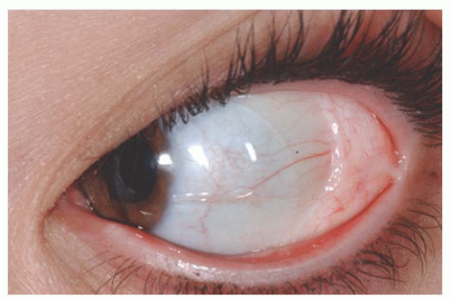
The complex choristoma has an association with the linear nevus sebaceous of Jadassohn (26) (Fig. 19.9). The nevus sebaceous of Jadassohn includes cutaneous features with sebaceous nevus in the facial region and neurologic features including seizures, mental retardation, arachnoidal cyst, and cerebral atrophy. The ophthalmic features of this syndrome include epibulbar complex choristoma and posterior scleral cartilage. The management of complex choristoma depends upon the extent of the lesion. Observation or wide local excision followed by mucous membrane graft reconstruction are options.


Epithelial Conjunctival Tumors

There are several benign and malignant tumors that can arise from the squamous epithelium of the conjunctiva.

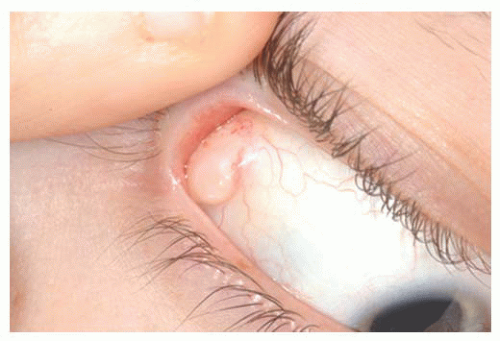
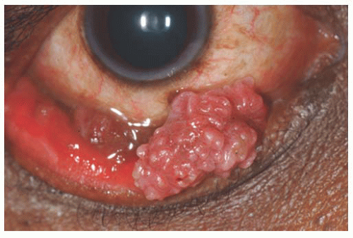
Papilloma Squamous papilloma is a benign tumor, documented to be associated with human papillomavirus infection of the conjunctiva (28,29). This tumor can occur in both children and adults. It is speculated that the virus is acquired through transfer from the mother’s vagina to the newborn’s conjunctiva as the child passes through the mother’s birth canal. Papillomas appear as a pink fibrovascular frond of tissue arranged in a sessile or pedunculated configuration (Fig. 19.10). The numerous fine vascular channels ramify through the stroma beneath the epithelial surface of the lesion. In children, the lesion is usually small, multiple, and located in the inferior fornix. Histopathologically, the lesion shows numerous vascularized papillary fronds lined by acanthotic epithelium.

There are several treatment options for small sessile papillomas. Sometimes observation allows for slow spontaneous resolution of this tumor. Larger or more pedunculated
tumors can lead to foreign body sensation, chronic mucous production, hemorrhagic tears, incomplete eyelid closure, and poor cosmetic appearance, requiring therapeutic intervention. Complete removal of the mass without direction manipulation of the tumor (no touch technique) is advisable to avoid spreading of the virus (30). Double freeze-thaw cryotherapy is applied to the remaining conjunctiva around the excised lesion in order to prevent tumor recurrence. In some instances, the pedunculated tumor is frozen alone and then excised while frozen. Topical or injection interferon and Mitomycin C have been employed for resistant or multiply recurrent conjunctival papillomas (31,32). For difficult recurrent lesions, oral cimetidine for several months following surgical resection can minimize recurrence by boosting the patient’s immune system and suppressing the virally stimulated mass (33).


FIGURE 19.10. Conjunctival papilloma.

Hereditary Benign Intraepithelial Dyskeratosis Hereditary benign intraepithelial dyskeratosis (HBID) is a rare, benign condition seen in an inbred isolate of Caucasian, African American, and Native Americans (Haliwa Indians), initially identified in North Carolina. It is an autosomal dominant disorder characterized by bilateral elevated fleshy plaques on the nasal or temporal perilimbal conjunctiva and on the buccal mucosa. It can remain asymptomatic or can cause redness and foreign body sensation. It is characterized histopathologically by acanthosis, dyskeratosis on the epithelial surface and deep within the epithelium, and prominent chronic inflammatory cells. HBID does not usually require aggressive treatment. Smaller, lesssymptomatic lesions can be treated with ocular lubricants and topical corticosteroids. Larger symptomatic lesions can be managed by local resection with mucous membrane grafting if necessary.

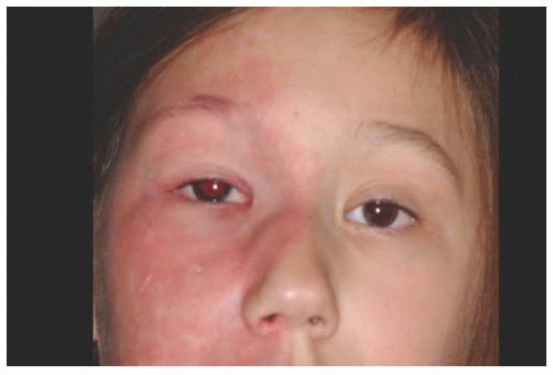
Squamous Cell Carcinoma/Conjunctival Intraepithelial Neoplasia Squamous cell carcinoma and conjunctival intraepithelial neoplasia (CIN) are malignancies of the surface epithelial cells. Intraepithelial neoplasia displays anaplastic cells within the epithelium, whereas squamous cell carcinoma displays extension of anaplastic cells through the basement membrane into the conjunctival stroma. Clinically, invasive squamous cell carcinoma is usually larger and more elevated than CIN. Leukoplakia can be seen with either condition.

Patients who are medically immunosuppressed from organ transplantation, those with HIV, or those with underlying DNA repair abnormalities like xeroderma pigmentosum are at particular risk to develop conjunctival squamous cell carcinoma and malignant melanoma (34). In these cases, the risk for life-threatening metastatic disease is greater.

The management of conjunctival squamous cell carcinoma varies with the extent of the lesion. Tumors in the limbal area require alcohol epitheliectomy for the corneal component and partial lamellar scleroconjunctivectomy with wide margins for the conjunctival component followed by freeze-thaw cryotherapy to the remaining adjacent bulbar conjunctiva. Extensive tumors or those tumors that are recurrent, especially with extensive corneal component, are treated with adjuvant topical Interferon, Mitomycin C, or 5-Fluorouracil (31,32,34,35).


Melanocytic Conjunctival Tumors

There are several lesions that arise from the melanocytes of the conjunctiva and episclera. The most important ones include nevus, racial melanosis, primary acquired melanosis (PAM), and malignant melanoma (Table 19.3). Ocular melanocytosis should be included in this section as its scleral pigmentation can masquerade as conjunctival pigmentation.

Jun 20, 2016 | Posted by in OPHTHALMOLOGY | Comments Off on Ocular Tumors of Childhood

Full access? Get Clinical Tree

Get Clinical Tree app for offline access