 Questions
1. Which of the following is true of a choristoma?
a. There is hypotrophic, immature tissue present at a normal location.
b. There is hyperplastic, mature tissue seen at a normal location.
c. There is mature tissue at an abnormal location.
d. A cavernous hemangioma is an example of a choristoma.
2. The most common intraconal orbital tumor is the
a. meningioma.
b. rhabdomyosarcoma.
c. neurofibroma.
d. cavernous hemangioma.
3. Which of the following is true regarding Russell bodies?
a. Russell bodies result from production of erythropoetin.
b. Russell bodies represent an aggregate of T cells.
c. Russell bodies originate from plasma cells.
d. Russell bodies occur only intracellularly.
4. All of the following are correct regarding corneal stromal wound healing except
a. Keratin sulfate and chondroitin sulfate are the corneal glycosaminoglycans.
b. Fibrovascular proliferation is a distinct aspect of the healing response.
c. Normal wound healing depends on both the epithelium and endothelium.
d. The subepithelial layer of the anterior, acellular stroma does not regenerate when incised.
5. Which of the following is true regarding anterior segment structures?
a. The glycosaminoglycan found in sclera is chondroitin sulfate.
b. Limbal wound healing involves scleral, episcleral, and corneal tissues.
c. Iris stroma regenerates after injury, but iris melanocytes do not regenerate after injury.
d. Retinal scars are produced by fibroblasts.
6. All of the following are correct regarding cyclodialysis except
a. There is disinsertion of the longitudinal portion of the ciliary muscle from the scleral spur.
b. Overproduction of aqueous humor and increased intraocular pressure are characteristic.
c. There is free access of the aqueous humor to the suprachoroidal space.
d. The condition is associated with a decreased blood supply to the ciliary body.
7. When obtaining a conjunctival biopsy for immunofluorescence studies in a patient suspected of having ocular cicatricial pemphigoid, what is the most appropriate solution to place the specimen?
a. formalin.
b. Zenker acetic fixative.
c. glutaraldehyde.
d. Michel fixative.
8. At approximately what rate does formalin diffuse through tissue?
a. 0.5 mm/h.
b. 1.0 mm/h.
c. 2.0 mm/h.
d. 5.5 mm/h.
9. A patient with lattice corneal dystrophy undergoes penetrating keratoplasty. All of the following tissue stains may be used to demonstrate the pathology in the corneal button specimen except
a. thioflavin T.
b. colloidal iron.
c. crystal violet.
d. Congo red.
10. What is the most helpful feature in distinguishing between a right and left eye specimen?
a. the position of the vortex veins.
b. the elliptical horizontal orientation of the cornea.
c. the insertion of the inferior oblique muscle.
d. the course of the long posterior ciliary artery and nerve.
11. Which of the following is true of traumatic angle recession?
a. A fusiform appearance of the ciliary body is seen acutely.
b. There is a plane of relative weakness between the oblique and circular muscle fibers of the ciliary body.
c. The ciliary processes are anteriorly displaced.
d. Damage to the ciliary body does not directly cause glaucoma.
12. All of the following are points of attachment of the uveal tract to the sclera except
a. scleral spur.
b. internal ostia of the vortex veins.
c. peripapillary tissue.
d. Schlemm’s canal.
13. The definition of phthisis bulbi includes all of the following, except
a. disorganization.
b. atrophy.
c. discomfort.
d. shrinkage.
14. A patient suspected of having a schwannoma undergoes orbitotomy. What immunohistochemical stain is most appropriate to positively confirm the lesion is a schwannoma?
a. HMB-45.
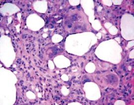
b. S100.
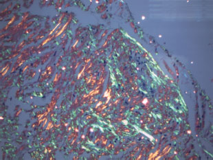
c. chromogranin.
d. Her2Neu.
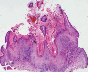
15. Which of the following is correct regarding the conjunctiva?
a. The plica semilunaris is medial to the caruncle.
b. The conjunctiva is formed of nonkeratinizing, stratified columnar epithelium and an underlying stroma.
c. The palpebral conjunctiva lines a portion of the eyelids and contains lymphoid follicles in young individuals.
d. Goblet cells are most numerous on the bulbar conjunctiva.
16. All of the following are true of dermolipomas, except
a. The lesion is classified as a choristoma.
b. There is often an absence of dermal adnexal structures within the lesion.
c. The lesions tend to occur in the fornix at the inferotemporal quadrant.
d. The lesion may extend into the orbit.
17. All of the following are correct regarding a pinguecula, except
a. The stromal collagen in the lesion shows fragmentation.
b. The stromal collagen in the lesion shows eosinophilic degeneration.
c. There may be dysplasia of the overlying conjunctival epithelium.
d. The lesion is caused by ultraviolet light.
18. All of the following are correct regarding a true conjunctival membrane, except
a. The underlying epithelium is intact.
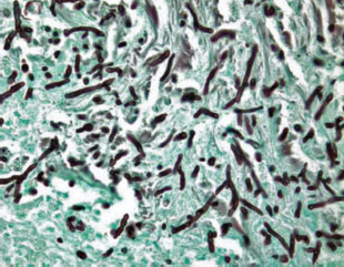
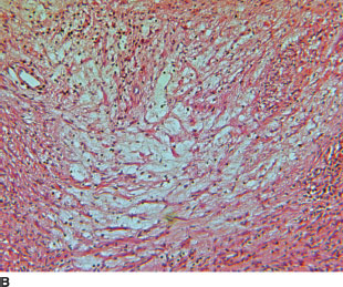
b. Granulation tissue is present.
c. Bleeding occurs when the membrane is removed.
d. Fibrinopurulent exudates may occur.
19. Which of the following is correct regarding conjunctival amyloidosis?
a. The condition is characterized by an eosinophilic, intracellular accumulation of hyalinelike material.
b. The lesion usually reflects a systemic condition.
c. There is an absence of dichroism when the specimen is stained with Congo red.
d. The amyloid material collects within the substantia propria.
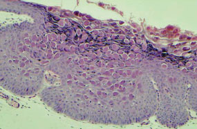
20. All of the following are true of hereditary benign intraepithelial dyskeratosis, except
a. The condition has an autosomal dominant inheritance pattern.
b. There may be lesions of the oral mucosa.
c. The condition typically begins in the fourth to fifth decade of life.
d. Dyskeratosis, acanthosis, and parakeratosis are characteristic.
21. All of the following may be helpful in characterizing a lymphoid lesion in a conjunctival biopsy, except
a. flow cytometric analysis.
b. polarization microscopy.
c. immunoperoxidase staining.
d. gene rearrangement studies.
22. What percentage of patients with primary acquired melanosis with atypia will progress to melanoma?
a. 0%.
b. 23%.
c. 46%.
d. 67%.
23. The corneal epithelium is composed of which of the following?
a. stratified, squamous, nonkeratinizing epithelium.
b. pseudostratified, columnar, nonkeratinizing epithelium.
c. stratified, cuboidal, nonkeratinizing epithelium.
d. bilayered, cuboidal, nonkeratinizing epithelium.
24. Which of the following anterior segment structures does not stain positively with the periodic acid-Schiff (PAS) stain?
a. basement membrane of the corneal epithelium.
b. Bowman’s layer.
c. Descemet’s membrane.
d. lens capsule.
25. The cornea is embryologically derived from which of the following?
a. surface ectoderm.
b. neural crest.
c. mesoderm.
d. surface ectoderm and neural crest.
26. All of the following are true of Descemet’s membrane, except
a. The membrane originates during fetal development.
b. The membrane is composed of type IV collagen.
c. The membrane gradually thins during adulthood.
d. The membrane is produced by the corneal endothelium.
27. Which of the following is true of posterior keratoconus?
a. Men are more often affected.
b. Most cases are sporadic.
c. Most cases are bilateral and nonprogressive.
d. Descemet’s membrane and the endothelium are usually absent in the area of the defect.
28. In comparing the two forms of congenital hereditary endothelial dystrophy, all of the following are correct, except
a. The autosomal recessive form is more common.
b. The autosomal dominant form appears later in life.
c. The autosomal recessive form remains stable.
d. The autosomal dominant form exhibits nystagmus.
29. Which of the following is correct regarding sclerocornea?
a. Histologically, there is no differentiation between sclera and cornea.
b. Women are more commonly affected.
c. Most cases are unilateral.
d. The condition is sporadic without a specific pattern of inheritance.
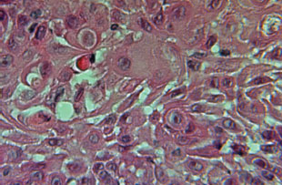
30. Which of the following organisms can be readily visualized using hematoxylin and eosin (H&E) stain preparations?
a. Actinomyces.
b. Fusarium.
c. Mucor.
d. Acremonium.
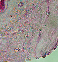
31. The photomicrograph (see image below) shows a corneal specimen. What is the most likely diagnosis for the patient?
a. herpes simplex keratitis.
b. Aspergillus infection.
c. bullous keratopathy.
d. Acanthamoeba infection.
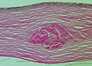
32. A patient with a corneal dystrophy undergoes penetrating keratoplasty. The corneal button is shown (see image, periodic acid-Schiff [PAS] stain, below). Which histologic stain would best demonstrate the patient’s condition?
a. Congo red.
b. Alcian blue.
c. Masson trichrome.
d. hematoxylin and eosin.
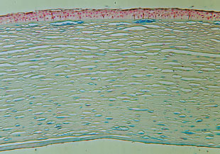
33. The corneal button of a patient is shown (see image, Alcian blue stain, below). Which of the following is true of the corneal dystrophy depicted?
a. The condition is autosomal dominant.
b. The lesions extend to the limbus.
c. The accumulated material is birefringent.
d. The abnormality has been mapped to chromosome 5q.
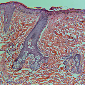
34. A lesion is removed from the limbus of a child (see image below). Which of the following is true?
a. The epithelium is nonkeratining.
b. The superotemporal quadrant is usually involved.
c. There are associated congenital syndromes.
d. There is an absence of skin adnexal structures.
35. The corneal lesion shown had a golden-brown appearance clinically (see image below). What is the etiology of the histopathologic lesion in the figure?
a. viral infection.
b. ultraviolet light.
c. chronic inflammation.
d. topical anesthetic abuse.
36. All of the following are potential causes of the histopathologic finding shown (see image below) of the posterior cornea, except
a. pellucid marginal degeneration.
b. endothelial dystrophy.
c. terrien marginal degeneration.
d. keratoconus.
37. What material does the von Kossa stain shown in the photomicrograph confirm (see image below)?
a. amyloid.
b. calcium.
c. iron.
d. hemosiderin.
38. What is the diagnosis of the patient in question 37?
a. pagetoid spread.
b. keratoconus.
c. Avellino dystrophy.
d. band keratopathy.
39. The trabecular meshwork is derived from which of the following?
a. endoderm.
b. neural crest.
c. mesoderm.
d. surface ectoderm.
40. Which of the following is one of the structures that define the anterior chamber angle?
a. ciliary body face.
b. Schlemm’s canal.
c. iris pigment epithelium.
d. external surface of the trabecular meshwork.
41. The photomicroph shows the corneal button of a patient that presented with a central opacity at birth (see image below). All of the following are typical of the condition, except
a. absence of Descemet’s membrane in the area of the defect.
b. absence of the endothelium in the area of the defect.
c. breaks in Bowman’s layer.
d. iridocorneal adhesions.
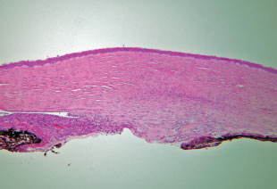
42. The condition of the patient in question 41 is characterized by which of the following?
a. predominance of unilateral presentation.
b. low risk of developing glaucoma.
c. potential for improvement of corneal opacity.
d. absence of associated syndromes.
43. The condition in the figure of question 41 is associated with all of the following, except
a. persistent fetal vasculature.
b. microphthalmia.
c. aniridia.
d. posterior polar cataract.
44. Abnormal proliferation of which of the following is a universal feature of all forms of the iridocorneal endothelial syndrome?
a. trabecular meshwork.
b. Descemet’s membrane.
c. corneal endothelium.
d. iris stroma.
45. Which of the following is true regarding exfoliation (also known as pseudoexfoliation) syndrome?
a. There is generalized depigmentation of the trabecular meshwork.
b. There is splitting of the lens capsule.
c. The accumulated material is PAS positive.
d. The iris pigment epithelium is spared.
46. All of the following have been associated with exfoliation syndrome, except
a. optic neuropathy.
b. myocardial infarction.
c. cerebrovascular events.
d. orthostatic hypotension.
47. The sclera is derived from which of the following?
a. surface ectoderm.
b. mesoderm.
c. neural crest.
d. endoderm.
48. Which of the following is true of nanophthalmos?
a. The eyes tend to have low to moderate myopia.
b. The lens is of normal size.
c. There is pathologic thinning of the sclera.
d. The condition is typically unilateral.
49. The lens is derived from which of the following?
a. surface ectoderm.
b. endoderm.
c. mesoderm.
d. neural crest.
50. In patients with homocystinuria, the condition is caused by a defect in
a. paired box gene 6 (PAX6).
b. glutathione-S-transferase.
c. cystathionine beta-synthase.
d. lysine alpha-ketoglutarate reductase.
51. The condition of phacoantigenic endophthalmitis is mediated by which immunoglobulin?
a. IgM.
b. IgG.
c. IgA.
d. IgE.
52. All of the following statements about lens-induced uveitis and glaucoma are true, except:
a. Phacoantigenic endophthalmitis is probably on the same spectrum as phacolytic glaucoma, simply being more severe.
b. Propionibacterium acnes may be an important contributor to lens-induced uveitis, particularly in pseudophakic eyes.
c. The classic pattern of inflammation in phaco-antigenic endophthalmitis is the zonal granuloma centered about a site of injury to the lens.
d. The stimulus for phacoantigenic uveitis appears to be lens cortical protein.
53. What is the average volume of the vitreous humor in adults?
a. 10 mL.
b. 8 mL.
c. 4 mL.
d. 2 mL.
54. The general characteristics of the secondary vitreous are which of the following?
a. vascular and relatively cellular.
b. avascular and relatively cellular.
c. avascular and relatively acellular.
d. vascular and relatively acellular.
55. All of the following may present as ocular inflammation (or pseudoinflammation), except
a. non-Hodgkin’s lymphoma.
b. uveal melanoma.
c. retinoblastoma.
d. melanocytoma.
56. Which of the following is true regarding the phakomatous choristoma?
a. The most frequent site of involvement is the eyelid.
b. Phakomatous choristoma may be associated with ocular colobomata.
c. There is frequently associated hyperpigmentation in phakomatous choristoma.
d. Acquired forms of phakomatous choristoma have been reported.
57. All of the following are conditions that may be associated with granulomatous ocular inflammation, except
a. sarcoidosis.
b. rheumatoid arthritis.
c. Behçet’s disease.
d. syphilis.
58. The earliest accumulations of cystoid macular edema (CME) fluid occur in the
a. outer plexiform layer.
b. nerve fiber layer.
c. subretinal space.
d. inner plexiform layer.
59. Due to variation in scleral thickness, at which of the following locations is globe rupture after blunt ocular trauma least likely to occur?
a. in an arc at the limbus opposite the site of impact.
b. at the insertion of the inferior oblique.
c. at the equator of the globe.
d. at the sclera immediately posterior to the lateral rectus insertion.
60. Which one of the following statements about retinal dialysis is false?
a. Many patients with nontraumatic retinal dialysis will have a family history of retinal detachment.
b. Most nontraumatic dialyses are superotemporal.
c. Unlike typical retinal tears, the posterior vitreous is frequently attached in the setting of retinal dialysis.
d. Retinal detachment caused by retinal dialysis is typically rapidly progressive.
61. All of the following are variations of the hyaloidolenticular vascular system, except
a. persistent pupillary membrane.
b. Mittendorf ‘s dot.
c. Bergmeister’s papilla.
d. persistent hyaloid artery.
62. The retinal circulation supplies all of the following layers, except
a. inner plexiform layer.
b. inner nuclear layer.
c. ganglion cell layer.
d. outer nuclear layer.
63. All of following are typical characteristics of the histopathology of diabetic eye disease, except
a. lacy vacuolization of the iris pigment epithelium.
b. thickening of the basement membrane of the ciliary epithelium.
c. thinning of the corneal epithelial basement membrane.
d. relative loss of pericytes.
64. Which of the following structures is the most sensitive to radiation?
a. optic nerve.
b. retina.
c. lens.
d. cornea.
65. Which of the following is true regarding the congenital tumor, phakomatous choristoma?
a. It results from hyperplasia of the accessory lacrimal glands.
b. Balloon cells are seen histologically.
c. The upper eyelid is commonly affected.
d. The material is PAS positive.
66. What is the etiology of the lesion shown in the photomicrograph (see image at the top of the right column)?
a. ultraviolet light exposure.
b. lipogranulomatous reaction.
c. viral infection.
d. hyperlipoproteinemic state.
67. What clinical finding may commonly seen in the patient shown above?
a. eyelid papules.
b. follicular conjunctivitis.
c. madarosis.
d. meibomian gland inspissation.
68. The most common organisms for preseptal cellulitis in children are
a. gram-positive rods.
b. gram-positive cocci.
c. gram-negative rods.
d. gram-negative cocci.
69. Which of the following best characterizes the clear spaces shown in the photomicrograph (see image below)?
a. The clear spaces result from fixation with neutral buffered formalin.
b. The clear spaces result from processing the tissue for histology.
c. The clear spaces are part of the cribriform pattern of a malignancy.
d. The clear spaces represent accumulations of mucin.
70. A biopsy is shown in the photomicrograph (see image below). What is the name of the sheet of configured protein in the condition?
a. beta.
b. lamda.
c. alpha.
d. kappa.
71. Which site of involvement has the highest association with a systemic disease process for the condition shown in question 70?
a. eyelid.
b. conjunctiva.
c. orbit.
d. lacrimal gland.
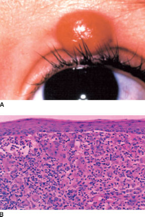
72. A 68-year-old patient presented with an eyelid lesion (see image at the top of the right column), which had grown over the last 6 weeks. Which of the following is true regarding this condition?
a. Mitotic activity is often present at the superficial apices of the lesion.
b. Perineural invasion is less likely than in squamous cell carcinoma.
c. The central crater contains sebaceous material.
d. Continued growth is likely over the next several months.
73. All of the following eyelid neoplasms are associated with systemic malignancies, except
a. actinic keratoses.
b. seborrheic keratoses.
c. sebaceous adenomas.
d. trichilemmomas.
74. What helpful diagnostic feature of squamous cell carcinoma is present throughout the photomicrograph (see image below)?
a. keratin pearls.
b. perineural invasion.
c. separation artifact.
d. intercellular bridges.
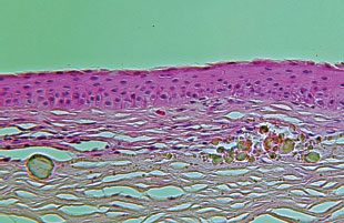
75. The photomicrograph shows a sagitally-oriented specimen depicting the orbit (right side of the figure) and superior half of the upper eyelid (left side of figure) (see image below). Which of the lettered anatomic sites (a–d) is incorrectly labeled?
a. preaponeurotic fat pad.
b. levator aponeurosis.
c. upper eyelid tarsus.
d. superior rectus muscle.
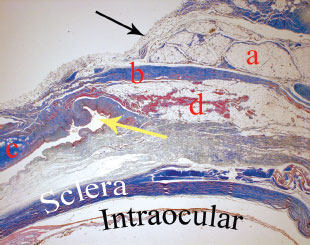
76. What structure does the black arrow mark in question 75?
a. orbital septum.
b. levator aponeurosis.
c. levator muscle.
d. superior transverse ligament.
77. What structure does the yellow arrow mark in the figure of question 75?
a. plica semilunaris.
b. caruncle.
c. canaliculus.
d. superior fornix.
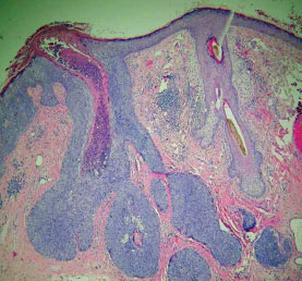
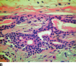
78. A 74-year-old patient presents with a 1-year history of a growth along the upper eyelid margin. The photomicrographs show the results of the eyelid biopsy (see image at the top of the right column). What anatomic site is the origin of the lesion shown?
a. meibomian glands of the tarsus.
b. accessory lacrimal glands.
c. basal layer of the epithelium.
d. glands of Zeiss.
79. The biopsy results of a patient with a flat indurated eyelid plaque are shown (see image below). Which of the following is the treatment of choice for this lesion?
a. Mohs micrographic surgery.
b. exenteration.
c. wide local excision.
d. cryotherapy.
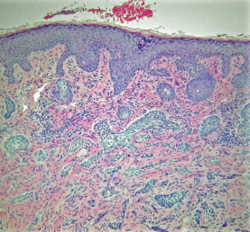
80. All of following are true, regarding the biopsy in the previous question (see image from question 79), except:
a. The deep margin is positive.
b. There is a desmoplastic reaction within the dermis.
c. Separation artifact is present.
d. Pagetoid spread is present.
81. A 40-year-old woman presents with a 2-month history of pain and diplopia. The histopathology of the lesion (see image below) is shown. Which of following is true of this condition?
a. The tumor is encapsulated.
b. The lesion may arise de novo or from a benign tumor.
c. The tumor contains a mixture of cellular and myxoid components.
d. The cribriform pattern portends a worse prognosis, than lesions without a cribriform component.
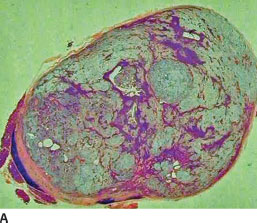
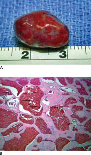
82. A 35-year-old man presents with a painless mass, which has been gradually enlarging over the last year. The histopathology of the lesion (see images A and B) is shown. All of the following are true regarding this condition, except:
a. The tumor may contain cartilage and bone.
b. The lesion comprises endothelial and stromal elements.
c. The lesion lacks a true capsule.
d. Tubules are lined by a bilayer of cells.
83. Which of the following is true regarding the condition shown in question 82?
a. Bony erosion is expected on computed tomography.
b. Adenoid cystic carcinoma may arise in the tumor.
c. Superonasal displacement of the globe is common.
d. Incisional biopsy is the preferred initial approach in management.
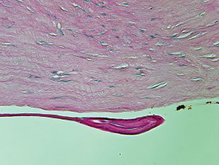
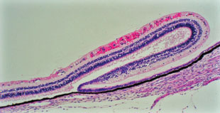
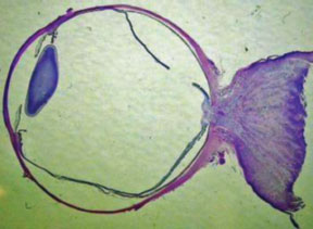
84. The autopsy eye of a child with retinal hemorrhages secondary to nonaccidental trauma is seen in the photomicroph (see image below). Which of the following best characterizes the fold of neural retina present at the ora serrata?
a. It occurs more often in adult eyes.
b. It is an artifact of fixation.
c. It represents a normal anatomic clinical variant.
d. It is seen in rhegmatogenous retinal detachment.
85. Mucosa-associated lymphoid tissue (MALT) lymphomas are generally characterized with which of the following immunophenotype?
a. CD5 positive, CD10 negative, and CD20 positive.
b. CD5 positive, CD10 positive, and CD20 negative.
c. CD5 negative, CD10 negative, and CD20 positive.
d. CD5 negative, CD10 positive, and CD20 negative.
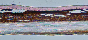
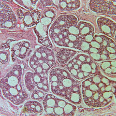
86. An 8-year-old child presents with large lightly pigmented ciliary body and iris mass (see image below). All of the following general statements are true of this condition, except
a. The extracellular material stains with Alcian blue.
b. Homer Wright rosettes can occur.
c. Cystic changes are characteristic on imaging.
d. Surgical resection is the treatment of choice.

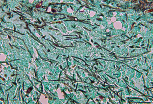
87. The sinus biopsy of a patient with nasal polyps and chronic sinusitis is stained with Gomori methenamine silver (see image at the top of the right column). An organism characterized by septate hyphae is seen. All of the following are likely to be true, except
a. The patient is immunocompetent.
b. The patient has peripheral eosinophilia.
c. CT imaging shows bone erosion.
d. Steroid therapy is contraindicated.
88. Which of the following orbital tumors is not encapsulated?
a. neurilemoma.
b. fibrous histiocytoma.
c. lymphangioma.
d. hemangiopericytoma.
89. A 36-old-patient presents with a well circumscribed retrobulbar mass causing gradually increasing proptosis. Histopathology of the mass shows a biphasic pattern in images A (below) and B (at the top of the next page in the left column). Immunohistochemical staining was positive for S100 protein. Which of the following is true of this condition?
a. The areas of nuclear palisading represent degeneration within the mass.
b. The tumor arises from histiocytes.
c. An association in children with neurofibromatosis exists.
d. Sensory corpuscles are present within the tumor.

90. Rhabdomyosarcoma of the conjunctiva, known as the botryoid variant, originates from which histologic type?
a. undifferentiated.
b. alveolar.
c. pleomorphic.
d. embryonal.
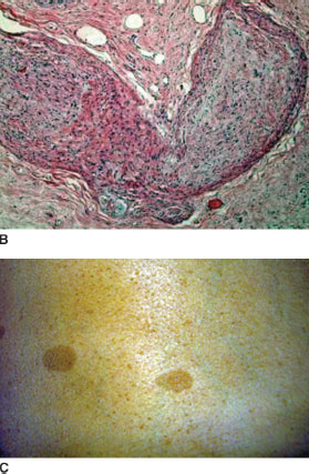
91. A child with an S-shaped deformity of the right upper eyelid undergoes surgical reconstruction as shown in images A (below) and B (at the top of the right column). Dermatologic examination of the patient is significant for multiple pigmented skin macules (see image C in the next column). Which of the following is the patient least likely to have?
a. posterior subcapsular cataracts.
b. pigmented iris hamartomas.
c. orbital encephalocele.
d. optic nerve tumor.
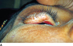
92. Which of the following best characterizes the eyelid lesion shown in the previous question (see images A and B)?
a. The lesion includes several cell types, one of which is the Schwann cell.
b. The lesion is encapsulated.
c. The lesion is relatively nonspecific for the patient’s systemic condition.
d. The lesion invariably feels like a bag of worms on palpation.
93. A 44-year-old woman presents with gradually increasing proptosis. MRI of the orbits shows a well circumscribed retrobulbar mass. Status post excision, the specimen is examined with gross and microscopic examinations (see images A and B at the top of next page). All of the following are true of this condition, except
a. The fibrous tissue setae stain with smooth muscle actin (SMA).
b. In adults, the tumor is the most common benign neoplasm of the orbit.
c. The cavernous spaces are lined by endothelium.
d. Pregnancy causes transient reduction of the tumor.
94. Fluorescein angiography is helpful in distinguishing choroidal melanoma from
a. metastatic tumors.
b. subretinal hemorrhage.
c. choroidal hemangioma.
d. large nevi.
95. All of the following are true of the tumor shown (see image at the top of the right column), except
a. In patients without neurofibromatosis, the tumor tends to invade the dura mater.
b. Hyperplasia of the arachnoid can mimic meningioma.
c. Deeply eosinophilic fibers representing degenerating cell processes are not pathognomonic.
d. Optic nerve biopsy is usually not necessary.
96. A 1-year-old child undergoes surgical excision of the lesion shown in the images A and B below. Which of the following statements is false?
a. Histopathology, as shown in the figure, demonstrates the presence of Touton’s giant cells.
b. The lesion usually spontaneously regresses in the child by the age of 5 years.
c. The lesion shown in the figure can undergo malignant transformation.
d. Topical corticosteroids can help control secondary glaucoma if it develops in these patients.
97. The histopathology of a sulfur granule from a patient with purulent canalicular reflux is shown (see image, GMS stain, below). What would you use to treat this patient?
a. metronidazole.
b. penicillin G.
c. valacyclovir.
d. amphotericin B.
98. Which of the following is the most common ophthalmic site of metastasis for primary non-ocular tumors in children?
a. orbit.
b. choroid.
c. retina.
d. optic nerve.
99. Among patients younger than 5 years of age in the United States, which of the following is the most common presenting sign of retinoblastoma?
a. hyphema.
b. iris heterochromia.
c. cataract.
d. ocular inflammation.
100. The condition of a retinal capillary hemangioma (von Hippel’s disease) is termed von Hippel’s syndrome, when associated with which of the following?
a. pancreatic cysts.
b. renal cell carcinoma.
c. cerebellar hemangioblastomas.
d. pheochromocytomas.
 Answers
1. c. Choice c is the definition of a choristoma. Dermoid cysts and dermolipomas are examples of choristomas. Choice b defines a hamartoma. Hemangiomas are a classic example of a hamartoma. Of note, Lisch nodules are iris hamartomas.
2. d. Cavernous hemangiomas are the most common benign neoplasm of the orbit in adults.
3. c. Russell bodies originate from plasma cells. Typically, Russell bodies are located intracellularly, but they can also occur extracellularly.
4. b. The cornea is avascular. Choice d refers to Bowman’s layer, which does not regenerate when incised.
5. b. The limbal healing response involves are three tissue types seen at the limbus.
6. b. In cyclodialysis, there tends to be an underproduction of aqueous humor. Consequently, the intraocular pressure is decreased rather than increased. The other choices are correct.
7. d. Fresh tissue is needed for immunofluorescence (IF). While the Michel fixative is commonly referred to as a fixative, technically it is a transport medium, which does not fix the tissue and allows IF studies.
8. b. The formalin fixes the tissue at a rate of 1 mm/h. Thus an average globe with a radius of 12 mm needs at least 12 hours of fixation.
9. b. Colloidal iron stains mucopolysaccharides. Along with Alcian blue, the stain is useful in macular corneal dystrophy and Schnabel’s cavernous optic atrophy. Thioflavin T, Crystal violet, and Congo red all stain amyloid, which is the substance seen in lattice corneal dystrophy.
10. c. Of the choices given, the insertion of the inferior oblique muscle is the only one specific for lateralizing a globe. The course of the long posterior ciliary artery and nerve and the oblong shape (in the horizontal meridian) of the cornea can facilitate differentiation of the horizontal and vertical axes of the globe, but not whether the eye is a right or left one.
11. d. Traumatic angle recession is an indirect indicator of potential damage to the trabecular mesh-work. Recession of the angle itself does not directly A cause glaucoma. The fusiform appearance of the U ciliary body occurs over time and is a late finding. A V plane of relative weakness exists between the longitudinal and circular muscles of the ciliary body. The ciliary processes are displaced posteriorly.
12. d. The uveal tract does not attach to the sclera in Schlemm’s canal. The remaining three choices are the points of attachment of the uveal tract to the sclera.
13. c. Shrinkage, atrophy, and disorganization define phthisis bulbi. Pain or ocular discomfort may be a symptom of phthisis bulbi, but it is not a defining characteristic.
14. b. The S100 stain is markedly positive in schwannoma. HMB-45 reacts with melanocytic lesions and would only rarely react in the case of a schwannoma (e.g., a rare, pigmented schwannoma). Chromogranin stains neuroendocrine lesions, such as metastatic carcinoid, Merkel cell tumor, and small cell carcinoma. Her2Neu is positive in certain metastatic breast carcinomas.
15. c. Choice c is correct. The plica semilunaris is lateral to the caruncle. The conjunctival epithelium is stratified squamous. Goblet cells are most numerous in the plica and fornices.
16. c. Dermolipoma tends to occur in the superotemporal quadrant. The remaining choices are correct. Unlike a dermoid, dermolipomas often lack dermal adnexal structures.
17. b. The degeneration of the stromal collagen in a pinguecula is known as elastotic or basophilic degeneration. The other choices are correct. Histologically, the lesion is identical to a pterygium with the exception that a pterygium grows over the cornea and destroys Bowman’s layer.
18. a. The underlying epithelium is absent in a true membrane. Because there is no underlying epithelium, clinically, once a true membrane is removed, bleeding occurs. The remaining choices are correct.
19. d. Amyloid collects within the substantia propria. The material is extracellular. Unlike in the orbit and eyelid, conjunctival amyloid usually reflects a localized process rather than a systemic condition. Less commonly, amyloid may occur in the setting of a chronic inflammatory condition, for example, trachoma, or a systemic condition, for example, multiple myeloma. When stained with Congo red, amyloid exhibits the properties of dichroism and birefringence.
20. c. Hereditary benign intraepithelial dyskeratosis (HBID) is an autosomal dominant condition. The rare condition was first described in a triracial population of Halifax County, North Carolina. The condition begins in early childhood. HBID is benign and does not undergo malignant transformation. Patients with HBID present with bilateral limbal conjunctival and oral mucosal plaques.
21. b. Polarization microscopy is useful for the assessment of amyloidosis and the detection of foreign material, but does not have a role in the evaluation of lymphoproliferative disease.
22. c. Nearly half of patients with primary acquired melanosis (PAM) with atypia will progress to melanoma. In contrast, PAM without atypia does not progress to melanoma.
23. a. The corneal epithelium is nonkeratinizing, stratified squamous epithelium and has a thickness of about four to six cells. Respiratory epithelium, such as that found in the nasolacrimal duct, is ciliated, pseudostratified, columnar.
24. b. Bowman’s layer is the anterior, acellular aspect of the corneal stroma. It is not a true membrane and does not stain with PAS.
25. d. The cornea is both surface ectoderm and neural crest derived.
26. c. Descemet’s membrane becomes gradually thicker as one ages.
27. b. Posterior keratoconus is typically sporadic, unilateral, and progressive. Women are more commonly affected. Descemet’s membrane and the endothelium are typically intact.
28. d. The more common autosomal recessive form presents with nystagmus, whereas the autosomal dominant form does not.
29. a. Ninety percent of patients with sclerocornea are affected bilaterally. Men and women are equally affected. Half of cases are sporadic; half exhibit either a dominant or recessive pattern of inheritance.
30. c. Mucor is a relatively large nonseptate fungal organism. Mucor differs from the other three organisms in that Mucor is visible on H&E stain.
31. d. Acanthamoeba cysts are visible within the corneal stroma in the photomicrograph.
32. a. Unlike macular and granular dystrophies, lattice dystrophy stains positive with PAS (as seen in the image in question 32), Congo red, and crystal violet stains.
33. b. A photomicrograph of macular dystrophy is shown. Macular dystrophy involves the entire cornea, including up to the limbus. Macular dystrophy is autosomal recessive, while granular and lattice dystrophies are autosomal dominant. Birefringent material collects in lattice dystrophy, but not macular. The genes for macular dystrophy and Avellino dystrophy are chromosomes 16q and 5q, respectively.
34. c. A corneal dermoid is covered by keratinizing stratified squamous epithelium and contains skin adnexal structures. A corneal dermoid is a choristoma and may be associated with congenital syndromes, for example, Goldenhar’s syndrome. The superotemporal quadrant is the most frequent site of involvement for dermolipomas.
35. b. A photomicrograph of spheroidal degeneration (also known as actinic or Labrador keratopa-thy) is shown. The condition has a golden-brown appearance clinically and is caused by ultraviolet light exposure.
36. b. A discontinuity in Descemet’s membrane is seen. Breaks in Descemet’s membrane occur in choices a, c, and d, but not in endothelial dystrophy. The finding can also occur in obstetric forceps injury and congenital glaucoma.
37. b. Calcium stains positively with the von Kossa stain.
38. d. Calcium deposition in the corneal epithelial basement membrane and anterior stroma (including Bowman’s’ layer) is known as band keratopathy.
39. b. The trabecular meshwork arises from the neural crest.
40. a. The boundaries of the anterior chamber angle are the posterior cornea, the internal surface of the trabecular meshwork, the ciliary body face, and the anterior iris.
41. c. The corneal button of a patient with Peter’s anomaly is shown. The abnormal posterior cornea and iris adhesions are seen. Bowman’s layer is in the anterior cornea and is unaffected in Peter’s anomaly.
42. c. The central opacity of Peter’s anomaly may improve over time.
43. d. Peter’s anomaly is associated with anterior cataracts (cortical or polar).
44. c. The corneal endothelium proliferates abnormally in all three forms of the iridocorneal endo-thelial syndrome.
45. c. The deposits stain positively with PAS and involve the anterior segment structures. There is increased pigment in the trabecular meshwork. Splitting of the lens capsule occurs in the rare true exfoliation syndrome, which is associated with exposure to infrared radiation, for example, glass blower’s cataract.
46. d. Hypertension, rather than hypotension is associated with exfoliation syndrome. Choices a, b, and c have been associated with exfoliation syndrome.
47. c. The sclera arises from the neural crest.
48. b. In nanophthalmos, the eye is smaller than normal. The lens, however, may be either normal or enlarged. The condition is typically bilateral, results in hyperopia, is associated with glaucoma, and predisposed to uveal effusion (likely secondary to a thickened sclera).
49. a. The lens arises from the surface ectoderm.
50. c. Homocystinuria is an autosomal recessive condition with a defect in cystathionine beta-synthase. Homocystinuria is one of a number of systemic conditions associated with ectopia lentis. Classically, the lens is dislocated down (or inferonasal) in homocystinuria, whereas in Marfan’s and Weill-Marchesani’s syndromes, the lens is dislocated up and superotemporally (or temporal), respectively.
51. b. Phacoantigenic endophthalmitis is mediated by immunoglobulin G (IgG) directed against lens protein. Other terms that have been used for the condition include lens-induced granulomatous endophthalmitis and phacoanaphylactic endophthalmitis.
52. a. Although some authors consider phacolytic (phacotoxic) uveitis to be a less severe form of phacoantigenic endophthalmitis, phacolytic glaucoma is not true active inflammation. Rather, there is an increase in intraocular pressure (IOP) caused by clogging of the trabecular meshwork (TM) by lens protein and macrophages. The lens capsule is typically not ruptured in phacolytic glaucoma. Phacoantigenic endophthalmitis describes lens-induced inflammation as a result of immunoglobulin G (IgG) directed against lens proteins. Also, because mast cells play no role in “phacoanaphylaxis,” phacoantigenic is a better descriptor.
53. c. The average volume of the vitreous humor in adults is 4 mL.
54. c. Postnatally (and continuing to adulthood) the main vitreous is comprised of the secondary vitreous, which is avascular and relatively acellular. There are a few cells, known as hyalocytes, present in the secondary vitreous.
55. d. Large-cell lymphoma (reticulum cell sarcoma) may present as a pseudovitritis. Retinoblastoma may present as a pseudohypopyon. Aggressive choroidal melanoma may become necrotic or cause necrosis of the overlying retina with secondary uveitis. Melanocytomas typically do not present as ocular inflammation.
56. a. The phakomatous choristoma (Zimmerman’s tumor) represents a congenital developmental anomaly (choristoma) with lens-like (hence, “phakomatous”) tissue forming a mass within the eyelid, usually lower. Such a growth has been reported nowhere else in or on the body. The lesion is rare and has no reported ocular associations, with surgical excision being the usual treatment.
57. c. Behçet’s disease is a nongranulomatous vasculitis that causes acute uveitis with hypopyon, aphthous stomatitis, and genital ulceration. Rheumatoid arthritis causes a zonal granuloma centered on scleral collagen. Sarcoidosis typically produces a discrete granulomatous reaction, whereas sympathetic ophthalmia and Vogt-Koyanagi-Harada syndrome (VKH) generally cause diffuse granulomatous inflammation.
58. a. Because of the oblique orientation of the axons in this layer of the fovea, fluid accumulates more easily here than in retinal layers where the neural and glial elements are oriented radially.
59. b. The sclera is very thin (0.2 mm) immediately posterior to the insertion of the rectus muscles. Unlike the tendonous insertions of the rectus muscles, the inferior oblique has a muscular insertion on the globe. The inferior oblique inserts onto the globe in the area overlying the macula and there is no associated thinning of the sclera in this location. The sclera is thickest (1.0 mm) at the posterior pole.
60. d. Retinal dialysis occurs because of mechanisms other than posteroanterior vitreous traction; thus, it is more common to discover dialyses in eyes with attached posterior hyaloid. Spontaneous dialyses, often in patients with a family history of retinal dialysis and/or detachment, occur most commonly in the inferotemporal quadrant. Subsequent detachment is typically slowly progressive and often silent clinically.
61. a. Persistent pupillary membrane is an incomplete regression of the anterior tunica vasculosa lentis. Bergmeister’s papilla is a nonpatent hyaloid artery remnant extending from the optic disc. If this structure is patent, it is called persistent hyaloid artery. Mittendorf ’s dot is an opacity on the inferonasal posterior lens capsule from connective tissue associated with the central hyaloid vasculature and posterior tunica vasculosa.
62. d. The retinal circulation supplies the nerve fiber layer, the ganglion cell layer, the inner plexiform layer, and the inner one-third of the inner nuclear layer. The choroidal circulation supplies the remainder of the neurosensory retina and retinal pigment epithelium (RPE).
63. c. The basement membrane of the corneal epithelium is thickened in diabetic patients. As a result, the corneal epithelium in diabetics is weakly adherent to the Bowman’s layer. Consequently, diabetic patients are more prone to corneal abrasions and poor corneal epithelial healing.
64. c. The lens is the most radiosensitive component of the eye, followed by the cornea, retina, and optic nerve.
65. d. A phakomatous choristoma is a rare tumor of the inferomedial eyelid composed of lens epithelial cells. Just as in the capsule of the crystalline lens, PAS-positive material is seen in phakomatous choristoma. Bladder cells similar to those seen in the histopathology of cataract may occur. Balloon cells refer to cells that occur in specific subtypes of conjunctival and choroidal nevi and melanoma.
66. c. Molluscum contagiosum lesion is shown, which is caused by a pox virus. Ultraviolet light exposure leads to actinic damage. The histopathology of a chalazion is lipogranulatous inflam-mation. Eyelid xanthelasma can occur in the setting of a hyperlipoproteinemia.
67. b. A follicular conjunctival reaction is a feature of molluscum contagiosum infection of the eyelid. Madarosis is a potential finding in eyelid malignancy, rather than molluscum. Meibomian gland inspissation occurs in blepharitis.
68. b. In children, the most common organisms for preseptal and orbital cellulitis are gram-positive cocci.
69. b. Processing of tissue for histology involves the use of organic solvents that dissolve lipids. Consequently, the lipid material in a chalazion (as shown in the image in question 69) appears as artifactually empty vacuoles on light microscopy.
70. a. Amyloidosis is seen in the photomicrograph. The beta-pleated sheet imparts the birefringence and dichroism staining properties of amyloid.
71. a. In ocular adnexal amyloid, eyelid involvement is most indicative of systemic disease.
72. b. A keratoacanthoma is seen in the photomicrograph. The lesion typically shows rapid growth within a period of 3 months and may spontaneously resolve. Keratoacanthoma characteristically has a cup shaped configuration, which contains keratin. Histopathologically, there is mitotic activity at the deep margin. The term well-differentiated squamous cell carcinoma is sometimes used to designate keratoacanthoma because of significant overlap between the two entities. However, perineural invasion and metastasis are characteristic of squamous cell carcinoma, not keratoacanthoma.
73. a. An actinic keratosis is a premalignant skin lesion unassociated with systemic malignancy. The sudden occurrence of multiple seborrheic keratoses can be associated with an internal malignancy, for example, Leser-Trélat sign (most commonly gastrointestinal adenocarcinoma). Sebaceous adenomas are associated with Muir-Torre syndrome (most commonly colorectal carcinoma). Multiple trichilemmomas is associated with visceral malignancy, for example, Cowden’s disease.
74. d. A photomicrograph of squamous cell carcinoma is seen at high power magnification. Numerous intercellular bridges are seen as fine projections between the tumor cells. Dyskeratosis in the form of keratin pearl formation and perineural invasion are not present, but are features of squamous cell carcinoma. Separation artifact is a histopathologic feature of basal cell carcinoma.
75. d. Choices a–c are correctly labeled. Choice d depicts Müller’s muscle. The superior rectus muscle tendon is seen attaching to the globe just posterior to the word “Sclera” in the photomicrograph.
76. a. The black arrow shows the orbital septum, which is anterior to the preaponeurotic fat (structure “a”).
77. d. The yellow arrow depicts the superior fornix.
78. d. Sebaceous carcinoma is seen arising from a gland of Zeiss. A normal pilosebaceous unit is seen to the right of the lesion.
79. a. A photomicrograph of the morpheaform (sclerosing) type of basal cell carcinoma is seen. Margin control with frozen sections or Mohs micrographic surgery is required.
80. d. Pagetoid spread is a characteristic of sebaceous carcinoma. Choices a–c are all present in the photomicrograph shown.
81. b. A photomicrograph of adenoid cystic carcinoma is shown. The tumor is unencapsulated and may arise de novo or from a pleomorphic adenoma. It is the basaloid pattern, rather than the cribriform pattern that carries a worse prognosis.
82. b. High and low magnification photomicrographs of a pleomorphic adenoma (benign mixed tumor [BMT]) are shown. BMT is the most common epithelial tumor of the lacrimal gland. Clinically, the tumor is more common in men (median age 35). The presentation is one of a painless lacrimal gland mass, which exhibits bony remodeling (rather than erosion) on CT imaging. The lesion can also occur in ectopic locations, accessory lacrimal gland sites, for example, Kraus or Wolfring, or in the salivary glands. Histologically, the lesion develops a capsule-like structure (pseudocapsule) from chronic compression of the surrounding tissue. The substance of the tumor is comprised of a mixture of epithelial and stromal components. The stroma may contain cartilage and bone. The epithelial aspect forms nests or tubules, which are lined by a bilayer of cells.
83. b. Please see discussion from previous question. In addition, inferonasal displacement is expected from a lacrimal gland mass. Complete removal, rather than incisional biopsy, is required to prevent tumor seeding.
84. b. A photomicroph of Lange’s fold is seen. The fold is an expected finding and represents an artifact of fixation in the eyes of infants and children. Lange’s fold is not present in unfixed eyes and does not occur in adult eyes.
85. c. MALT lymphomas arise from B cells and are generally CD5–, CD10–, and CD20+.
86. d. A photomicrograph of a medulloepithelioma (diktyoma) is shown. Medulloepithelioma is a rare, congenital tumor that arises from the nonpigmented ciliary epithelium. The tumor typically presents during the first decade of life. Clinically, the mass appears lightly pigmented or amelanotic. Ultrasound imaging may reveal large, highly reflective cysts posterior to the iris that are characteristic of the tumor. Histologically, the tumor secretes hyaluronic acid, which stain positively with Alcian blue. In addition, Homer Wright rosettes may be seen with light microscopy.
87. d. The photomicrograph shows Aspergillus organisms. Aspergillus are septate hyphae with a 45° angle branching pattern. The patient described has allergic aspergillosis sinusitis, which occurs in immunocompetent individuals. Peripheral eosinophilia occurs in patients with allergic aspergillosis sinusitis. CT imaging may show bony erosion or remodeling. The condition is treated with steroid therapy and potentially surgical debridement.
88. c. Choices a, b, and d are all encapsulated. Lymphangioma is unencapsulated.
89. c. A neurilemoma (schwannoma) is seen in the photomicrographs. The tumor arises from Schwann cells. Two histologic patterns are seen. Figure (A) shows the Antoni A pattern, which has tightly configured spindle cells and palisading nuclei (may form Verocay bodies). Figure B shows the Antoni B pattern, which is characterized by myxoid appearance and loosely arranged cells. The areas of Antoni B correspond with tumor degeneration. Histologically, the lesion is markedly S100 positive with immunohistochemical staining. A schwannoma of the eighth cranial nerve is known as an acoustic neuroma and is associated with neurofibromatosis type 2. Sensory corpuscles are unrelated to schwannomas.
90. d. The botryoid variant originates from the embryonal type of rhabdomyosarcoma.
91. a. The patient has neurofibromatosis type 1 (NF1). NF2 (not NF1) is associated with posterior subcapsular cataracts. Choices b–d are associated with NF1. Lisch nodules are iris hamartomas. Orbital encephalocele may occur in the setting of dysplasia of the greater wing of the sphenoid bone. Optic nerve gliomas (and much less commonly meningiomas) also have associations with NF1.
92. a. A plexiform neurofibroma, which is composed of Schwann cells, axons, and endoneural fibroblasts, is shown. The tumor is unencapsulated. Plexiform neurofibromas occur in about 30% of patients with NF1 and are highly specific for NF1. Although widely described, in actuality, the bag-of-worms feel occurs infrequently.
93. d. The patient presents with a cavernous hemangioma. Pregnancy may accelerate the growth of the tumor.
94. b. There are no fluorescein angiographic signs that are pathognomonic for choroidal melanoma (or for any intraocular tumor, for that matter). Subretinal hemorrhage (most frequently from choroidal neovascularization) can be differentiated from melanoma by the absence of late hyper-fluorescence characteristic of melanoma.
95. a. An optic nerve glioma is shown. In patients without neurofibromatosis, the tumor tends to remain confined to the optic nerve. In patients with neurofibromatosis, the tumor often proliferates in the subarachnoid space. Rosenthal fibers are not unique to optic nerve glioma.
96. c. The lesion shown is a xanthogranuloma. It is characterized by the presence of Touton’s giant cells (shown in the image B in question 96). Patients with iris lesions secondary to juvenile xanthogranulomas are at risk for spontaneous hyphema and consequent secondary glaucoma.
97. b. Actinomyces is a gram-positive non– acid-fast anaerobic bacteria. The bacteria also stain positively with GMS. The bacteria form long, fine branching filaments. The organism can grow as a cluster, known as a sulfur granule. Appropriate treatment consists of removal of the canalicular concretions and antibiotic therapy. A number of antibiotics are effective in the treatment of acti-nomyces; however, metronidazole does not have any effect on the organism.
98. a. In adults, the choroid is the most common site of ocular/periocular metastasis. In children, however, the orbit is more commonly involved.
99. d. For children under 5 years of age, in the United States (in order of frequency), the three most common presenting signs of retinoblastoma are leukocoria (54%–62%), strabismus (18%–22%), and ocular inflammation (2%– 10%). Choices a–c also occur, but are not in the top three.
100. c. Retinal capillary hemangiomatosis associated with cerebellar hemangioblastoma is termed von Hippel-Lindau syndrome. Other lesions, including renal cell carcinoma and pheochro-mocytoma, are associated but are not a part of the definition of the syndrome. Multiple retinal angiomas (without cerebellar tumors) are referred to as von Hippel’s disease.
Stay updated, free articles. Join our Telegram channel

Full access? Get Clinical Tree


