
Fig. 5.1
Types of NOE fractures: (a) anatomy of the NOE region. (b) Telescopic displacement of nasal bones into the interorbital space. (c) Trajectory of a NOE fracture. (d) Complete bilateral type I NOE fracture. (e, f) Incomplete unilateral comminuted type I NOE fracture. (g) Test for motility of the central fragment
5.2 Definition of a Naso-Orbito-Ethmoid Fracture
NOE fractures in essence are broken nasal bones and cartilages telescoped backward into the interorbital space (Fig. 5.1b) usually as a result of an assault or a motor vehicle accident [6, 7]. The force vector resulting in a NOE fracture is usually transmitted through and thus fracturing five sutures, the frontal process of the maxilla in the place where it joins the internal angular process of the frontal bone, then the medial orbital wall, the infraorbital rim, the lateral nasal wall, and the nasomaxillary suture of piriform aperture (Fig. 5.1c) [8]. The resulting segment (namely, the frontal process of the maxilla, forming the lower two-thirds of medial orbital rim) is the central fragment of the NOE fracture, to which the medial canthal tendon (MCT) is attached. Formation of one or several movable fragments of the medial orbital rim with the attached MCT is the key factor in pathogenesis of NOE fracture.
5.3 Classification of NOE Fractures
Classification of NOE fractures is based on the integrity of the central fragment [9]. According to Markowitz et al. [10], there are three types of fractures:
Type I – isolated fracture resulting in one large fragment which is also the central fragment (Fig. 5.1d–f).
Type II – fracture of the central fragment resulting in comminuted fragments with fracture lines going around the MCT attachment site so that the latter remains intact (Fig. 5.2a, b).

Fig. 5.2
Types of NOE fractures: (a, b) the unilateral (a) and bilateral (b) type II NOE fractures. (c, d) Unilateral (c) and bilateral (d) type III NOE fractures. (e) Shortening of the palpebral fissure and widening of the nasal bridge due to displacement of central fragment. (f) Combination of a NOE fracture and a zygomatic orbital fracture. Materials from www.aofoundation.org were used for this illustration
Type III – fracture of the central fragment involves comminuted fragments with destruction of the MCT attachment site to the extent of its avulsion (Fig. 5.2c–e).
5.4 Clinical Presentation of Type I NOE Fractures
This group of fractures accounts for 18 % of all fractures in this region [11]. Complete bilateral type I NOE fractures resulting in an isolated central fragment, detached from the surrounding osseous structures by all five fracture lines, are more typically an exclusion rather than a rule. It is usually a low-energy unilateral “greenstick” fracture located in the site of the junction of the frontal process of maxilla and the internal angular process of the frontal bone above the MCT attachment site (Fig. 5.1e) [11].
As the central fragment moves downward, it affects the medial palpebral commissure, which causes lengthening of the palpebral fissure and prolapse of the medial canthus. Sagging of the inner infraorbital rim alongside deformation of the piriform aperture is very likely, but it is usually disguised by edema and hematoma of the soft tissues. Injury of the lateral nasal wall causes ipsilateral face asymmetry and obstruction of the lacrimal pathways. Meanwhile, the length of the nasal bridge and intercanthal distance usually do not change, which may give an illusion of the intact NOE complex. In such case, palpating the MCT attachment site or testing central fragment for flexibility under general anesthesia makes diagnosis considerably easier (Fig. 5.1g). Crepitation or flexibility of the bone fragment unmistakably indicates a fracture that requires open repositioning or rigid fixation.
5.5 Clinical Presentation of Type II and III NOE Fractures
These are moderate-energy fractures that comprise 72 % of all the fractures in this region [11].
Since the only difference between type II and type III fractures is the condition of bones around the MCT attachment site, the respective symptoms are very similar. The detailed clinical presentation of a typical comminuted NOE fracture of both types includes:
Symptoms determined by lateral displacement of the central fragment caused, in turn, by the orbicularis oculi strain (flattening and widening of the nasal bridge, shortening of the palpebral fissure and rounding of its medial angle, and the increase in intercanthal distance – traumatic telecanthus) (Fig. 5.2e)
Symptoms determined by telescopic displacement of fractured nasal bones (saddle nose deformity, epicanthus caused by displacement of nasal skin on the medial palpebral commissure, epiphora caused by obstruction of the lacrimal pathways with bone fragments, epistaxis, anosmia, and obstruction of nasal passages) [12, 13]
Fractures can be either unilateral (the so-called hemi-NOE-fracture) or bilateral (Fig. 5.2b, d). The latter, observed in two-thirds of injured patients, is often asymmetrical and combines types I and II.
Only 10 % of NOE fractures are isolated; more commonly a NOE fracture is a part of the extensive fracture that engages other facial bones or the skull base (Fig. 5.2f) [5, 13, 14]. Fragments of the vomer, ethmoid, and nasal bones may penetrate into the cranial cavity as they are telescoped backward. As a consequence, 50 % of the time this type of fracture involves brain injury; in 40 %, cerebral spinal fluid (CSF) leak; and in 30 %, vision-threatening injuries of the eyeball and optic nerve.1
A CSF leak is usually caused by propagation of the fracture to the walls of the frontal sinus associated with dura mater rupture. The leak can be detected through visual examination; sometimes a patient himself/herself senses a metallic taste in the nasopharynx. CSF fluid may also gather under the periosteum of the orbital wall either as palpable fluctuating formation or intermittent swelling of orbital tissues worsening at straining and coughing or squeezing of the jugular veins [6].
In 4.5 % of cases, a high-energy fracture of NOE complex is accompanied by a circular fracture of both orbits (3–4 walls), types I and III Le Fort fractures of the zygomatic bones maxilla and mandible that lead to lateral transposition, increase in orbital volume, and divergence of orbits.2 Widening of the face, lateral dislocation of both eyeballs, increase in interorbital, interpupillary, and intercanthal distances are the classic signs of traumatic hypertelorism. While this condition accounts for only 1.5 % of all midfacial traumas, the incidence is probably much higher because of the high mortality rate secondary to severe brain injury and other life-threatening injuries caused by the original trauma. In every second patient, injury of the optic nerve causes bilateral blindness. Half of the patients surviving this trauma have bilateral blindness secondary to optic nerve damage. Ruptured globes are often found in these traumas as well [10].
5.6 Diagnosis of NOE Fractures
It would not seem difficult to diagnose a NOE fracture for its pathognomonic symptoms such as flattened nasal bridge and telecanthus. However, the difficulty is that in the early days following injury, the obvious signs of fracture are disguised by swelling, ecchymosis, and emphysema of midfacial soft tissues [14, 16].
CSF leak, epistaxis, and epiphora are typical, yet not pathognomonic symptoms. This is where a clinician should be especially suspicious. As bones of the NOE complex endure the load of up to 30 g/cm2, any nasal fracture may be a part of a more extensive injury [7]. That is why every midfacial trauma should be treated as a potential NOE fracture.
When making the final diagnosis, CT scanning of 1.5-mm sections is very important (Fig. 5.3) [5, 17]. Axial CT signs indicating a NOE fracture are as follows: spread of the nasomaxillary suture, asymmetrical nasolacrimal ducts, shadowing and destruction of ethmoid air cells, depression and displacement of nasal bones, displaced fracture of the medial orbital wall accompanied by displacement of segments, and orbital emphysema. Coronal CT scans can reveal both inferomedial spread of the nasomaxillary suture and fracture of the infraorbital rim with posterior displacement.


Fig. 5.3
CT scan of a NOE fracture: (a, b) telescopic displacement of broken nasal bones backward into the interorbital space (denoted with arrows). (c) Fracture line crosses both nasolacrimal ducts (long arrows). (d) Unilateral (hemi-) NOE fracture. (e) Unilateral disruption of the nasomaxillary suture in an axial scan (long arrow). Short arrow indicates zone of diastasis of the zygomaticomaxillary suture, verifying that the patient has a combination of NOE and maxilloorbital fractures. (f) The same combination of two fractures. The nasolacrimal duct is destroyed (long arrow), a fracture of the zygomatic arch (short arrow). (g) Combination of a bilateral (long arrows) NOE fracture and depressed fracture of anterior wall of maxillary sinus (short arrows). (h) 3D reconstruction of the same injury of facial bones. (i, j) 3D reconstruction of a combination of NOE (long arrows) and zygomatic orbital (short arrows) fractures
5.7 Treatment of NOE Fractures
Considering that the overwhelming majority of NOE fractures are very complex, treating them often requires the multidisciplinary approach involving a neurosurgeon, a maxillofacial surgeon, and an ophthalmologist [4, 13].
The treatment begins with stabilization of vital signs and evaluation of the neurological status. The surgical treatment of a NOE fracture can be started only after the risk of penetrating brain injury or open globe injury has been eliminated [18]. In the situation where there is either open brain injury or an open globe, neurosurgical and ophthalmic surgical interventions are performed first, followed by reduction of the NOE fracture.
On condition that the patient’s neurological status is stable, a CSF leak should not prevent early fracture repositioning, because the intervention may stop the leak [6].
The goal of the treatment is to reconstruct the initial appearance of the palpebral fissure and nose, which involves restoration of the intercanthal distance, height, and contour of the nasal bridge and symmetry of medial palpebral commissures [19].
A key to success is an experienced surgeon knowledgeable of the complex midface anatomy, the use and interpretation of appropriate ancillary tests, as well as training in the surgical repair of midfacial trauma [20–23].
5.8 Main Stages of the Surgery
5.8.1 Surgical Approach
Five incisions are used to give proper exposure of the NOE region: subciliary, upper gingivobuccal, coronal, limited median vertical, and the gull-wing approach (Figs. 5.4a–c and 5.5a–c).
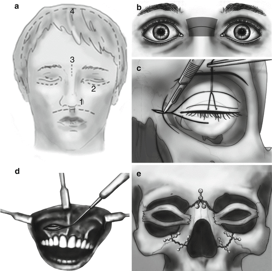


Fig. 5.4
Treatment of NOE fractures: (a–c) approaches to the NOE region: 1 – upper gingivobuccal (a detailed image is given in figure “d”), 2 – subciliary, 3 – limited median vertical, 4 – coronal. (b, c) glabellar, (b) and extended glabellar (c) approaches. (e–h) Fixation of central fragment at complete bilateral (e), incomplete unilateral, (f, g) and complete (h) type I NOE fractures
Fig. 5.5
Stages of surgical treatment of a NOE fracture: (a–c) degloving of comminuted fracture through gull-wing incision. (d–e) Fixation of fragments with wire (d) or titanium microplates (e). Comminuted fracture requires placing some plates in the immediate proximity to the medial canthal tendon, which is an undesirable but compulsory measure
The subciliary approach exposes the infraorbital rim and the orbital floor; upper gingivobuccal incision provides access to stabilize the nasomaxillary suture and piriform aperture. Gull-wing incisions give the best exposure of the entire NOE region; coronal incisions are essential for fractures extending to the frontal sinus, anterior, and lateral orbital walls. A nasal or glabellar injury provides an additional approach to the fracture and is used in about one third of the cases [26, 27].
Choice of the incision (or their combination) is determined by the characteristics of the fracture (uni- or bilateral, coarse or comminuted, isolated or extended) [5, 10, 28].
Subciliary and gingivobuccal incisions will suffice to deglove a unilateral type I NOE fracture with inferior displacement [11]. All other cases (superior dislocation of the central fragment, type I bilateral fractures, comminuted fractures) require a combination of the superior and inferior (subciliary and gingivobuccal) approaches. A coronal incision is used for extended fractures, and median vertical and the gull-wing incisions for isolated fractures.
Identification of the MCT and central fragment sometimes poses a serious challenge, as there is a risk of complete avulsion of the former from the central fragment if one is not careful. In order to avoid this iatrogenic complication, one should start the surgical dissection at the nasal bones to identify the anatomy. The Eyelash traction test is another way to evaluate this situation (Furnas and Bircoll 1973); it is used to determine whether MCT is detached by means of pulling the eyelashes of the upper eyelid.
Restoration of the medial orbital rim via open repositioning and rigid fixation of the central fragment 3 is the key stage of surgery whose technique is defined by the fracture type [10, 21, 23].
In patients with complete bilateral types I NOE fractures, the central fragment which is displaced posteroinferiorly is fixed with 1.5- and 2-mm titanium microplates to the supraorbital rim and piriform aperture (Fig. 5.4e) Lateral displacement of the fragment can be effectively treated by a transnasal reduction by the technique presented below.
Stay updated, free articles. Join our Telegram channel

Full access? Get Clinical Tree


