High-output chylous leak beyond 5 to 7 days of conservative medical treatment should be treated promptly to avoid the risk for nutritional and imunologic depletion. Given the effectiveness and low morbidity of this minimally invasive treatment, this is a reasonable first option before surgical repair of thoracic duct leak not responsive to conservative medical treatment.
Anatomy
When operating in the inferior neck, injury to delicate lymphatic vessels may occur . The earliest reports of chyle leak are attributed to Cheever in 1875 and Allen and Briggs in 1901 The thoracic duct returns chyle into the venous circulation, traveling from the chest into the left neck to terminate at the left internal jugular vein at its junction with the left subclavian vein. The course of the thoracic duct may be somewhat variable, and branching has been described in approximately 10% of cases. The duct courses along the medial border of the anterior scalene muscle; therefore, dissection between the phrenic nerve and the carotid sheath places the thoracic duct at risk for injury. Importantly, an accessory duct may exist in the right neck. Reports suggest up to 25% of chyle fistulae occur on the right.
Physiology
Ingested long chain triglycerides are carried via the thoracic duct as chylomicrons and delivered to the systemic circulation. Medium chain triglycerides, however, are carried through the portal circulation. Approximately 2 to 4 L of chyle are transported daily. The composition of chyle varies, containing 2% to 4.5% protein and 1% to 3% fat.
Consequences of chyle fistula
Most obviously, chlye leak may result in prolongation of hospital stay. A leak of up to 1 L per day may be tolerated for 1 to 2 days before resulting in electrolyte disturbance. Prolonged leaks also may result in protein loss. Rarely, chyle leak in the neck can be associated with development of chylothorax. Accumulation of chyle within the neck leads to dermal irritation, erythema, and dehiscence of surgical flaps.
Management
Opinions vary regarding the optimal management and timing of interventions regarding chyle leak postoperatively. Management may involve (1) intraoperative recognition of injury with repair, (2) conservative postoperative interventions, (3) management using interventional radiology, or (4) re-exploration of the wound with repair. It has been suggested that half of all chyle leaks respond to conservative measures.
Conservative management
Chyle leak is recognized postoperative by flap elevation, erythema, and milky increased drain output. Often, this becomes evident once a patient begins enteral feeds. Chyle leak can be confirmed by laboratory analysis of drain output for presence of increased triglycerides. The basic conservative management consists of decreasing chyle production. This is accomplished by intravenous nutrition or tube feeds using only medium chain triglycerides (eg, Vivonex, Novartis International AG, Basel, Switzerland). Also, continued suction drains and pressure dressings are used. A variety of reports discuss infusion of sclerosing agents or other compounds via drain tubing; however, there is a lack of evidence suggesting efficacy. Surgical opinions vary regarding how long to continue with conservative measures. If leak output exceeds 200 to 300 mL per day for 3 days, however, it is unlikely to stop with conservative measures alone.
Surgical management
When high-output chyle leak persists, definitive management may require return to the operating room for wound re-exploration and ligation of the injured ducts. The identification of a source of leak is aided by giving cream via feeding tube before exploration, which stimulates high output of milky chyle. Also, placing patients in Trendelenburg position is useful. The visualization of thoracic duct branches may be aided by the use of surgical loupes or an operating microscope. Vessels are ligated or oversewn using 4-0 silk suture. Occasionally, placement of a muscle flap over the area helps in establishing a source of pressure over the area of leakage. Postoperatively, conservative measures (discussed previously) are continued for 2 to 3 days.
Imaging of thoracic duct leak
Thoracic duct injury during surgery if not suspected at the time of surgery should be considered when there is persistent wound drainage or persistent or increasing pleural effusion. Because chylous fluid is not always milky, for example when fasting or when mixed with blood, fluid analysis may be needed to confirm its presence. Imaging does not play a significant role in this initial diagnosis. A standard chest radiograph and decubitus film demonstrate a pleural effusion. CT scanning offers little diagnostic help as chylous fluid appears as water density and, therefore, similar to other causes of pleural effusion. Only rarely does it have a fat fluid level. The role of imaging initially is after the progress during conservative management.
If conservative treatment fails and more aggressive intervention is contemplated then further imaging is warranted. This is aimed at defining the site and extent of chylous accumulation. Ultrasound and CT scanning can define the presence of fluid accumulation in the neck, chest, and abdomen. The exact site or sites of leak from the thoracic duct cannot be accurately imaged, however, without opacification of the lymphatic system. This may be performed using standard lymphangiography (discussed later) or combined with CT scanning. The addition of scanning probably offers little to the detection of the sites of leak .
Standard pedal lymphangiography opacifies the thoracic duct and demonstrates foci of leak more frequently in high-output states. It may fail to show a right thoracic duct leak. If this is suspected from the time of surgery then a right upper limb lymphangiogram is necessary to demonstrate the right duct. The technique of peripheral lymphangiography is technically difficult and may not be available except in certain centers. Other noninvasive methods are described to image the thoracic duct. These are direct opacification of the thoracic duct using an oral mixture of ethiodized oil (Savage Laboratories, Melville, New York) and a 50% fat emulsion (Magnacal, Novartis International AG) .
The authors’ limited evaluation of this technique has proved inconsistent in demonstrating the thoracic duct.
Another imaging technique is lymphoscintigraphy . This is achieved by injecting technetium 99m–labeled filtered sulfur colloid into the feet or hand and imaging the body using a gamma camera. The technique can confirm a leak by identifying regional areas of leak. The results can help in surgical planning especially if lymphangiography is not possible or available. The technique suffers from inadequate anatomic detail.
Percutaneous lymphangiography-guided cannulation and embolization of the thoracic duct
An effective and minimally invasive alternative to open surgical intervention for chylous leaks unresponsive to conservative management is percutaneous lymphangiography-guided cannulation and embolization of the thoracic duct.
Beginning in the mid-1990s, Constantin Cope published several articles on the feasibility of thoracic duct cannulation and embolization in animals with hopes of using those techniques in humans to manage thoracic duct injuries .
In 1998, Cope described a technique for the percutaneous lymphangiography-guided cannulation and embolization of the thoracic duct as treatment of high-output chylothorax in humans. That technique has been duplicated and modified only slightly by other investigators .
Percutaneous lymphangiography-guided cannulation and embolization of the thoracic duct represents a minimally invasive treatment procedure that can be used in the management of thoracic duct leaks unresponsive to conservative treatment.
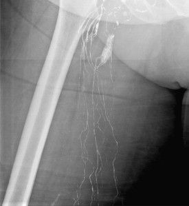
Initially a pedal lymphangiogram is performed to opacify the cisterna chyli and thoracic duct. This procedure was first described by Kinmonth and colleagues in 1955 and by others throughout the 1960s . Although slight variations have been proposed and used, the basic technique has changed little over the past 50 years.
Briefly, patients are fasted and given a suitable broad-spectrum antibiotic. A noncontrast CT scan of the abdomen is helpful in preliminary planning and may demonstrate particular hazards, such as aortic aneurysms, the position of the right renal artery, or prior surgical changes, that require a modification of the needle puncture approach. The cisterna chyli often can be identified with CT scan and with abdominal MRI .
The authors generally choose the right foot if only one side is to be accessed for lymphangiography. Bipedal lymphangiography sometimes is needed. The dermal foot lymphatics are opacified using an intradermal injection of methylene blue and after a small skin incision a lymphatic is cannulated with a lymphangiographic needle (Cook, Bloomington, Indiana).
Once the needle is secured, oil-based contrast (Ethiodol, Savage Laboratories) carefully is injected through a glass syringe at a rate of 7 to 10 μL per hour . Dosages above 20 μL total should be avoided because of pulmonary oil embolism syndrome . Patients who have pre-existing pulmonary disease should be treated with as low a dose as is feasible to obtain adequate opacification . The time to reach the retroperitoneal lymphatics may be 60 to 90 minutes ( Figs. 1 and 2 ). Fluoroscopic evaluation can be used to trace the flow of contrast to the cisterna chyli. Once the lower retroperitoneal lymphatics are identified, the abdomen should be prepped and draped in preparation for the percutaneous access of the cisterna chyli and thoracic duct ( Fig. 3 ).