Migraine and Other Headaches


Migraine and Other Headaches


Kathryn B. Freidl

B. Todd Troost

Mark L. Moster


When you’re lying awake With a dismal headache And repose is taboo’d by anxiety, I conceive you may use Any language you choose To indulge in without impropriety. “Iolanthe” by W.S. Gilbert

Headache is one of the most common presenting complaints for clinicians and encompasses a diverse spectrum of maladies. The International Headache Society (IHS) has revised their classification of headache diagnoses, and this is summarized in Table 16-1.1 This chapter addresses the neuro-ophthalmologic aspects of migraines and provides a brief review of other common headaches, facial and ocular pains.

Migraine is a periodic, paroxysmal, protean disorder whose prevalence varies by race and geographic region. In the United States, observed prevalence is lowest among Asian Americans, intermediate in African Americans, and highest among Whites, independent of demographic covariates.2 Similarly, an international meta-analysis found that the prevalence was lowest in Africa and Asia and higher in Europe and Central/South America.3 The highest estimates were found in North America.

In the United States, migraine affects an estimated 18% of women and 6% of men.4,5 It has been estimated that nearly one in four US households had someone with migraine.6 Higher income and education levels are associated with lower rates of migraine.2

One of the first names for this disorder was the descriptive term hemicrania; this was later contracted by the French in the 13th century to the word “migraine.” Although descriptions of migraine-like symptoms and conditions were found in Babylonian writings dating back to 3000 BC and in Egyptian papyri from 1550 BC, the first modern description of migraine and its possible causes is attributed to Thomas Willis about 300 years ago. Famous historical migraine sufferers included Julius Caesar, Emmanuel Kant, Alexander Pope, Isaac Newton, and Sigmund Freud. Throughout the 18th and 19th centuries, descriptions of the clinical phenomena and suggestions for therapy appeared in the writings of many prominent men in the medical professions. Edward’s masterful treatise On Megrim, Sick Headache, and Some Allied Disorders (1873, as detailed by Sacks) is an unequaled description of the disorder.7 Further detailed clinical descriptions are found in the writings of Gowers.8

In contemporary medicine, Dalessio, Goadsby, Raskin, Sacks, Silberstein, Lipton, Stewart, Saper, and Welch have all contributed significantly to the study of migraines. A constant tenet for all is that migraine cannot solely be defined by a unilateral (hemicranial) headache. “It is necessary to state that headache is never the sole symptom of a migraine, nor indeed is it the necessary feature of migraine attacks… Migraine is diagnosed by the entire history, not by physical findings or by the presence of headache alone.”7,9

Unfortunately, many have limited their concept of migraine to a stereotyped syndrome of visual disturbance followed by unilateral throbbing headache, which can be diagnosed by the response to ergot preparations. In actuality, migraine includes several well-recognized syndromes as well as a variety of “equivalents” less commonly classified as migraine. The symptom-complexes or syndromes of migraine include migraine without aura, migraine with aura, ophthalmoplegic migraine, retinal migraine, as well as the others listed in Table 16-1. The clinical features of migraine will be discussed according to the formal criteria published by the IHS in 2004.1

Other conditions and syndromes discussed in this chapter include cluster headache, trigeminal neuralgia, atypical facial pain, temporal arteritis, and the headaches produced by intracranial mass lesions, muscle contraction, trauma, vascular anomalies, and ocular lesions.


TABLE 16-1. New IHS Classifications of Headaches


1.


Migraine


1.1


Migraine without aura


1.2


Migraine with aura


1.3


Childhood periodic syndromes


1.4


Retinal migraine


1.5


Complications of migraine


1.6


Probable migraine


2.


TTH


2.1


Infrequent episodic TTH


2.2


Frequent episodic TTH


2.3


Chronic TTH


2.4


Probable TTH


3.


Cluster headache and other trigeminal autonomic cephalalgias


3.1


Cluster headache


3.2


Paroxysmal hemicrania


3.3


SUNCT


3.4


Probable trigeminal autonomic cephalalgia


4.


Other primary headaches


4.1


Primary stabbing headache


4.2


Primary cough headache


4.3


Primary exertional headache


4.4


Primary headache associated with sexual activity


4.5


Hypnic headache


4.6


Primary thunderclap headache


4.7


Hemicrania continua


4.8


New daily-persistent headache


5.


Headache attributed to head and/or neck trauma


5.1


Acute posttraumatic headache


5.2


Chronic posttraumatic headache


5.3


Acute headache attributed to whiplash injury


5.4


Chronic headache attributed to whiplash injury


5.5


Headache attributed to traumatic intracranial hematoma


5.6


Headache attributed to other head and/or neck trauma


5.7


Postcraniotomy headache


6.


Headache attributed to cranial or cervical vascular disorder


6.1


Headache attributed to ischemic stroke or transient ischemic attack


6.2


Headache attributed to nontraumatic intracranial hemorrhage


6.3


Headache attributed to unruptured vascular malformation


6.4


Headache attributed to arteritis


6.5


Carotid or vertebral artery pain


6.6


Headache attributed to cerebral venous thrombosis


6.7


Headache attributed to other intracranial vascular disorder


7.


Headache attributed to nonvascular intracranial disorder


7.1


Headache attributed to high CSF pressure


7.2


Headache attributed to low CSF pressure


7.3


Headache attributed to noninfectious inflammatory disease


7.4


Headache attributed to intracranial neoplasm


7.5


Headache attributed to intrathecal injection


7.6


Headache attributed to epileptic seizure


7.7


Headache attributed to Chiari malformation type I


7.8


Syndrome of transient headache and neurologic deficits with CSF lymphocytosis


7.9


Headache attributed to other nonvascular intracranial disorder


8.


Headache associated with a substance or its withdrawal


8.1


Headache induced by acute substance use or exposure


8.2


MOH


8.3


Headache as an adverse event attributed to long-term medication


8.4


Headache attributed to substance withdrawal


9.


Headache attributed to infection


9.1


Headache attributed to intracranial infection


9.2


Headache attributed to systemic infection


9.3


Headache attributed to HIV/AIDS


9.4


Chronic postinfection headache


10.


Headache attributed to disorder of homeostasis


10.1


Headache attributed to hypoxia


10.2


Dialysis headache


10.3


Headache attributed to arterial hypertension


10.4


Headache attributed to hypothyroidism


10.5


Headache attributed to fasting


10.6


Cardiac cephalalgia


10.7


Headache attributed to other disorder of homeostasis


11.


Headache or facial pain attributed to disorder of cranium, neck, eyes, ears, nose, sinuses, teeth, mouth, or other facial or cranial structures


11.1


Headache attributed to disorder of cranial bone


11.2


Headache attributed to disorder of neck


11.3


Headache attributed to disorder of eyes


11.4


Headache attributed to disorder or ears


11.5


Headache attributed to rhinosinusitis


11.6


Headache attributed to disorder of teeth, jaws, or related structures


11.7


Headache or facial pain attributed to a temporomandibular joint disorder


11.8


Headache attributed to other disorder of cranium, neck, eyes, ears, nose, sinuses, teeth, mouth, or other facial or cervical structures


12.


Headache attributed to psychiatric disorder


12.1


Headache attributed to somatization disorder


12.2


Headache attributed to psychotic disorder


13.


Cranial neuralgias and central causes of facial pain


13.1


Trigeminal neuralgia


13.2


Glossopharyngeal neuralgia


13.3


Nervus intermedius neuralgia


13.4


Superior laryngeal neuralgia


13.5


Nasociliary neuralgia


13.6


Supraorbital neuralgia


13.7


Other terminal branch neuralgias


13.8


Occipital neuralgia


13.9


Neck-tongue syndrome


13.10


External compression headache


13.11


Cold-stimulus headache


13.12


Constant pain caused by compression, irritation, or distortion of cranial nerves or upper cervical roots by structural lesions


13.13


Optic neuritis


13.14


Ocular diabetic neuropathy


13.15


Head or facial pain attributed to herpes zoster


13.16


Tolosa-Hunt syndrome


13.17


Ophthalmoplegic “migraine”


13.18


Central causes of facial pain


13.19


Other cranial neuralgia or other centrally mediated facial pain


14.


Other headache, cranial neuralgia, central or primary facial pain


The International Classification of Headache Disorders. Published on behalf of the International Headache Society. Cephalalgia. 2004;24(suppl 1):1.


DESCRIPTION OF THE MIGRAINE ATTACK

Blau10 has divided the migraine attack into five phases: the prodrome, occurring hours or days before the headache; the aura, which come immediately before the headache; the headache itself; the headache termination; and the postdrome. Most sufferers experience more than one phase, but no one particular phase is required for the diagnosis of migraine.11


Prodrome

Premonitory phenomena occur in approximately 60% of migraineurs, often hours to days before the onset of headache. These phenomena can have psychological, neurologic, constitutional, and/or autonomic features. Psychological symptoms include depression, euphoria, irritability, restlessness, mental slowness, hyperactivity, fatigue, and drowsiness. Neurologic phenomena include photophobia, phonophobia, and hyperosmia. The generalized or constitutional symptoms include a stiff neck, a cold feeling, sluggishness, increased thirst, increased urination, anorexia, diarrhea, constipation, fluid retention, and food cravings. Some patients just report a poorly characterized premonition that a migraine attack is coming.


Aura

An aura refers to the appearance of focal neurologic symptoms that precede or even accompany an attack of migraine. Approximately 20% of migraine sufferers experience auras. Most aura symptoms develop over a course of 5 to 20 minutes and usually last less than 60 minutes. The aura can be characterized by visual, sensory, or motor phenomena and may also involve language or brain stem disturbances. When a headache follows, it most often occurs within 60 minutes of the end of the aura. Isolated aura without headache is termed migraine dissociée. The most common aura is visual, previously termed classic migraine. It usually has a distribution in the right or left homonymous hemifields. Sensory disturbances involve one side of the body and are described as numbness or tingling on the face and in the hand. Further neurologic symptomatology is discussed under the heading of migraine with prolonged aura and migrainous infarction.


Headache Phase

The typical migraine headache is unilateral and described by most patients as throbbing.12 However, it should be noted that the pain may be bilateral and constant at first and become throbbing later and that a throbbing headache is described in other types of headache as well.13 The pain of migraine is almost always accompanied by other features such as anorexia. Nausea occurs in up to 90% of patients and vomiting occurs in approximately one-third of migraineurs.12

Many patients experience photophobia, phonophobia, and osmophobia, and seek seclusion in a dark, quiet room. Additional generalized symptoms include blurry vision, nasal stuffiness, anorexia, hunger, tenesmus, diarrhea, abdominal cramps, polyuria (followed by decreased urinary output after the attack), facial pallor (or, less commonly, redness), sensations of heat or cold, and sweating.11 Localized edema of the scalp, the face, or the periorbital regions may occur; tenderness may occur and be particularly prominent. There may also be tenderness of the scalp, a special prominence of a vein or artery in the temple, or stiffness/tenderness of the neck. Impaired concentration is common; memory impairment occurs less frequently. Depression, fatigue, anxiety, nervousness, and irritability are common. A sensation of faintness may be experienced, and syncopal migraines have been described.14 The IHS selects particular associated features as cardinal manifestations for diagnosis.


Termination and Postdrome

In the termination phase, the pain diminishes. Thereafter the patient may be “postictal”: listless, tired, or “washed out” for 24 to 48 hours. Rarely patients feel unusually refreshed or euphoric after an attack—it is more common to note depression and malaise.11


GENETICS IN MIGRAINE

Migraines frequently run in families, strongly suggesting that hereditary factors are involved.15,16 However, a polygenic, multifactorial inheritance coupled with environmental factors has hindered efforts to pinpoint causative mutations. Analysis of the monogenic familial hemiplegic migraine (FHM), a rare subtype of migraine with aura, has provided some insight into the origin of the migraine. FHM is an effective model for the common forms of migraine because, apart from the hemiparesis, the headache and aura features are identical.17 Furthermore, two-thirds of FHM patients also have attacks of common nonhemiplegic migraine.18

Thus far there are three identified FHM genes, each of which can be produced from various amino acid sequence mutations. The first gene identified, FHM1, was the CACNA1A gene located on the chromosome 19p13, which codes for the pore-forming subunit Cav2.1 of P/Q-type sodium channels.19 The FHM2 gene is ATP1A2, located at 1q21-31 gene, which encodes the Na+/K+-ATPase a2 subunit.16 The final known FHM gene (FHM3) is SCN1A encoding the voltage-gated sodium channel gene on chromosome 2q24.20

The FHM genes appear to cause increased levels of glutamate and potassium in the synaptic cleft. This in turn may lead to an increased propensity for cortical spreading depression (CSD), a self-propagating depolarization of neurons followed by a long-lasting inhibition of spontaneous and evoked neuronal activity21 As will be discussed later, there is a considerable body of clinical evidence that CSD is the likely basis of the migraine aura.


PATHOPHYSIOLOGY

Migraine is a neurovascular process.22 It appears to be a polygenetic “channelopathy” of ion channels in the brain stem nuclei that modulate sensory input.23 An abnormal fluctuation of ions leads to electrophysiologic changes that subsequently cause changes in cerebral blood flow (CBF). Initially, there is a brief decrease in CBF, followed by a rebound vasodilation and increased CBF for several minutes, then finally an extended period of decreased blood flow (several hours).24 The initial electrophysiologic changes, termed CSD, are the likely basis of migraine aura.25,26 The headache was initially thought to be the solely result of painful vasodilation, but it is now thought to also be caused by activation of the trigeminovascular system (TGVS). The TGVS is superficial cortical and meningeal blood vessels innervated by the trigeminal nerve. It projects to the trigeminal caudal nucleus in the brain stem, which in turn projects to higher order pain centers, causing headache.24 It is unclear if CSD also plays a role in activation of the TGVS.

The role of vascular phenomena in the migraine headache was recognized largely by the work of Wolff and colleagues.27 Research suggests that the migraine attack starts as a neuronal phenomenon with secondary hemodynamic consequences.28,29,30,31,32 Wolff divided the migraine attack into four phases: preheadache, headache, late headache, and postheadache. The preheadache phase is characterized by the constriction of certain blood vessels that supply the brain. Then, the beginning of the headache phase is characterized by vascular dilatation, particularly involving branches of the external carotid such as the temporal, occipital, and middle meningeal arteries. Local tenderness of the scalp ensues, and the scalp vessels may become rigid. The nature of the headache then changes from a pulsatile type to a more constant dull ache. Alleviation of the early headache phase with vasoconstrictors (e.g., ergotamine) supports vasodilation as a source of pain. However, vasodilatation may occur without pain, and additional factors are involved in the production of the headache. Local tissue changes take place (e.g., vessel edema, scalp swelling, and conjunctival chemosis) that may continue after vasodilatation has ceased.

Various substances have causative roles in the production of large and small vessel dilatation as well as local tissue changes. Among the substances most frequently cited are the kinins (neurokinin and bradykinin), acetylcholine, histamine, serotonin, and reserpine. Migraine, then, may result from dysfunction of brain stem or diencephalic nuclei involved in nociceptive modulation of afferents from the trigeminal vascular system.28

Positron emission tomography has detected activation in the brain stem during attacks of migraine.33,34 Sicuteri35 hypothesized that the following sequence occurs: the initial event is a local release of catecholamines (with vasoconstriction and increased urinary excretion of vanillylmandelic acid); during subsequent reactive hyperemia serotonin is released (documented by plasma serotonin decrease36 and increased urinary 5-hydroxy indoleacetic acid37), presumably from platelets or mast cells, which sensitizes cranial pain receptors perhaps also affected by the kinins.

Additional evidence suggests that there are nervous system connections between the trigeminal ganglia and cerebral blood vessels, the TGVS.38 Trigeminovascular neurons and their peripheral unmyelinated nerve fibers contain the neurotransmitter peptide, substance P. Stimulation of this system by various mechanisms would cause the release of substance P, which is postulated to increase vascular permeability and dilate cerebral blood vessels. The role of this system in the generation of human vascular headache may account for the effects of hormones or other circulating substances that change the receptive field properties of trigeminal ganglion cells.

Individuals prone to chemically induced headaches from ingestion of tyramine, alcohol, phenylethylamine, monosodium glutamate, nitroglycerine, wine, or chocolate also experience spontaneous headaches.39 Extensive studies of the reactivity of blood vessels in migraine40 and CBF30,32,41,42 suggest that abnormal vasomotor responses may be present in patients with migraine between, as well as during, migraine attacks.

There are several lines of indirect evidence that suggest a relationship between serotonin and migraine, making the understanding of the pharmacology of serotonin important for understanding the pharmacology of the new serotonin agonist in migraine therapy11 The serotonin or 5-HT receptors consist of at least three distinct types of molecular structures: guanine nucleotide G proteincoupled receptors, ligand-gated ion channels, and transporters. At least five 5-HT1 receptor subtypes are present in humans. Headaches resembling migraine can be triggered by serotonergic drugs such as reserpine (a 5-HT releaser and depleter) and m-chlorophenylpiperazine (a serotonin agonist).43,44

Other metabolic and endocrine factors also influence migraine attacks. According to Friedman and Merritt,45 80% of pregnant women previously prone to migraines either lose the headaches or experience improvement. However, Callaghan46 found an increase in the severity of migraine in pregnancy. The use of oral contraceptives appears to increase the incidence and severity of migraine.47,48 Whitty et al.49 felt that migraine might be precipitated by withdrawal of progesterone, whereas Somerville50 found from a study of three women with regular menstrual migraine that their attacks were related to estradiol withdrawal rather than to decreasing levels of progesterone. Tyramine has also been invoked as a precipitating factor, especially in the so-called allergic migraine;51 however, only approximately 5% of migraine subjects notice headaches precipitated by food. However, some patients are unusually sensitive to chocolate or alcohol, particularly red wines. Recent therapeutic trials with dietary therapy designed to avoid hypoglycemia suggest that glucose and/or insulin metabolism may play a role in the generation of vascular headache.

The role of trauma in the production or exacerbation of a preexisting migrainous tendency is still incompletely defined. Many individuals experience vascular headaches of the common migraine type after even minor head trauma.52 A previously well-controlled migraineur can experience a recrudescence of prior symptomatology after a head trauma. Such exacerbations are usually short-lived with a return to the preinjury status in weeks to months. However, there are some patients who experience post-traumatic migraine headaches for years after a head injury. Other triggering events preceding migraine attacks include bright light, especially sunlight reflected from water, exercise or exertion, and high altitude. Vascular headache of the migraine type may also follow orgasm.53 The role of stress is less clear. It appears more likely that migraine headache follows a period of psychological stress than occurring during the time of stress.

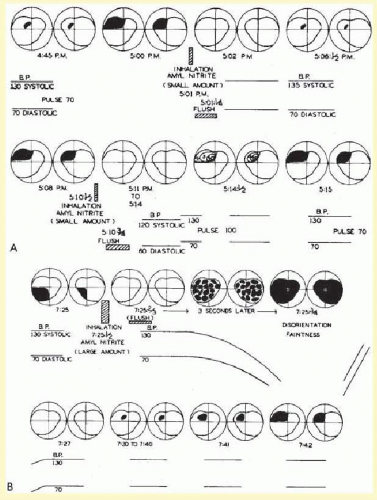
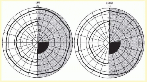
The pathophysiology of the migraine aura itself also has been studied extensively. Wolff showed that the use of a potent vasodilator, amyl nitrate, could abort the migraine scotoma (Fig. 16.1), supporting the vasoconstrictor hypothesis. Milner54 suggested that the scotomas of migraine and the neurophysiologic phenomenon, Leão spreading depression, may be related. The spreading depression progresses across the cortex at approximately 3 mm per minute, similar to the slow evolution of the visual phenomenon that had been detailed by Lashley,55 and estimated to spread over the occipital cortex at a rate of 3 mm per minute.

It is currently believed that the aura of migraine may be the human counterpart of the animal phenomenon of Leão spreading depression.18 The aura is characterized by a wave of decreased blood flow or oligemia passing across the cortex30,56,57,58 at a slow rate (2 to 6 mm per minute) consistent with the spread of the visual phenomenon through the visual cortex, as mentioned above.19 There is a short phase of hyperemia preceding the oligemia that may be a correlate of the scintillating scatoma, also a characteristic of migraine with aura.59 However, persistent oligemia is probably a response to depressed neuronal function and is present when the headache starts, as noted by Goadsby.58,60,61 Such findings coupled with the direct evidence of adequate local oxygen supply62 vitiate the theory that migraine is just a vascular headache.61


FIG. 16.1 A: Effect of inhalation of small amount of amyl nitrite on preheadache scotomas in migraine subject. The amount was insufficient to cause a drop in blood pressure or “light-headedness.” B: With inhalation of a large amount of amyl nitrite, a drop in pressure occurred with amblyopia and faintness. (From Wolff HG. Headache and Other Head Pain. 2nd ed. New York, NY: Oxford University Press; 1963, with permission.)

Three cardinal factors are important in the pathogenesis of migraine, according to Goadsby.61 These include the cranial blood vessels, the trigeminal innervation of these vessels, and the reflex connection of the trigeminal system with a cranial parasympathetic outflow. The pain sensitive structures within the cranium, such as large blood vessels or the dura mater, are innervated by branches of the ophthalmic division of the trigeminal nerve,63 and the posterior fossa structures are innervated by branches of C2 nerve roots.64 Involvement of the ophthalmic division of the trigeminal nerve and the overlap with structures innervated by C2 explain the common distribution of the pain of migraine in the frontal and temporal regions, as well as involvement of parietal occipital and high cervical regions, by referred pain.61 Peripherally, the trigeminal afferents are activated in migraine by the release of calcitonin gene-related peptide (CGRP) a vasodilator,65 and while the mechanism of pain generation is not entirely clear, animal studies suggest that pain is caused by a sterile neurogenic inflammation in the dura mater.66 This may, in part, explain the prevention of migraine pain by substances such as botulinum toxin type A, which inhibit the release of CGRP.67 The pain may be a combination of an altered perception—as a result of peripheral or central sensitization—of craniovascular input that is not usually painful61,68 and the activation of feed-forward neurovascular dilator mechanism that is functioning specific to the first ophthalmic division of the trigeminal nerve.69 Again, the effect of botulinum toxin type A in reducing migraine pain may interfere with this peripheral activation and, therefore, functions to provide peripheral desensitization.67

Eighty percent of migraineurs experience photophobia, or exacerbation of the headache by what is normally a tolerated level of light, during migraine attacks.70 This photophobia is not a product of the traditional image forming pathway of the visual system, for example, the photoreceptors, retinal ganglion cells (RGCs), lateral geniculate nucleus, and visual cortex.71

Instead, it is a nonimage forming system of vision that originates in intrinsically photosensitive RGCs (ipRGCs) that appears to be the origin of photophobia. These ipRGCs contain the photoreceptor melanopsin and function without any synaptic input from the rods and cones.71 Their peak spectral sensitivity lies in the blue range at approximately 480 nm.72 The axons of these ipRGCs project via the optic nerve to the suprachiasmatic nucleus, intergeniculate leaflet, and the olivary pretectal nucleus.72 It is through these projections that the melanopsin system mediates such nonimage forming visual functions like the pupillary light response and circadian rhythms. Both of circadian rhythm and pupillary light response are preserved in some blind patients. That is because the melanopsin-containing ipRGCs are spared in some blinding eye conditions, for example, mitochondrial optic neuropathies (Leber and dominant optic atrophy) and peripheral retinal degenerations (retinitis pigmentosa); however, they tend to be preferentially targeted in glaucoma.72


TABLE 16-2. Migraine Without Aura


1.1


Migraine without aura


Previously used terms: common migraine, hemicrania simplex


Description


Recurrent headache disorder manifesting in attacks lasting 4-72 h. Typical characteristics of the headache are unilateral location, pulsating quality, moderate or severe intensity, aggravation by routine physical activity and association with nausea and/or photophobia and phonophobia


Diagnostic criteria


A.


At least five attacks fulfilling criteria B-D


B.


Headache attack lasting 4-72 h (untreated or unsuccessfully treated)


C.


Headache has at least two of the following characteristics:


1.


Unilateral location


2.


Pulsating quality


3.


Moderate or severe pain intensity


4.


Aggravated by causing avoidance of routine physical activity (e.g., walking or climbing stairs)


D.


During headache at least one of the following:


1.


Nausea and/or vomiting


2.


Photophobia and phonophobia


E.


Not attributed to another disorder


Likewise, migrainous photophobia is preserved in some blind patients. Noseda et al.71 not only found that light modulated the activity of a subset of trigeminovascular thalamic neurons but also that most retinal projections to the thalamus consisted of axons of ipRGCs. They therefore posit that the melanopsin system carries the photic signal from the retina to the nociceptive thalamic TGVS, and thereby exacerbates migraine headache pain.


MIGRAINE WITHOUT AURA

The IHS classification has improved the diagnosis of headaches. It has also facilitated clinical research on migraine. In order to establish a diagnosis of migraine without aura, five attacks are needed (Table 16-2). Each attack must last 4 to 72 hours and have two of the following four pain characteristics: unilateral location, pulsating quality, moderate to severe intensity, and aggravation by routine physical activity. In addition, the attacks must be associated with at least one of the following: nausea, vomiting, or photophobia and phonophobia. With these criteria, no single characteristic is mandatory for a diagnosis of migraine. A patient who has severe pain aggravated by routine activity, photophobia and phonophobia, meets these criteria as does the more typical patient with unilateral throbbing pain and nausea.

Migraine usually lasts several hours or the entire day. When the migraine persists for longer than 3 days, the term “status migrainosus” is used. Frequency of attacks varies widely from a few per lifetime to several per week.11 The average migraineur experiences from one to three headaches per month.5 A precise location ascribed to migraine, such as unilateral or temporal, is misleading, for as Wolff40 wrote,


The sites of migraine are notably temporal, supraorbital, frontal, retrobulbar, parietal, postauricular, and occipital… They may as well occur in the malar region, in the upper and lower teeth, at the base of the nose, in the median wall of the orbit, in the neck, and in the region of the common carotid arteries and down as far as the tip of the shoulder.

The prodromes of common migraine are vague, preceding the attack by hours or days, and include psychic disturbances (such as depression or hypomania), gastrointestinal manifestations and changes in fluid balance. Usually the onset of the common migraine headache is unilateral, but the pain often becomes holocephalic. In an individual patient, the headache is commonly more prominent on a single side, with occasional or rare alternation. Some individuals always experience a unilateral headache, while in approximately one-third the headache is diffuse from onset. Traditionally, the character of the headache is described as throbbing, but this may be a feature only at onset, with the discomfort soon changing to a steady ache. The victim can often relieve unilateral headache by carotid artery or temporal artery compression, only to experience resurgence of the pain after release.

Nausea in some degree almost always accompanies common migraine. Vomiting can occur at the height of an attack, sometimes with relief of the headache, but more often heralds an intensifying phase of the episode, which continues for many minutes or hours. Usually the migraine sufferer becomes pallid and seeks seclusion, darkness, quiet, and a cold towel or ice bag for the head. Frequently at the time of nausea with vomiting, a diuretic phase with polyuria ensues, the consequence of fluid retention that occurred in the hours or days preceding the acute headache.

Ocular signs and symptoms may occur in common migraine, such as conjunctival injection, periorbital swelling, excessive tearing, foreign body sensation, and photophobia; however, these phenomena are more prominent in cluster headache.


MIGRAINE WITH AURA (CLASSIC)

The new term for classic migraine, that is migraine with aura, requires at least two attacks with any three of the following four features (Table 16-3): one or more fully reversible aura symptoms, aura developing over a course of more than 4 minutes, aura lasting less than 60 minutes, and headache after aura within 60 minutes. Migraine with aura refers to a more well-defined clinical constellation than does migraine without aura. The episodes are characterized by definite prodrome or aura, which is usually a visual sensation; however, sometimes motor or other sensory phenomena precede the headache. The headaches of classic migraine tend to be more compact and intense, rarely lasting more than 12 hours; most often 2 to 3 hours.

Many general characteristics are shared by common and classic migraine. Both varieties affect men and women and can occur at any age, often seemingly triggered by a significant event such as puberty, school graduation, or marriage. A family history is usually present both in classic and common migraine, and there may be an earlier history of colic as a baby or car illness as a small child. The full history of a complete migraineur would include migraine with aura in the teens, migraine without aura with nausea and vomiting in the second and third decades, followed by simple periodic headache or isolated migrainous auras in later life.

Migraine with aura is subclassified into migraine with typical aura (homonymous visual disturbance, unilateral numbness or weakness, or aphasia), migraine with prolonged aura (or lasting longer than 60 minutes), FHM, basilar migraine, migraine without headache, and migraine with acute-onset aura.

The primary feature of migraine with aura is the visual aura. Extensive reviews of this phenomenon are found throughout the literature.7,8,40,73,74,75 Although many variations occur, the following description by Richards50 summarizes the most common type of visual phenomena (Figs. 16.2 and 16.3):


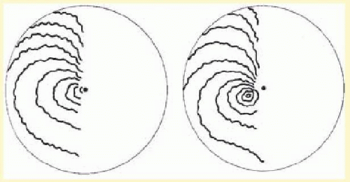
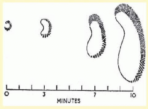
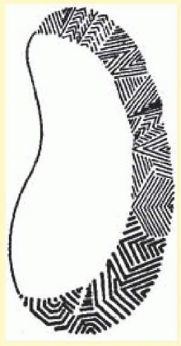
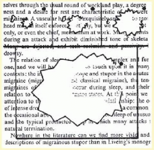
The visual disturbance usually precedes the headache … [it] begins near the center of the visual field as a small gray area with indefinite boundaries. If this area first appears during reading, as it often does, then the migraine is first noticed when words are lost in a region of “shaded darkness.” During the next few minutes the gray area slowly expands into a horseshoe with bright zigzag lines appearing at the expanding outer edge. These lines are small at first and grow as the blind area expands and moves outward toward the periphery of the visual field.

One important aspect of the visual disturbance just described is that it expands slowly over 10 to 20 minutes. The initial region of visual abnormality is most often near fixation and then, as described by Lashley,55 with increase in size the disturbed area moves or “drifts” across the visual field so that its central margin withdraws from the macular region as its peripheral margin invades the temporal; the area may be totally blind (negative scotoma), amblyopic, or outlined by scintillations.


TABLE 16-3. Migraine with Aura


1.2 Migraine with aura


Previously used terms: classic or classical migraine; ophthalmic, hemiparesthetic, hemiplegic, or aphasic migraine, migraine acompania, complicated migraine


Description


Recurrent disorder manifesting in attacks of reversible focal neurologic symptoms that usually develop gradually over 5-20 min and last for <60 min. Headache with features of migraine without aura usually follows the aura symptoms. Less commonly, headache lacks migrainous features or is completely absent


Diagnostic criteria


A.


At least two attacks fulfilling B


B.


Migraine aura fulfilling criteria B and C for one of the subforms 1.2.1-1.2.6


C.


Not attributed to another disorder*


1.2.1 Typical aura with migraine headache


Description


Typical aura consisting of visual and/or sensory and/or speech symptoms. Gradual development, duration no longer than 1 h, a mix of positive and negative features and complete reversibility characterize the aura which is associated with a headache fulfilling criteria 1.1 Migraine without aura


Diagnostic criteria


A.


At least two attacks fulfilling criteria B-D


B.


Aura consisting of at least one of the following, but no motor weakness:


1.


Fully reversible visual symptoms including positive features (e.g., flickering lights, spots or lines) and/or negative features (i.e., loss of vision)


2.


Fully reversible sensory symptoms including positive features (i.e., pins and needles) and/or negative features (i.e., numbness)


3.


Fully reversible dysphasic speech disturbance


C.


At least two of the following:


1.


Homonymous visual symptoms and/or unilateral sensory symptoms


2.


At least one aura symptom develops gradually over ≥5 min and/or different aura symptoms occur in succession over ≥5 min


3.


Each symptom last ≥5 and ≥60 min


D.


Headache fulfilling criteria B-D for 1.1 Migraine without aura within 60 min


E.


Not attributed to another disorder


* History and physical and neurologic examinations do not suggest any of the disorders listed in groups 5-12, or history and/or physical and/or neurologic examinations do suggest such disorder but it is ruled out by appropriate investigations, or such disorder is present but attacks do not occur for the first time in close temporal relation to the disorder.

Additional loss or blurring of central vision may occur.

History and physical and neurologic examinations do not suggest any of the disorders listed in groups 5-12, or history and/or physical and/or neurologic examinations do suggest such disorder, but it is ruled out by appropriate investigations, or such disorder is present but attacks do not occur for the first time in close temporal relation to the disorder.


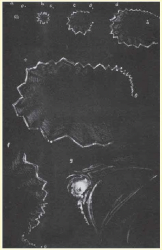
The scintillations surrounding the negative scotoma make “fortification” figures or spectrums, so called by the appearance of a “map of the bastions of a fortified town.”55 The scintillations are brilliant, with the intensity of a bright fluorescent bulb flickering at a rate of 5 to 10 cycles per second (Figs. 16.4 and 16.5).

Gowers
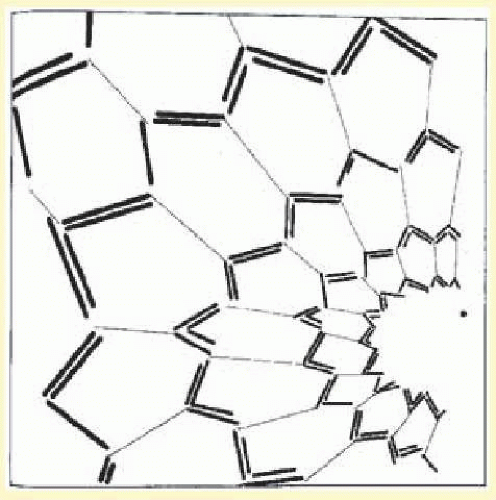
76 was particularly impressed with the intensity of the visual sensation. Many migraine sufferers can precisely recall their own vivid visual experiences well enough to describe them precisely or even on occasion to paint them (Figs. 16.6 and 
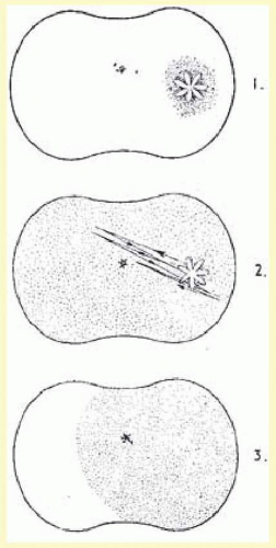
16.7). Not all migraine visual disturbances begin near the fixation point; some patients consistently experience scotomas starting eccentrically in the visual field, and these sensations can appear alternately or simultaneously in both hemifields (Fig. 16.8). Other less dramatic visual auras also occur: just the sensation of peripheral brightness or awareness of a rhythmicity or pulsating character in the intensity of the ambient light. The duration of these visual symptoms is measured in minutes rather than the brief few seconds of flashing, bright moving spots, or transient flickering phenomena characteristic of occipital epileptic discharges.9,76 Additional visual disturbances are categorized by Klee and Willanger,74 consisting of metamorphopsia, diplopia, polyopia, and apparent movement of stationary objects. Variations in the scotomas of migraine, including their occurrence in patients with acquired blindness, are well described.74,77 The auras of migraine, although most commonly only visual, have many other associated manifestations, such as hemihypesthesias, perioral anesthesia, vertigo, and transient aphasia. The aura or prodromes of classic migraine may be precipitated by intense stimuli: bright lights, loud noises, head trauma, or the intake of certain foods in susceptible individuals.


FIG. 16.2 Successive arcs expand across half of visual field, as shown in two diagrams based on airy. The spectra may take 20 to 25 minutes to expand from a fuzzy gray area near the fixation point (dot) to the outer limit of the visual field. (From Richards W. The fortification illusions of migraines. Sci Am. 1971;224:88, with permission.)


FIG. 16.3 Emerging honeycomb pattern from plotting data derived from visual phenomena in migraine subjects. Honeycomb and tendency for inner angle between lines to approximately 60° suggest a hexagonal organization of occipital cortical cells. (From Richards W. The fortification illusions of migraine. Sci Am. 1971;224:88, with permission.)


FIG. 16.4 Successive maps of a scintillating scotoma to show characteristic distribution of the fortification figures. (Modified from Lashley KS. Patterns of cerebral integration indicated by scotomas of migraine. Arch Neurol Psychiatry. 1941;46:333. Copyright © 1941, American Medical Association.)


FIG. 16.5 Variations in fortification figures. Coarser and more complicated figures are generally in lower part of field. (From Lashley KS. Patterns of cerebral integration indicated by scotomas of migraine. Arch Neurol Psychiatry. 1941;46:333. Copyright © 1941, American Medical Association.)

In the usual sequence of migraine with aura the sensory prodrome precedes the onset of the headache (in accord with the traditional concept of vasoconstriction followed by vasodilatation). The visual disturbance rarely may have a simultaneous onset with headache or, once having disappeared, may recur following the onset of headache. Such unusual patterns, or strict unilaterality for all attacks, should increase suspicion of a mass lesion or vascular malformation. As opposed to definite periodicity with symptom-free intervals and predictable circumstances, as in migraine without aura, migraine with aura may occur “out of the blue” and in multiple attacks over a few days.

Migraine-with-aura attacks tend to diminish in the third and fourth decades. Although most migraine patients experience a stereotyped clinical pattern, there is a well-recognized group in which both classic and common migraine attacks are admixed.78 Some patients with classic migraine may lose the headache component eventually and suffer only isolated auras thereafter. This monosymptomatic pattern stresses the importance of accurate history-taking when confronted by a patient with isolated visual phenomena (migraine dissociée). Haas79 emphasized the occurrence of “migraine aura status.” The differential diagnosis should include consideration of vertebro-basilar transient ischemic attacks. Symptomatology that favors migraine has been reviewed by Fisher80 and includes luminous visual images, build-up of images, progression from one aura to another, and benign outcome.


FIG. 16.6 Left-sided fortification spectrum of migraine. Illustration by Dr. Hubert Airy of his own scotomas. A bright stellate object (A) appeared suddenly below and to the left side of fixation (o). It rapidly enlarged, first as a circular zigzag, but on the inner side the zigzag was faint (B); as arc increased in size, it was broken centrally (C). In (D), original circular outline had become oval. Rectangular lines that made up the fortification spectrum became longer as the process extended peripherally. When spectrum had extended through greater portion of the field (E), upper portion also began to expand (F). At this time, the lower part of spectrum disappeared. The phenomenon ended in a whirling focus of light (G) 20 minutes after it began. At this time, a headache appeared on the right side. (From Gowers WR. Visual sensations in migraine. In: Subjective Sensations of Sight and Sound: Abiotrophy and Other Lectures. London, UK: Churchill; 1907, with permission.)


FIG. 16.7 Inhibitory character within angled oval of an expanding fortification spectrum. Outside the limiting line, vision is preserved; within it, vision is lost. This occurs at first over the whole area; afterwards, when the sphere is broken and has become oval, loss is most intense close to the limiting line and becomes less toward the middle. (From Gowers WR. Visual sensations in migraine. In: Subjective Sensations of Sight and Sound: Abiotrophy and Other Lectures. London, UK: Churchill; 1907, with permission.)


MIGRAINE WITH PROLONGED AURA AND MIGRAINOUS INFARCTIONS

In the new classification of migraine,1 subtype 1.6 indicates complications of migraine. This would include all of the permanent defects discussed in this section.

Focal symptoms and signs of the aura may persist beyond a headache phase. In the previous classification, this was termed complicated migraine. It is now defined by the IHS classification with two labels that increase specificity. If the aura lasts for longer than 1 hour but less than 1 week, it is termed migraine with prolonged aura. If the signs persist for more than 1 week or a neuroimaging procedure demonstrates a stroke, a migrainous infarction has occurred. As pointed out previously, in mid or later life the aura may occur independent of a headache and has been termed migraine accompagnée or migraine associée. Migraine with aura (classic) in early reports was sometimes referred to as “ophthalmic migraine” (to be differentiated from ophthalmoplegic migraine, a subtype of migraine with aura). Migraine with aura is further reviewed in sections Cerebral, Ophthalmoplegic, Retinal, Basilar, and Other Varieties.


FIG. 16.8 Radial movement of a visual stellate object that itself remained unchanged throughout the episode. Stellate form appeared near edge of right half of field just below the horizontal and consisted of approximately six pointed leaf-like projections alternately red and blue. It appeared on a small area of darkness, moved slowly toward the left and upward, passing above the fixation point to beyond the middle line. Then it returned to its starting place, retraced this path once or twice and passed to the right edge of the field, suddenly disappearing at the spot where it began. (From Gowers WR. Visual sensations in migraine. In: Subjective Sensations of Sight and Sound: Abiotrophy and Other Lectures. London, UK: Churchill; 1907, with permission.)


Cerebral

Various cerebral symptoms may occur in migraine with aura, including motor, visual, and other sensory defects. As pointed out previously, if the aura lasts for more than 1 hour but less than 1 week, it is called migraine with prolonged aura. However, if the signs persist for more than 1 week or a neuroimaging procedure shows a stroke, it is termed as migrainous infarction. The IHS classification of migraine-related stroke is presented in Table 16-4. Welch29 has classified migraine-related stroke into four subtypes. These are described briefly.


1. Coexisting stroke and migraine

A clearly defined clinical stroke syndrome must occur remotely in time from a typical migraine attack. Stroke in the young is rare; in contrast, migraine is common. According to Welch, the two conditions should coexist without migraine being a contributing risk factor for stroke.


2. Stroke with clinical features of migraine

A structural lesion that is unrelated to migraine pathogenesis presents with clinical features of a migraine attack. Subtype A is symptomatic migraine, and in these patients, established structural central nervous system (CNS) lesions or cerebral vessels cause episodic symptoms typical of migraine with neurologic aura, although infrequently. Cases of cerebral arteriovenous malformation (AVM) exemplify this concept and may masquerade as migraine with aura.9,81

Welch’s second subtype is a migraine mimic. In this category, stroke caused by acute and progressive structural disease is accompanied by headache and a constellation of progressive neurologic signs and symptoms. These situations are difficult to distinguish from those of migraine, hence the term migraine mimic. The diagnosis can be most difficult in patients who continue to have migraine late in life, when the incident of cerebrovascular disease increases.29


TABLE 16-4. Classification of Migraine-Related Stroke


1.5.4 Migrainous infarction


Description


One or more migrainous symptoms associated with an ischemic brain lesion in appropriate territory demonstrated by neuroimaging


Diagnostic criteria


A.


The present attack in a patient with 1.2 Migraine with aura is typical of previous attacks except that one or more aura symptoms persists for >60 min


B.


Neuroimaging demonstrates ischemic infarction in a relevant area


C.


Not attributed to another disorder


Comments


Ischemic stroke in a migraine suffer may be categorized as cerebral infarction or other cause coexisting with migraine, cerebral infarction of other cause presenting with symptoms resembling migraine with aura, or cerebral infarction occurring during the course of a typical migraine with an aura attack. Only the last fulfills criteria for 1.5.4 Migrainous infarction


Increased risk for stroke in migraine patients has been demonstrated in women under age 45 in several studies. Evidence for an association between migraine and stroke in older women and in men is inconsistent


3. Migraine-induced stroke

Migraine-induced stroke must meet the following criteria: the neurologic deficit must be identical to the migraine symptoms of previous attacks; the stroke must occur during the course of a typical migraine attack; and all other causes of stroke have been excluded, although stroke risk factors may be present.


4. Uncertain classification

Welch recognized that many migraine-related strokes cannot be categorized with certainty. For example, the IHS definition of migraine-induced stroke does not prevent the diagnosis in patients with migraine without aura. In addition, there are occasional cases of migraine-like symptoms and persistent neurologic deficit associated with cerebrospinal fluid (CSF) protein and pleocytosis.82,83 Other rare syndromes and migraine-related strokes include migraine associated with mitochondrial encephalopathies84 and “migraine coma.”85 Intracerebral hemorrhage has been reported,86 with most cases being migraine mimics according to Welch’s classification. Whether there is a role of antiphospholipid antibodies is still to be determined.87 Permanent homonymous visual field defects have been well documented in migraine patients.75,88,89,90 The defects almost always occur in patients who have previously had migrainous attacks with transient scintillating scotomas. Computed cranial tomography (CT) or magnetic resonance imaging (MRI) has now documented several cerebral infarctions usually in the occipital and parietal regions. Rothrock and colleagues91 evaluated 22 patients with migraine-associated stroke finding that 91% were women and 23% had a history of presumed migrainous stroke. They concluded that extracranial and intracranial vasospasm played a major role in some cases that they were able to document angiographically. One controlled study of migraine with aura reported that 91% of patients who had stroke during an attack had no arterial lesions. This was as opposed to 9% of migraine with patients with aura who suffered stroke remote from a migraine attack and 18% of patients with stroke without a migraine history.92 In a rigorous case controlled study, no overall association between migraine and ischemic stroke was found, but among women younger than 45 years, migraine and stroke were significantly associated; the risk was increased 4-fold, and it became even greater in women who smoked.93

According to Hollenhorst,94 approximately 4% of patients who have a typical sequence of visual aura followed by hemicranial headache experience transient hemianopia lasting up to 15 minutes. Much rarer are patients with permanent hemianopia. Bilateral upper quadrantic defects have been reported.95,96

Other sensory disturbances such as paresthesias particularly involve hands, fingers, and lips.95,96,97 Various aspects of cerebral migraine with aura are illustrated by the following case history.


FIG. 16.9 Visual field defect in complicated migraine. Defect cleared completely. Visual acuity 20/20 OU. Relative homonymous hemianopia (2, 5/1,000 W; scotoma out to 30/1,000 W).


A 26-year-old woman was seen with a complaint of difficulty with vision. She had a history of migraine since the age of 12, characterized by an aura of “black spots” slowly spreading over the field of vision for 20 minutes, occasionally accompanied by numbness in the right hand and arm. Thereafter throbbing headache would occur that was left-sided 90% of the time. Her father had a history of classic migraine as a young man. Ten days prior to first being seen, she had a typical attack of migraine but with persistent difficulty in vision after the episode. Examination was entirely normal except for a congruous right homonymous visual field defect (Fig. 16.9). Ten days later she again developed her visual aura, but with a moderate right hemiparesis as well, during her headache. Brain scan and cerebral arteriography were normal. During the next week all neurologic abnormality including her visual field defect cleared completely.

Frequently a disturbance of language occurs with migraine. This was described by Sir George Airy in 1865, who related his own inability to speak during an attack.


While reading, I become aware that I am unable to understand what I have just read. After rereading a paragraph two or three times, I begin to realize that I cannot understand the sense of words. The letters can be identified but the words are unintelligible … At this point a numb feeling occurs in my right hand and I finally realize that I am at the start of another migraine attack.

A wide variety of language difficulties, sensory defects, and motor abnormalities have been described (most often transient but rarely permanent) from presumed cerebral infarction. Caplan et al.98 reported 12 patients with transient global amnesia and prior migraine. In three patients, the classic migrainous phenomenon accompanied the amnestic attack.

Variable electroencephalographic (EEG) findings occur.97,99,100 There is a general lack of agreement as to the incidence and significance of abnormalities in the EEGs of patients with migraine. Some authors report a normal EEG,101 but various abnormal patterns have been recorded.99,100 A detailed review by Hockaday and Whitty101 indicated an incidence of EEG abnormality in 61% of 560 migraine patients. The highest frequency of abnormality occurred in patients with transient lateralized motor or sensory auras.

Friedman102 first reported the results of angiography during an attack of migraine; there were no abnormalities. Although the majority of patients in reported cases show normal arteriograms, others have demonstrated some abnormality during an attack (see Cluster Headache). However, angiography is considered by some to have increased risk in patients with migraine75,103 and has not yet provided useful information on the pathophysiology of complicated migraine.104,105,106

Hemiplegic migraine occurs both sporadically and as a familial syndrome. This entity is defined as a “vascular headache” featuring sensory and motor phenomena that persist during and (for a brief time) after the headache.107 A narrower view would be to use the term hemiplegic migraine when only motor involvement (i.e., weakness or paralysis) occurs.

The first mention of transient hemiparesis during an attack of migraine was by Liveing,7 and multiple reviews and case reports have appeared since.108,109 Heyck110 reviewed the neurologic complications of 980 of 3,890 patients with migraine. The majority of these patients complained of unilateral tingling or numbness that invariably involved the hand and sometimes spread to the arm, face, tongue, and, rarely, to the leg. The symptoms seldom lasted more than 30 minutes and could occur before or at the peak of the headache. Twelve of these patients had unilateral motor disturbances ranging from minimal loss of function to complete paralysis. There have been few permanent sequelae attributed to hemiplegic migraine; progressive dementia was noted by Symonds, and permanent hemiplegia has been reported.97,104,110

Reports of hemiplegic migraine in the literature seem to indicate that most cases are familial. However, Heyck104 pointed out the tendency to report familial cases; most cases do not occur in families with hemiplegic migraine but rather in “families with ordinary migraine as often as common or classic migraine.” FHM is well documented,108,110,111,112 at times concomitant with associated neuro-ophthalmologic findings such as retinal degeneration and nystagmus.113 One interesting report is that of Dooling and Sweeney,114 who describe a blind woman whose attacks were precipitated by breast feeding her infant. This led to the speculation that oxytocin (chemically similar to ergotamine) could exercise a complex effect on cerebral vessels predisposed to vasospasm.


Ophthalmoplegic

So-called ophthalmoplegic migraine has now been changed in the official Classification of Headache of the International Headache Society.1 This is because many reports have shown abnormalities of the oculomotor nerve using MRI in children with recurrent painful ophthalmoplegia fulfilling the previous criteria for ophthalmoplegic “migraine.”115,116 It is now believed that all modern cases show MRI enhancement of the third nerve, which may represent a type of inflammatory cranial neuropathy and, therefore, is more characteristic of what is seen with facial nerve palsy than with any form of migraine.117 In this rare variety of what was formerly termed “complicated migraine,” the headaches were associated with oculomotor nerve palsies.118,119 Usually the ophthalmoplegia is transient, however, it can become permanent especially after repeated attacks. Major controversy has surrounded the diagnostic and nosologic position of ophthalmoplegic “migraine” since its initial recognition in the mid-1880s. Until the 1930s and 1940s when angiography was introduced and practiced, it was impossible in many cases to rule out aneurysms and other lesions in the vicinity of the cavernous sinus. Multiple etiologies were cited as underlying causes of ophthalmoplegic “migraine” including aneurysm, basilar arachnoiditis, and tumors; indeed, many physicians believed that no separate clinical syndrome of ophthalmoplegic migraine existed but that all patients had specific organic lesions.

Carlow described125 reported cases of ophthalmoplegic migraine with MRI abnormalities at the midbrain exit. Those who received contrast all showed enhancement during the acute phase (20 patients), and all imaged with contrast in the quiescent phase (17 cases) demonstrated residual enhancement at the nerve’s exit from the brain stem. The patients who underwent noncontrast imaging showed focal thickening in the same area in both active and quiescent phases.118

In ophthalmoplegic “migraine” the third nerve is most frequently involved. Abducens palsy occurs with one-tenth the frequency of third-nerve palsy, and even rarer is affliction of the fourth nerve.75 In most cases, positive family history is not present. The typical clinical syndrome is a child or young adult with periodic headache has ophthalmoplegia involving all functions of the third nerve, beginning at the height of an attack of cephalgia, which is primarily unilateral and in the orbital region; the paresis lasts for days to weeks after the cessation of headache; recovery is gradual and tends to be less complete after repeated attacks. Below is an exemplary case report.


A 3-year-old boy presented with a complete left oculomotor palsy. The day before he had complained of headache was lethargic and went to bed early. The following morning he awakened with complete ptosis of the left upper lid, but his headache was gone. On examination, the left pupil was 6 mm and slightly reactive to light; all muscles supplied by the left third nerve were profoundly affected (Fig. 16.10). The neurologic examination and plain skull X-rays were entirely normal. The child recovered completely in 3 weeks’ time. An exactly similar episode occurred 20 months later, also with rapid spontaneous resolution, and a third episode occurred 1 year after that. The child is now well and suffers only occasional headaches.

The differential diagnosis of this condition should include aneurysm, tumor, diabetes, and sphenoid sinus mucocele. The age at onset, a negative glucose tolerance test, and radiologic studies will usually rule out these listed possibilities. Other clinical entities confused with ophthalmoplegic “migraine” include myasthenia gravis and the Tolosa-Hunt syndrome. Myasthenia is ruled out if the pupil is involved (and actually should not be considered in the presence of pain) and with response to edrophonium chloride (Tensilon); Tolosa-Hunt should be considered if the pain persists. On rare occasions only limited involvement of the third nerve occurs.


FIG. 16.10 Third-nerve paresis in 3-year-old boy with ophthalmoplegic migraine. Pupil was sluggishly reactive to light. Note failure of elevation, abducted position of left eye, and ptosis.

Reports of transient, otherwise unexplained unilateral pupillary mydriasis have been tentatively attributed to migraine in young patients.120,121 One should be careful to exclude intermittent angle-closure glaucoma in cases of mydriasis. Sarkies and colleagues122 reported a 31-year-old man with an 18-month history of episodic periorbital pain who, during an attack, noted blurred vision and a dilated pupil. He was found to have a sector palsy of the upper nasal quadrant of the left iris, an intraocular pressure of 16 mm Hg between attacks and on gonioscopy, and narrow angle with a plateau-type iris. During an attack his intraocular pressure increased to 26 mm Hg. After a provocative dark-room test, the patient developed a typical headache and was found to have an intraocular pressure of 45 mm Hg and a closed-angle on gonioscopy.

Rarely, ophthalmoplegic “migraine” may occur without headache. Durkan et al.123 described two children with isolated recurrent painless oculomotor palsy in whom neurodiagnostic investigations were all normal.

Concern should be raised in (1) the absence of a migraine history, (2) severe persistent headache with total ophthalmoplegia, (3) onset after age 20, and (4) symptoms and signs of subarachnoid hemorrhage. However, ophthalmoplegic “migraine” is a diagnosis of exclusion, and noninvasive imaging tests such as MRI or magnetic resonance angiography (MRA) should be performed in all cases to exclude the possibility of aneurysm.119 The finding of an entirely normal MRI, except for nerve enhancement, in a child with a third cranial nerve palsy after a 4-day history of headache, who is otherwise well, should complete the workup; aneurysmal third-nerve palsies are extremely rare in children under age 14.124 However, in third-nerve palsy involving pupillomotor function, serious consideration should be given to angiography. The usual cause will be a posterior communicating artery aneurysm (PCom A aneurysm). Newer techniques such as MRA or CT angiography (CTA) should detect nearly all aneurysms; however, reliable detection is also dependent on the training and accuracy of the interpreting radiologist.125

Only gold members can continue reading. Log In or Register to continue

Stay updated, free articles. Join our Telegram channel

Jul 11, 2016 | Posted by in OPHTHALMOLOGY | Comments Off on Migraine and Other Headaches

Full access? Get Clinical Tree

Get Clinical Tree app for offline access