Management of Posterior Segment Trauma


Management of Posterior Segment Trauma


James M. Osher

Eric D. Weichel

Michael M. Lai

Tarek S. Hassan


Ocular trauma is a significant cause of visual impairment in the United States. Approximately 2 million eye injuries occur annually, resulting in an estimated $200 million per year of hospital charges.1, 2 Worldwide, 2.3 million people have bilateral visual impairment due to ocular trauma, while an additional 19 million cases of monocular blindness (20/200 or less) are due to eye injuries.3 Vision is lost because of primary mechanical damage to vital ocular structures and secondary complications, such as infectious endophthalmitis and retinal detachment due to intraocular fibrous proliferation and contracture. Contemporary vitreoretinal surgical techniques play a vital role in the prevention and management of secondary complications of ocular trauma. The appropriate assessment and management of such cases are essential to preserving the eye and restoring vision.

Several technologic advances have facilitated the repair of complex traumatic eye injuries. Improved posterior segment visualization via wide-angle contact and noncontact viewing systems assist the repair of peripheral, anterior pathology. Silicone oil, heavy liquids, and intraocular gases serve as valuable surgical adjuncts in the repair of complex traumatic retinal detachments, and surgical instrumentation continually evolves. High-speed vitreous cutters and sutureless transconjunctival small-incision surgery have changed the face of vitreoretinal surgery, and are vital tools for the acute and secondary management of nonpenetrating and penetrating ocular trauma.

We describe the role of vitreoretinal surgery in the management of lens subluxation or dislocation, vitreous hemorrhage, retinal detachment, intraocular foreign bodies (IOFBs), traumatic macular hole, and posttraumatic endophthalmitis. Particular attention is paid to the mechanisms of acute and chronic traumatic injury, diagnostic evaluation, timing of surgical intervention, surgical principles, and results of current therapeutic modalities.

In 1996, a consortium named the Ocular Trauma Classification group created a standard terminology for ocular trauma to allow for improved diagnosis, study, and communication about trauma.4 The salient features are detailed in Table 66-1.


TABLE 66-1 Ocular Trauma Terminology


Term


Definition


Eye wall


Sclera and cornea


Closed globe injury


The eye wall does not have a full-thickness wound


Open globe injury


The eye wall does have a full-thickness wound


Rupture


Full-thickness eye wall wound caused by a blunt object; the impact results in momentary increase of the IOP and an inside-out injury mechanism


Laceration


Full-thickness wound of the eye wall, usually caused by a sharp object; the wound occurs at the impact site by an outside-in mechanism


Penetrating injury


Single laceration of the eye wall, usually caused by a sharp object


Intraocular foreign body


Retained foreign object(s) causing body injury entrance laceration(s)


Perforating injury


Two full-thickness lacerations (entrance plus exit) of the eye wall, usually caused by a sharp object or missile


IOP, intraocular pressure.


CLOSED GLOBE (NONPENETRATING) INJURIES

Nonpenetrating injuries are more common and cause more cases of visual impairment than penetrating injuries.3 Contusion injuries lead to a rapid shortening of the globe’s anterior-posterior axis causing acute traction along the vitreoretinal interface and vitreous base.
Subsequent vitreous base contracture may lead to traumatic injury, vitreous hemorrhage, retinal tears or detachment, or traumatic macular hole formation. Contusion injury may cause the lens to subluxate or dislocate.


Subluxation or Dislocation of the Lens

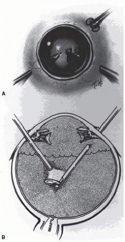
The presence of phakodenesis or iridodenesis on initial evaluation should alert the examining physician to suspect minor lens subluxation. Vitreous in the anterior chamber indicates that zonular rupture is present. In the absence of cataractous changes and related visual impairment, treatment is not indicated. A subluxated, cataractous lens can be removed by aspiration-irrigation or phacofragmentation through a limbal incision. However, a limbal approach may lead to posterior dislocation of the lens or lens fragments, vitreous prolapse and incarceration, and vitreous aspiration with resultant vitreous base traction and retinal tear formation. In addition, visualization of the anterior vitreous by coaxial illumination is poor compared with fiberoptic halogen or xenon endoillumination. These challenges to performing anterior segment surgery make pars plana vitrectomy and lensectomy a potentially safer alternative. Bimanual techniques permit fixation and simultaneous removal of the lens by the vitreous suction cutter, if the lens is soft, or phacofragmentation if it is sclerotic (Fig. 66.1A). Posteriorly dislocated fragments can be removed safely, with minimal vitreous traction, using the same incisions and instruments (Fig. 66.1B). With endoillumination, prolapsed and juxtalenticular vitreous is readily identified and excised with the vitrectomy probe.

Pars plana lensectomy is the preferred method for removing a completely dislocated lens. The technical aspects of dislocated lens and lens fragment removal are described elsewhere in these volumes.


Vitreous Hemorrhage

Blood in the vitreous may arise from tears in the iris, ciliary body, choroid, or retina. Hemorrhage from choroidal rupture accumulates beneath the neurosensory retina, and may then pass through the retina into the vitreous without necessarily causing a retinal break. Vitrectomy is indicated for vitreous hemorrhage caused by ocular contusion when a retinal detachment is suspected. Additional indications for vitrectomy in the setting of vitreous hemorrhage include suspicion of an undiagnosed scleral rupture, sudden additional loss of vision, retinal detachment detected through a window in the hemorrhage, retinal break or retinal detachment diagnosed by ultrasound, or lack of improvement after a reasonable period of observation.


FIG. 66.1 Pars plana lensectomy. A: Infusion cannula in lower temporal quadrant. Lens fixated with myringotomy blade. Vitrectomy probe or phacofragmentor inserted into lens through opposite pars plana. B: Posteriorly dislocated lens fragment removed by standard two-hand technique. (From Sternberg P. Trauma: principles and techniques of treatment. In: Ryan SJ, ed. Retina. Vol 3. St Louis, MO: CV Mosby; 1989:472, with permission.)

Preoperative contact A-scan and B-scan ultrasonography is helpful for detecting posterior vitreous detachment and differentiating it from retinal detachment, though other clinical indicators should be assessed when such a distinction is being made. It can be misleading to rely
completely upon the accuracy of ultrasound. Tissue presumed to be the posterior hyaloid should be approached with caution until it can be identified with certainty.

A three-port vitrectomy technique is the standard approach for nonclearing vitreous hemorrhage. We prefer 23G or 25G, sutureless, transconjunctival vitrectomy if the blood is not particularly dense and no extremely peripheral pathology is suggested. If the blood is dense, and visualization of the infusion port is a concern, a 6-mm infusion port, an illuminated infusion port (preferably with a xenon light source), or an illuminated infusion instrument may be substituted for the standard 4-mm port to facilitate visualization, and larger 20G vitrectomy techniques are preferred. Assuming that the anterior chamber is fairly clear, a central core of opaque vitreous is initially removed, where the tips of the cutter and endoilluminator can be visualized and seen to be safely away from both the retina and the lens. The excision is carried posteriorly, and successive layers of hemorrhagic and fibrinous vitreous are removed until the anticipated plane of the posterior hyaloid is approached. A constant surveillance is maintained for a gray membrane containing radially oriented vessels (undiagnosed detached retina). A small opening is made in the detached posterior hyaloid, through which unclotted blood is aspirated by active suction from a softtipped cannula or vitreous cutter. Once the retina has been visualized, it is best to remove as much retrohyaloid blood as possible to prevent dispersion into the vitreous cavity with consequent loss of visual control.

If the posterior vitreous cortex is not detached, it can be separated from the retina by gentle suction with a softtipped cannula or vitreous cutter at the edge of the optic disk. In cases where the hyaloid is difficult to detach from the retina, such as in children, a small amount of preservative-free triamcinolone acetonide can be introduced into the vitreous. We prefer to have the infusion off during the instillation of the drug, to prevent diffuse spread. The vitrector can then be placed in the eye with the infusion on, and the free triamcinolone can be removed. The residual triamcinolone may adhere to the hyaloid, highlighting its anatomy and facilitating the creation of a posterior vitreous separation.

After establishing the plane between the hyaloid and the retina, the surgeon attempts to remove the entire cortical vitreous except for the firmly attached portion at the anterior vitreous base. Cortex that does not separate with gentle manipulation is isolated from surrounding vitreous to eliminate traction on the retina. It is important to remove the cortical vitreous from areas on and adjacent to retinal breaks. Failure to do so may result in subsequent tangential traction and retinal detachment. When possible, retinopexy is applied to the posterior edge of the tear, and the flap of the tear is amputated. As discussed below, a scleral buckle should be considered if retinal breaks cannot be freed from surrounding vitreous cortex, and in some cases it may be considered when there is a greater risk for the development of future anterior peripheral base contraction.

The placement of sclerotomies close to the 3- and 9-o’clock positions facilitates maximal removal of the hemorrhagic anterior vitreous skirt, which thereby improves visualization of the peripheral retina and pars plana. Aided by coaxial illumination and scleral depression, the peripheral vitreous on the temporal side of the globe is trimmed with the cutter placed through the temporal sclerotomy to reach both the superior and inferior quadrants, after which it is transferred to the nasal sclerotomy, and the process is repeated. The fiberoptic endoilluminator may damage the lens if used internally to illuminate the peripheral vitreous on the opposite side of the globe. However, the cone of light from the probe may be directed externally through the cornea to augment or replace the internal coaxial light source. Alternatively, xenon-powered chandelier lights or lighted infusion cannulas can be utilized to increase the amount of illumination and provide diffuse, wide-field light to allow the surgeon to perform simultaneous vitrectomy and scleral depression without the aid of an assistant. Hemorrhagic retrolenticular vitreous can be stripped from the posterior lens capsule with gentle aspiration into the cutting port followed by withdrawal of the probe and simultaneous activation of the cutting mode. This technique may be dangerous when used in young children because the retrolenticular vitreous is adherent to the lens, which is sufficiently pliable to be aspirated into the port with consequent cataract formation. When the lens is clear, the process of removing peripheral and retrolenticular vitreous is less important than preserving lens integrity in most cases.

It is important to expose the peripheral retina and vitreous base as most retinal breaks caused by ocular contusion are located in this area.5 The use of wide-angle contact and noncontact viewing systems such as the BIOM (Binocular Indirect Ophthalmo Microscope, Insight Instruments Inc., Stuart, Florida) can greatly facilitate visualization of the peripheral fundus.6 All retinal breaks should be treated. Endolaser is used for posterior breaks, whereas peripheral breaks are treated with either a curved endolaser probe, indirect laser assisted by scleral depression, or transscleral cryoretinopexy. The xenon-illuminated endolaser probes are needed to treat retina pathology when the view is poor from anterior segment opacification either from catararact formation, corneal edema, or stromal scarring. Cryotherapy is preferred when residual opaque vitreous partially obscures the targeted break.


Encircling scleral buckles are not necessary after vitrectomy for nonclearing vitreous hemorrhage caused by ocular contusion when a clear view of the fundus periphery reveals no peripheral retinal tears or signs of traction. Similarly, the support of a buckle is usually not needed for treated retinal breaks without retinal detachment. A segmental scleral buckle element may be used to support areas with residual traction on a peripheral break. An encircling scleral buckle should support the peripheral retina when traction on breaks in the oral zone persists or the periphery is poorly visualized because of residual opaque vitreous.


Retinal Breaks

Retinal breaks are created at the time of nonpenetrating blunt injuries in 10% to 20% of eyes.7, 8, 9 and 
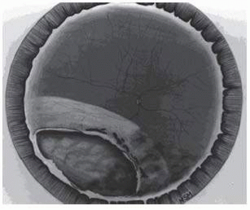
10 Retinal dialyses are most frequent, seen mostly in the lower temporal (
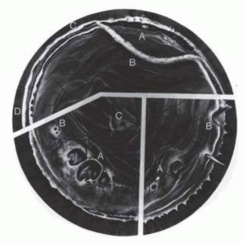
Fig. 66.2) and upper nasal periphery (Fig. 66.3).5, 11 Large irregular breaks at the point of impact of blunt trauma are less common but equally characteristic of nonpenetrating injuries (Fig. 66.3).5, 12 Horseshoe and operculated tears of the equatorial retina (Fig. 66.3) are characteristically seen in 25% of eyes.5 Small round holes in atrophic retina at the point of traumatic impact and macular holes (Fig. 66.3) are infrequently observed after ocular contusion.5, 12

Prophylactic treatment is indicated for most traumatic retinal breaks. Breaks at the point of impact are one exception because they are frequently self-sealing. The surrounding necrotic retina and choroid often unite in a common scar without prophylaxis. It is wise, however, to treat these large tears when scleral depression reveals a slight elevation and movement of their edges and the surrounding retina.


FIG. 66.2 Large lower temporal dialysis at the point of impact of blunt trauma. (From Cox MS. Retinal breaks caused by blunt nonperforating trauma at the point of impact. Trans Am Ophthalmol Soc. 1980;78:418, with permission.)


FIG. 66.3 Top: Traumatic vitreous base traction. Upper nasal dialysis (A). Avulsed vitreous base (B). Dialysis at anterior vitreous base border (C). Tenting-up of retina and pars plana epithelium (D). Bottom left: Retinal breaks without vitreoretinal attachments. Large irregular breaks at the point of impact of blunt trauma (A). Small round holes in atrophic retina (B). Macular hole (C). Bottom right: Horseshoe and opercular tears. Of the equatorial retina (A). At the posterior vitreous base border (B).


Retinal Detachment

Retinal detachment may be seen in the setting of closed globe eye injury. Trauma may cause a rapid shortening of anteroposterior axis of the globe that is followed by equatorial elongation, leading to peripheral retinal tears and dialyses. Traumatic injuries are more commonly observed in a younger patient population. The vitreous gel is normally dense in young patients and may tamponade the retinal break. Later liquefaction may lead to retinal detachment months following the initial injury.5 The detachment is typically shallow and slowly progressive because the large volume of formed vitreous gel in younger eyes inhibits the bullous retinal elevation that commonly occurs in older patients with nontraumatic retinal detachments.

Closed globe injury may also lead to traumatic retinal dialyses that are classically seen in the inferotemporal or superonasal quadrants.11 Careful preoperative and
intraoperative indirect ophthalmoscopy with scleral depression is important to identify the etiologic rhegmatogenous sites for the successful treatment of detachments caused by traumatic retinal dialyses. Small dialyses at the vitreous base borders may be difficult to identify, particularly in the superonasal quadrant, and breaks of the pars plana epithelium, at the anterior vitreous base border, are less apparent than retinal tears at the posterior edge of the vitreous base. During scleral depression, small dialyses may be closed, and thus they may be more easily seen on the lateral slopes of the scleral indentation than on its crest. Transscleral cryotherapy is diagnostically helpful. It is not unusual to discover breaks at the vitreous base border for the first time when the edges of the tear are whitened by freezing a suspicious area.

It is prudent to treat the entire zone of vitreous base pathology with cryotherapy under direct visualization (Fig. 66.3). In this way, treatment of all retinal breaks is assured. The anterior, posterior, and lateral limits of the treated zone are carefully localized and then supported by a broad scleral buckle to relieve traction to the entire area. The posterior edge of the dialysis should fall on the crest of the buckle, which must be sufficiently broad to support the anterior edge as well to prevent a recurrent detachment from anterior tracking of subretinal fluid. Segments of grooved solid silicone tires and an encircling band are positioned on the great circle of the globe to minimize anterior or posterior migration of the buckle. The band is anchored by a nonabsorbable mattress suture or scleral belt loop in each of the quadrants not occupied by the tire segment. A high encircling buckle promotes posterior gaping or “fish-mouthing” of the dialysis and is thus avoided.

The retinal detachment is often shallow. To avoid retinal perforation or incarceration, subretinal fluid is released through a sclerotomy in an area of sufficient retinal elevation, as determined by intraoperative indirect ophthalmoscopy with scleral depression. Viewed in profile, the scleral indentation helps gauge the distance between retina and retinal pigment epithelium. To avoid retinal incarceration and blowout, the drainage sclerotomy should be left unsutured when made in the bed of the buckle, and securely sutured if made posterior to the buckle, particularly if additional manipulation of the buckle or an intravitreal gas injection is anticipated. Nondrainage scleral buckling procedures are also acceptable in cases without retinal incarceration.

Retinal dialyses in the lower temporal quadrant are often large, with gaping posterior edges located well behind the equator (Fig. 66.2). They are caused by injuries to the inferotemporal globe that result in the dissolution and disappearance of retinal tissue.12 In contrast to nontraumatic giant retinal tears with rolled-over retinal edges, they respond favorably to scleral buckling without vitrectomy. A scleral buckle is indicated for smaller dialyses that can be closed with an explant of reasonable size. Very large or posterior breaks, as illustrated in Figure 66.2, are best treated with vitrectomy, gas or silicone oil tamponade, and laser, as recommended for nontraumatic giant retinal tears, rather than with a very large scleral buckle.

Vitrectomy is often used to repair traumatic rhegmatogenous retinal detachments, particularly when there is an associated vitreous hemorrhage, giant retinal tear, or proliferative vitreoretinopathy. Intraocular gases, silicone oil, and perfluorocarbon liquids may all aid in the repair.


Traumatic Macular Hole

First described by Herman Knapp13 in 1869, traumatic macular hole is a well-recognized complication of closed globe ocular trauma. It is thought to result from tangential traction to the posterior pole occurring with globe deformation from the blunt trauma.14 Outward expansion of the equator is followed by flattening and subsequent posterior displacement of the posterior pole. The trampoline-like movement of the posterior pole creates tangential tractional forces on the retinal surface that lead to the formation of the macular hole. Spontaneous closure of the hole is not uncommon; therefore, a period of observation for several weeks following injury is appropriate prior to surgical repair.15 Optical coherence tomography is a useful diagnostic tool to follow the progress of traumatic macular holes and diagnose vitreomacular traction.16, 17 The presence of vitreomacular traction and/or the failure of the macular hole to close after a sufficient period of observation are indications for surgical repair.

Small gauge 23G or 25G transconjunctival vitrectomy may be used to repair the traumatic macular hole. A core vitrectomy, followed by removal of the posterior hyaloid is performed. The vitrectomy may then be carried anteriorly as per standard macular hole surgery technique. While the importance of routinely removing the internal limiting membrane (ILM) during the repair of idiopathic macular holes is controversial, we prefer to remove the ILM during the repair of traumatic macular holes.18, 19 We typically utilize 0.2 cm3 of 0.125% indocyanine green (ICG) dye to stain the ILM. We instill the dye with the infusion off, and allow it to sit on the surface of the retina for several seconds before removing the dye with aspiration. The ICG may highlight residual vitreous unintentionally left behind but will generally stain the ILM. The ILM is then pinched down upon using an end-grasping or ILM forceps, and removed from the entire macula in a
rhexis maneuver. Medium- to long-term gas tamponade, combined with facedown positioning, is performed in each case. More recent studies demonstrate a high rate of macular hole closure with facedown positioning of 3 days’ duration.20 In pediatric patients, 0.4 IU of intravitreally injected plasmin enzyme may facilitate the induction of a posterior vitreous detachment during surgery.21


OPEN GLOBE (PENETRATING) INJURIES

Although less common than contusion, penetrating injuries lead more often to severe visual impairment. Penetrating injuries result from a single outside-in laceration of the eye wall. Globe ruptures from blunt trauma are often inside-out disruptions of the eye wall. The diagnosis of an open globe injury is determined by a comprehensive eye exam and a combination of computed tomography (CT) scan and B-scan ultrasound if media opacity obscures a view to the anterior or posterior segment.22 The visual prognosis is favorable when the primary mechanical damage caused by sharp penetration is limited to the anterior segment of the eye.23 Modern microsurgical techniques permit better wound closure and reconstruction of the anterior ocular structures. Penetrating injuries of the posterior segment carry a less favorable prognosis. The primary mechanical damage of vital structures by such injuries may be so great that the potential for useful vision is instantly destroyed. Predictors of poor vision following penetrating trauma include initial visual acuity of light perception or no light perception, wounds extending posterior to the rectus muscle insertion plane, and wound length greater than 10 mm.24 In a study conducted prior to the era of pars plana vitrectomy, only 6% of patients with open globe injury between 1952 and 1970 achieved visual acuity of 5/200 or better.25 The advent of modern vitreoretinal microsurgical techniques has resulted in improvement of both anatomic and visual outcomes following open globe injury. A retrospective review of patients with similar open globe injuries between 1970 and 1981 found that 55% of patients achieved visual acuity of 5/200 or better.26 Similar visual outcomes were found in a more recent study that examined patients from the same institution with open globe injuries between 1985 and 1993.27 In addition, the incidence of enucleation was found to be lower than in previous studies.27 The Ocular Trauma Score (OTS) can be used to predict visual outcome. The OTS uses ocular injury variables such as presenting visual acuity, rupture, endophthalmitis, perforating injury, retinal detachment, and afferent pupillary defect to stratify final visual acuity.28


Acute Effects of Closed Globe Injury

Penetration of the eye by relatively blunt objects causes compression of the globe with resultant iridoparesis, iridodialysis, subluxation and dislocation of the lens, traumatic cataract, choroidal rupture, and retinal breaks at the vitreous base borders.29, 30 Ruptures of the uvea can lead to anterior chamber, choroidal, subretinal, and vitreous hemorrhage with high intraocular pressure. Massive blunt trauma may cause corneal and scleral ruptures and possibly avulse the optic nerve.31


Acute Effects of Ocular Penetration

Penetrating objects cause lacerations of the cornea, iris, lens, sclera, ciliary body, choroid, retina, and optic nerve. Anterior chamber, choroidal, subretinal, and vitreous hemorrhages result from lacerations of uveal and retinal blood vessels. Perforation of the corneoscleral wall permits prolapse and incarceration of the lens, uvea, retina, and vitreous.


Early Secondary Complications of Ocular Penetration

The secondary complications of ocular penetration are important causes of the visual impairment that results from such injuries. Examples include the toxic effects of intraocular foreign material, such as copper, and the introduction of bacteria and fungi with consequent infectious endophthalmitis. Chronic inflammation caused by retained disrupted lens material, vitreous hemorrhage, and incarcerated vitreous and uvea play an important role in the stimulation of intraocular fibrocellular proliferation.32, 33 and 34


Intermediate Secondary Complications of Ocular Penetration

Cleary and Ryan32 reported experimental evidence that blood and, to a lesser extent, lens material in the presence of a large scleral wound caused fibrocellular proliferation and transvitreal membranes, the contraction of which produced tractional retinal detachments (Fig. 66.4). The clinical consequences of fibrocellular proliferation and membrane contraction are well established and include traction retinal detachments, retinal breaks, rhegmatogenous retinal detachments, proliferative vitreoretinopathy, cyclitic membranes, ciliary body detachments, hypotony,
and phthisis bulbi.35 A recent study has published similar visual outcomes of retinal detachments secondary to both open and closed globe trauma.36


FIG. 66.4 Fibrocellular proliferation and membrane formation cause tractional retinal detachment. (From Cleary PE, Ryan SJ. Method of production and natural history of experimental posterior penetrating eye injury in the rhesus monkey. Am J Ophthalmol. 1979;88:219, with permission.)


Late Secondary Complications of Ocular Penetration

Tractional and rhegmatogenous retinal detachments secondary to recurrent fibrocellular proliferation and proliferative vitreoretinopathy are common late complications of penetrating injuries.37 Macular pucker is a similar complication of lesser magnitude. Fungal endophthalmitis and sympathetic ophthalmia are infrequent late complications of ocular penetration that carry potentially devastating consequences.38


Primary Surgical Repair


Exploration of the Globe

A careful exploration of the globe is required to determine the presence and extent of defects in the eye wall whenever ocular penetration is established or suspected. Posterior segment exploration is deferred until anterior lacerations or ruptures are repaired so that manipulations required to expose the posterior segment cause no additional prolapse of ocular contents through open anterior wounds. A complete conjunctival peritomy is performed and all quadrants of the globe inspected. Traction on temporary sutures looped around the rectus muscles, combined with the retraction of conjunctiva and Tenon fascia, facilitates visualization of the sclera beneath the extraocular muscles and posteriorly. When passing sutures or a muscle hook beneath rectus muscles, care must be taken to avoid inadvertent globe penetration through an undetected defect in the sclera.


Wound Closure

Restoration of the structural integrity of the globe by careful anatomic reapposition of the wound edges is the goal of the primary surgical repair of penetrating injuries. Corneal lacerations are closed with deeply placed interrupted 10-0 nylon sutures. Interrupted 9-0 nylon sutures are used for a limbal closure, and interrupted 8-0 nylon sutures are used to repair scleral wounds to withstand the stress of subsequent vitrectomy should a second operation become necessary. Homologous corneal or scleral grafts (Tutoplast®) or cyanoacrylate tissue glue may be used to close large defects caused by the loss of tissue. Posterior exit wounds are usually self-sealing and not repaired unless they are large.39 Prolapsed lens material is removed and vitreous excised preferably with the vitreous cutter or alternatively with Weck-Cel spears and scissors. Unless necrotic and exposed for more than 24 hours, prolapsed uveal tissue is reposited. Extruded retina is also reposited, while great care is taken to avoid incarceration.


Reconstruction of the Anterior Chamber

Reconstruction of the anterior chamber and restoration of the pupil are important goals of the primary surgical repair. Iris and vitreous incarceration in a corneal laceration
causes chronic inflammation, peripheral anterior synechiae, and closure of the anterior chamber angle. Incarcerated vitreous provides a scaffold for fibrous ingrowth that may create membranes, the contraction of which could cause tractional retinal detachments and retinal tears. A cyclodialysis spatula is used to reposit the iris through the wound or sweep it free from the wound via a limbal incision on the opposite side of the globe. Alternatively, sodium hyaluronate may be used to reform the anterior chamber, protect a clear lens if present, and reposit tissues. An anteriorly displaced cataractous lens or lens fragments are removed with a vitrectomy probe through the limbus or, if large choroidal detachments have been excluded, the pars plana. Phacofragmentation may be needed to remove hard lens material from the eyes of elderly patients, in which case suction must be carefully monitored to avoid vitreous aspiration and consequent traction. An anterior vitrectomy is performed, and air may be placed in the anterior chamber to prevent recurrent iridocorneal adhesions and reincarceration of the vitreous.


Removal of Foreign Material

Foreign material should be removed from the eye during the primary repair of penetrating injuries. The management of IOFBs will be discussed later. If infection is present, a vitrectomy is performed during the primary operation to remove organisms, toxins, and inflammatory debris.

In most cases, a traumatic cataract is removed during a second operation unless its presence compromises visualization. The removal of blood from the anterior chamber is also best deferred until a secondary repair is undertaken, unless visualization of the posterior segment is necessary at the time of the primary repair.


Prophylactic Cryopexy

The value of placing prophylactic cryopexy around the sutured wound is controversial. All perforations of the eye wall that extend posterior to the ora serrata lacerate the retina, but many retinal lacerations are self-sealed by incarcerated vitreous and subsequent surrounding chorioretinal scarring. Furthermore, experimental studies indicate that cryopexy breaks down the blood-retina barrier and stimulates cellular proliferation and migration, potentially contributing to membrane formation and tractional retinal detachment.40 On the other hand, retinal lacerations were responsible for 20% of the rhegmatogenous retinal detachments reported in one study of detachments caused by penetrating injuries.41 We currently treat retinal lacerations with ophthalmoscopically monitored light cryoretinopexy whenever possible, but not if blood in the vitreous prevents their visualization.


Prophylactic Antibiotics

Penetrating ocular injuries are complicated by infectious endophthalmitis in 2% to 7% of cases.42, 43 The presence of infection may be masked by the pain and inflammation of the injury, which may result in a disastrous delay in diagnosis. A recent study identified several risk factors associated with the development of posttraumatic endophthalmitis, including a dirty wound, presence of a retained IOFB, lens capsule breach, and delay in primary repair.44 However, a multivariate analysis performed by other investigators found rupture of the lens capsule to be the only independent risk factor for the development of endophthalmitis following penetrating ocular trauma.45 Interestingly, this series found that the presence of an IOFB did not significantly increase the risk of endophthalmitis, in contrast to the National Eye Trauma Study and other studies, which did find a statistically significant correlation between the presence of an IOFB and an increased risk of posttraumatic endophthalmitis.46, 47 Approximately 25% of cases of endophthalmitis after penetrating injuries are caused by Bacillus species, the virulence of which results in a poor prognosis.48, 49

Systemic prophylaxis with intravenous antibiotics has long been given in cases of penetrating trauma, though the low drug level obtained within the vitreous cavity makes this practice of questionable value. If intravenous prophylaxis is to be used, we prefer vancomycin for the coverage of increasingly prevalent strains of penicillinand cephalosporin-resistant gram-positive organisms. A third-generation cephalosporin (ceftazidime) is used for gram-negative coverage instead of parenteral aminoglycosides, which may cause renal complications. Intravenous prophylaxis may be continued for 3 to 5 days depending on the level of concern generated by the nature of the penetrating injury. The fluoroquinolone antibiotic ciprofloxacin may be of prophylactic value because of its reported penetration of the eye with systemic administration.50, 51 and 52 We now typically utilize an oral fluoroquinolone in trauma patients because of its ease of use, broad spectrum of antimicrobial activity, and significant ocular penetration.53 Prophylactic antibiotics may be intravitreally injected in eyes at particularly high risk for infection during the primary surgical repair. For example, foreign bodies contaminated by soil cause approximately 90% of cases of posttraumatic Bacillus endophthalmitis.54 These virulent organisms are sensitive to intravitreal vancomycin. Although gram-negative organisms are rarely the cause of posttraumatic endophthalmitis, they are covered more safely by ceftazidime than by gentamicin.55, 56 We recommend the intravitreal injection of 1 mg of vancomycin and 2.25 mg of ceftazidime when the history and clinical findings suggest such a high risk of infection.

Only gold members can continue reading. Log In or Register to continue

Stay updated, free articles. Join our Telegram channel

Jul 11, 2016 | Posted by in OPHTHALMOLOGY | Comments Off on Management of Posterior Segment Trauma

Full access? Get Clinical Tree

Get Clinical Tree app for offline access