M
M cell See cell, ganglion; cell, M.
M Units See acuity, near visual.
MacAdam ellipses See ellipses, MacAdam.
Mach bands When a light area is separated from a dark area by a transition zone in which the brightness increases or decreases regularly and rapidly, two bands are seen: one light band next to the dark area and another dark band next to the light area. The appearance of these two bands, known as Mach bands, is attributed to lateral inhibition processes occurring in the retina. The phenomenon is usually demonstrated with a rotating disc with black and white areas separated by a zone of brightness gradient. Syn. Mach rings.
macrocornea See keratoglobus.
macropsia Anomaly of visual perception in which objects appear larger than they actually are. It may occur as a result of abnormal accommodation (less than required for the fixation distance) or because of various retinal anomalies in which the visual receptors are crowded together, or because of the recent wear of either base-in prisms or a presbyopic correction, etc. Syn. megalopsia.
See dysmegalopsia; micropsia; metamorphopsia.
macula See fovea centralis; macula lutea.
macula, ectopia of the Anomaly characterized by displacement of the macula, which can be either of acquired or congenital origin. It may follow some forms of retinal scarring, retinal detachment surgery, previous inflammation, etc. It may result in reduced acuity, metamorphopsia or strabismus. Syn. dystopia of the macula; heterotopia macula.
macula, false The retinal area of the deviating eye of a strabismic subject which corresponds to the fovea of the fixating eye.
See retinal correspondence, abnormal.
macula lutea An oval area of the retina 3–5 mm in diameter, with the foveal depression at its centre, slightly below the level of the optic disc and temporal to it (its centre lies 3.5 mm from the edge of the disc). The side wall of the depression slopes gradually towards the centre where the fovea centralis is located and where the best photopic visual acuity is obtained. Around the fovea, the ganglion cells are much more numerous than elsewhere, being arranged in five to seven layers. The outer molecular layer is also thicker than elsewhere and forms the outer fibre layer of Henle and there is a progressive disappearance of rods so that at the foveola only cones are found. The area of the macula lutea is impregnated by a yellow pigment (macular pigment) in the inner layers and for that reason is often called the yellow spot. Syn. area centralis (although that area is considered to be slightly larger, about 5.5 mm in diameter); punctum luteum.
See entoptoscope, blue field; fovea centralis; pigment, macular.
macula, sparing of the See macular sparing.
macular cyst A swelling in the macular area in which fluid has accumulated after injury to the eye or cystoid macular oedema. It may eventually burst into the vitreous producing a macular hole.
macular degeneration, age-related (ARMD, AMD) A common, chronic degenerative condition found in a large percentage of elderly patients (and sometimes middle-aged ones) characterized by loss of central vision. There are two main forms of the condition: non-neovascular (dry, atrophic) AMD, which is the most common, and exudative (wet, neovascular) AMD in which the loss of vision is the most severe. The main features of dry AMD are the presence in the macular region of small, yellowish-white spots (hard drusen) and large, poorly defined, coalescing soft drusen, focal hyperpigmentation of the retinal pigment epithelium (RPE) and at a later stage geographic atrophy of the RPE and depigmentation exposing choroidal vessels. Visual acuity becomes markedly reduced, there is metamorphopsia and the condition usually becomes bilateral over several years. The condition is managed essentially by the use of low vision aids.
Exudative AMD has a similar clinical picture initially but is followed by choroidal neovascularization (CNV), which gives rise to subretinal fluid, haemorrhages, exudation, RPE detachment and subretinal fibrosis in the macular region resulting in severe loss of central vision. If detected early (usually with an Amsler chart), treatment with laser photocoagulation will reduce the risk of further visual loss. Photodynamic therapy (PDT) is another method of reducing the risk of visual loss. It allows selective destruction of the choroidal neovascularization with minimal damage to the overlying retinal tissue. It consists of injecting a photosensitizing agent (e.g. verteporfin) that is taken up by the abnormal vessels and when activated by a laser light of a given wavelength (e.g. 689 nm) it damages and shrivels up the vessels. Recent drug therapies, such as the anti-VEGF ranibizumab and bevacizumab, which are injected intravitreally at regular intervals and designed to stop the leakage and the growth of blood vessels, not only reduce loss of vision but improve visual acuity in a significant percentage of cases of wet AMD. Syn. senile macular degeneration.
See angiography, fluorescein; disciform scar; drusen; dystrophy, macular; lipofuscin; maculopathy, age-related; oxidative stress; pigment, macular; rule, Kollner’s; test, photostress; VEGF.
macular epiretinal membrane See fibrosis, preretinal macular.
macular hole A condition in which there is a partial or full thickness absence of the retina in the macular area. It may occur as a result of trauma, degeneration, old age, preretinal macular fibrosis or pathological myopia. It appears ophthalmoscopically as a round or oval, well defined, reddish spot at the macula. There is metamorphopsia, loss of visual acuity and a central scotoma. An operculum of retinal tissue may overlie the hole. The vitreous in front of the hole eventually condenses and separates from the retina. In partial macular hole a layer of photoreceptors may still be attached to the retinal pigment epithelium (lamellar hole), as in cystoid macular oedema. Treatment usually consists of reattaching the retina, if detached, and possibly vitrectomy.
See fibrosis, preretinal macular; ophthalmoscope, confocal scanning laser; retinal break; retinopathy, solar; tomography, optical coherence.
macular oedema See oedema, cystoid macular; retinopathy, background diabetic.
macular pigment See pigment, macular.
macular pucker See fibrosis, preretinal macular.
macular sparing Retention of macular function in spite of losses in the adjacent visual field as, for example, in homonymous hemianopia due to a cortical lesion (e.g. an interference in the blood flow in the middle cerebral artery). This is due to the fact that most cortical lesions are not large enough to affect the whole extensive cortical area representing the macula, thus leaving some of the area unaffected.
See magnification, cortical.
macular star Deposits of hard exudates material, mainly lipids, in Henle’s fibre layer radiating out in a star-like pattern. It can occur in neuroretinitis, hypertensive retinopathy, etc.
maculopathy, age-related A condition in which there are large whitish-yellow soft drusen in the macular area and hyperpigmentation or depigmentation of the retinal pigment epithelium associated with the drusen. Small hard drusen may also be present. It occurs most commonly in individuals over 50 years of age and represents the early stage of age-related macular degeneration. The risk of progression of this condition may be decreased by dietary supplements of vitamin C and E, antioxidants (carotenoids such as lutein and zeaxanthin) and minerals (zinc and cupric acid).
maculopathy, bull’s eye An ocular condition in which degeneration of the retinal pigment epithelium in the macular area causes alternating ring-like light and dark zones of pigmentation, as in a target. It may result from drug toxicity or hereditary conditions (e.g. cone dystrophy, Laurence–Moon–Bardet–Biedl syndrome). The main symptoms are a loss of visual acuity, reduced colour vision and aversion to bright sunlight.
maculopathy, diabetic A pathological disorder of the macula which frequently develops in diabetic patients. It is characterized by oedema, hard exudates, microaneurysms and ischaemia in the macular area. If the oedema is severe visual acuity will be reduced, but a blue-yellow colour vision defect is usually noted before the loss of acuity. Management involves laser photocoagulation.
See retinopathy, background diabetic.
madarosis Loss of, either or both, the eyebrows and the eyelashes. It may occur in blepharitis, in the presence of hair follicle mites, trauma from rubbing, in alopecia, in leprosy, or as a complication of acquired syphilis.
Maddox cross A scale for measuring the angle of heterophoria and heterotropia consisting of one horizontal and one vertical line in the form of a cross with a light source placed at the centre of intersection. The lines are graduated in prism dioptres or degrees and calibrated for use at a given distance (usually 6 metres). Syn. Maddox tangent scale.
Maddox double prism See bi-prism, Fresnel’s; test, double prism.
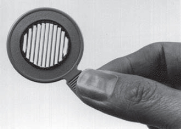
Maddox rod This is not a rod but a series of cylindrical grooves ground usually into a coloured piece of glass and mounted in a rim. (Originally it consisted of a single cylindrical rod.) It is used to measure heterophoria by placing it in front of one eye of a subject viewing a spot of light binocularly. The Maddox rod and eye together form a long streak of light perpendicular to the axis of the grooves and this retinal image is so unlike the image formed in the other eye that the fusion reflex is not stimulated. The eyes will then stay in the passive position. If there is a phoria the streak of light will not intersect the spot of light. For horizontal phorias the rod axis is placed horizontally and for vertical phorias, vertically. The amount and type of the phoria can be quantified by placing a prism of appropriate power and direction in front of either eye such that the streak appears superimposed on the spot of light. Alternatively, the angle of the phoria could be determined using a Maddox cross and placing a rod in front of one eye; the phoria can be read directly by the patient who indicates where the streak of light appears to cross the scale. The Maddox rod is also used to detect or measure cyclophoria (Fig. M1)
See position, passive; test, Maddox rod; test, Thorington.
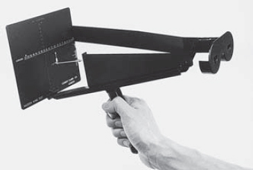
Maddox wing Handheld device used to measure heterophoria at near. It consists of a septum and two slit apertures, one for each eye. One eye sees a double tangent scale (vertical and horizontal) calibrated to read in prism dioptres, while the other eye sees a white arrow pointing upward and a red arrow pointing horizontally to the left. As the two retinal images are quite different there is no attempt at fusion and the eyes stay in the passive position. The arrows seen by the left eye point to the numbers seen by the right eye. The numbers represent the vertical and horizontal components of the phoria, which can be read directly by the observer (Fig. M2).
magenta 1 . Hue produced by the additive mixture of red and blue. 2. Hue evoked by any combination of wavelengths which act as the complement of a wavelength of 515 nm. See colour, complementary.
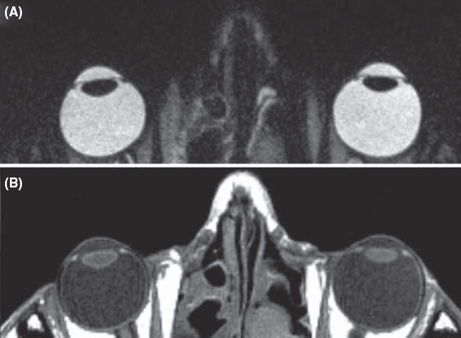
magnetic resonance imaging (MRI) Non-invasive method of imaging part of the body to facilitate diagnosis and therapy. Unlike other radiological methods, this technique does not expose the patient to ionizing radiations. It depends instead on a strong magnetic field which induces the spins of atomic nuclei within the body (e.g. hydrogen atoms) to align themselves along the axis of the magnetic field. When exposed to a pulse of electromagnetic energy at a specific radio frequency, the nuclear spins tilt momentarily then regain their original orientations thus re-emitting an electromagnetic signal at the same radio frequency, which can be detected and analysed to generate a three-dimensional image. The rate at which the radio frequency signal decays can be used to characterize the properties of different tissues, both normal and abnormal. The rates are referred to as T1 and T2 and each yields different contrasts: T1-weighted images are best for anatomical details and T2-weighted images are best for pathological details. This technique provides better image contrast than X-ray computerized tomography in many instances (e.g. the patches of demyelination in the white matter of patients with multiple sclerosis), while the reverse is true in other instances (e.g. a meningioma in the posterior visual pathway). Usage includes the detection of optic nerve disease (e.g. glioma, optic neuropathy), retrobulbar neuritis, lesions in the chiasma and orbital tumours. In general, this procedure takes longer than X-ray computerized tomography (Fig. M3).
functional MRI (fMRI) A non-invasive method used to map the various areas of the cortex, as well as brain tumour mapping, by observing brain activity in response to performing a task that engages a specific behaviour (e.g. tests of colour vision, face perception, motion perception, solving a mathematical problem). This is accomplished, within a conventional MRI scanner, by detecting the change in magnetic susceptibility of blood haemoglobin (and thus blood oxygenation) in various areas of the brain. During task execution when a brain region is active there is an increase in blood flow, which is more oxygenated and has a lower concentration of haemoglobin. This leads to a focal increase in MR image intensity, of around 1–5%, which can be detected using appropriate statistical methods.
See areas, visual association; radiology; tomography, positron emission.
magnification 1. An increase in the apparent size of an object. Syn. enlargement. 2. Specification of a magnifying device to form an enlarged image.
angular m. Magnification expressed as the ratio of the angle α’ subtended at the eye by the image to the angle α subtended at the eye by the object (assuming small angles)
Syn. angular enlargement. Note: low vision practitioners consider this type of magnification in which no specific distance is specified as a synonym of apparent magnification.
apparent m. Magnification produced by a viewing instrument or lens expressed as the ratio of the angle w’ subtended at the nodal point of the eye by the image, to the angle w subtended at the nodal point by the object, when placed at a standard (reference) distance called the least distance of distinct vision’ from the unaided eye. It is conventional to take this distance as 250 mm and to place the object in the anterior focal plane of the magnifying device. The magnification M is, then, equal to (assuming small angles)

where f and F are the second focal length (in mm) and power (in dioptres) of the magnifying device, respectively (Fig. M4). In this object location the magnification (and therefore the retinal image size) is constant and independent of the distance between the magnifier and the eye, but the field of view decreases as the distance between the eye and the magnifier increases. Syn. conventional magnification; effective magnification; loupe magnification; nominal magnification; relative magnification; standard magnification.
If the object is closer to the magnifying device than its anterior focal plane so that its image is formed at the least distance of distinct vision (25 cm), and assuming that the eye is so close to the magnifier as to ignore the distance separating them and that the patient has an accommodation (or a near addition) of +4.00 D, the magnification M is, then, equal to
Example: a lens of +16.00 D provides, in these conditions, a magnification of 5×. Syn. iso-accommodative magnification; magnifying power; trade magnification.
See magnification, iso-accommodative; magnification, lateral; power, equivalent viewing.
axial m. The ratio of the distance along the optical axis between two points in image space l’ to the distance along the optical axis between the corresponding two points in object space l, i.e. l’/l. The axial magnification is approximately equal to the square of the lateral magnification when the object is far away from the optical system. This magnification is useful when considering an image in its three dimensions. Clinically, it is important when assessing the thickness of a retinal lesion in indirect ophthalmoscopy. Syn. longitudinal magnification.
Table M1
Apparent magnification (or conventional magnification) of microscopic lenses of various powers used in the correction of low vision and their corresponding reading distance (assuming emmetropia or correction for distance and that no accommodation is exerted)
| magnification | lens power (D) | reading distance (cm) |
| 1× | +4.00 | 25 |
| 1.5× | +6.00 | 16.7 |
| 2× | +8.00 | 12.5 |
| 2.5× | + 10.00 | 10 |
| 3× | + 12.00 | 8.3 |
| 4× | + 16.00 | 6.25 |
| 5× | +20.00 | 5 |
| 6× | +24.00 | 4.2 |
| 8× | +32.00 | 3.12 |
| 10× | +40.00 | 2.5 |
combined m. The product of the individual values of each type of magnification used in combination with each other. Example: if a patient uses a CCTV monitor to provide a magnification of 5× viewed at a distance of 50 cm, and then views the same screen at a distance of 25 cm, thus producing a relative distance magnification of 50/25 = 2×, the total magnification is 5 x 2 = 10×. Syn. total magnification.
conventional m. See magnification, apparent.
cortical m. Term referring to the fact that the amount of cortical area devoted to processing visual information from the central area of the retina far exceeds the amount devoted to the peripheral retina. It is estimated that about 25% of the cells in the visual cortex are devoted to processing the central 2.5° of the visual field. Syn. magnification factor.
See area, visual; macular sparing.
distance m. See magnification, relative distance.
effective m. See magnification, apparent.
electronic m. Magnification obtained using an electronic vision enhancement system (EVES), such as a closed-circuit television (CCTV). It is equal to the ratio of the size of the image on the screen to the size of the original object being viewed. Example: an object 2cm in height measures 6 cm on the screen, the magnification is 6/2 = 3×. Syn. real image magnification; transverse magnification.
m. factor See magnification, cortical.
iso-accommodative m. The magnification of a lens (or lens system) when the distance of the image from the eye (or spectacle plane) formed by a magnifier is equal to the distance of the object from the eye viewed without the magnifier. Thus the same amount of accommodation (or near addition) is required with or without the magnifier. It is equal to
where F is the power of the magnifier (assumed to be so close to the eye as to ignore the distance separating them) and D the object vergence. The special case in which the object distance from the eye is 25 cm (D = 4.00 D) is the trade magnification.
See magnification, apparent.
lateral m. Magnification of a lens or of an optical system, expressed as the ratio the size of the image h’ to the size of the object h. It is usually denoted by
Table M2
Approximate lateral magnification (in %) corresponding to various changes in spectacle lens distance and for various lens powers
| distance change | lens power (D) | |||||
| 1 | 2 | 4 | 6 | 8 | 10 | |
| 1 mm | 0.1 | 0.2 | 0.4 | 0.6 | 0.8 | 1.0 |
| 2 mm | 0.2 | 0.4 | 0.8 | 1.2 | 1.6 | 2.0 |
| 3 mm | 0.3 | 0.6 | 1.2 | 1.8 | 2.4 | 3.0 |
| 4 mm | 0.4 | 0.8 | 1.6 | 2.4 | 3.2 | 4.0 |
| 5 mm | 0.5 | 1.0 | 2.0 | 3.0 | 4.0 | 5.0 |
| 10 mm | 1.0 | 2.0 | 4.0 | 6.0 | 8.0 | 10.0 |
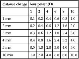
minus lens: if moved closer to the eye magnification increases; if moved further from the eye, magnification decreases plus lens: if moved closer to the eye magnification decreases; if moved further from the eye, magnification increases

where l’ and l are the distances of the image and object, respectively from the principal plane of the lens (or lens system) and L and L’ the object and image vergences, respectively. Syn. enlargement ratio (ER); linear magnification; transverse magnification.
(Note: some authors consider this last term a synonym of electronic magnification.)
See power, equivalent viewing.
linear m. See magnification, lateral.
longitudinal m. See magnification, axial.
loupe m. See magnification, apparent.
negative m. See minification.
nominal m. See magnification, apparent.
m. power See magnification, spectacle.
real image m. See magnification, electronic.
relative m. See magnification, apparent.
relative distance m. The magnification that results from decreasing the distance between an object and the eye. It is expressed as

where x and x’ are the initial distance and the new distance, respectively. Example: if the viewing distance is decreased from 60 cm to 20 cm, Md = 60/20 = 3×. Syn. distance magnification; relative distance enlargement.
relative size m. The magnification which results from increasing the actual size of an object viewed. Examples: a larger TV screen; a larger print book than one used previously. It is expressed as
Ms = h2/h1
where h2 and h1 are the sizes of the enlarged object and the initial object, respectively. Syn. size magnification; relative size enlargement.
relative spectacle m. (RSM) The ratio of the retinal image size in the corrected ametropic eye to that in a standard emmetropic eye.
shape m. Magnification resulting from a variation in the curvature of the front surface and thickness of an ophthalmic lens. In the treatment of aniseikonia it may be necessary to alter the magnification of a lens while leaving its dioptric power unchanged. Syn. shape factor.
See lens, aniseikonic; magnification, spectacle.
size m. See magnification, relative size.
spectacle m. The ratio of the retinal image of a distant object in the corrected ametropic eye to the blurred or sharp image formed in the same eye when uncorrected. It is greater than unity in the hyperopic eye, and less than unity in myopia. With a contact lens, though, this magnification is nearly equal to unity whatever the refractive error. Spectacle magnification SM depends both on the shape of the spectacle lens (i.e. the power of its front surface and its thickness) and on the power of the lens. Thus
where F1 is the power of the front surface, F’v the back vertex power of the lens, t its thickness, n the index of refraction and d the distance from the back surface of the lens to the entrance pupil of the eye. The first term in the formula represents the shape factor (shape magnification) and the second term the power factor (power magnification). However, since the shape factor is very small for most common ophthalmic lenses (except for high plus lenses), it is often ignored in the above formula.
telescopic m. Magnification obtained with a telescope, such as a galilean telescope which gives an erect image or an astronomical telescope in which an erecting system is used. The magnification is
Table M3
Approximate spectacle magnification of lenses of various back vertex powers (F’v in D) assuming d = 15 mm and the parameters of a typical spectacle lens of that power
| F’v | shape factor (S) | power factor (P) | spectacle magnification (SM) | percentage magnification | ||
| +12 | 1.08 | 1.22 | 1.32 | 32% increase | ||
| +10 | 1.06 | 1.18 | 1.25 | 25% ” | ||
| +8 | 1.04 | 1.14 | 1.18 | 18% ” | ||
| +6 | 1.03 | 1.10 | 1.13 | 13% ” | ||
| +4 | 1.02 | 1.06 | 1.09 | 9% ” | ||
| +2 | 1.01 | 1.03 | 1.04 | 4% ” | ||
| 0 | 1.01 | 1.00 | 1.01 | 1% ” | ||
| –2 | 1.00 | 0.97 | 0.97 | 3% decrease | ||
| –4 | 1.00 | 0.94 | 0.94 | 6% ” | ||
| –6 | 1.00 | 0.92 | 0.92 | 8% ” | ||
| –8 | 1.00 | 0.89 | 0.89 | 11% ” | ||
| –10 | 1.00 | 0.87 | 0.87 | 13% ” | ||
| –12 | 1.00 | 0.85 | 0.85 | 15% ” | ||
| –14 | 1.00 | 0.83 | 0.83 | 17% ” | ||
| –16 | 1.00 | 0.81 | 0.81 | 19% ” | ||
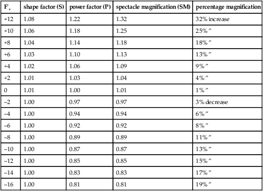
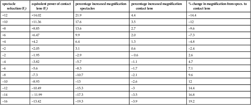
Table M4
Approximate spectacle and contact lens magnification assuming d = 15 mm (vertex distance 12 mm plus 3 mm between cornea and entrance pupil) and negligible lens thickness. The percentage change in magnification going from spectacles to contact lenses was calculated using (Fs/Fc – 1) × 100
| spectacle refraction (Fs) | equivalent power of contact lens (Fc) | percentage increased magnification spectacles | percentage increased magnification contact lens | % change in magnification from specs. to contact lens |
| +12 | +14.02 | 21.9 | 4.4 | –14.4 |
| +10 | +11.36 | 17.6 | 3.5 | –12 |
| +8 | +8.85 | 13.6 | 2.7 | –9.6 |
| +6 | +6.47 | 9.9 | 2.0 | –7.3 |
| +4 | +4.2 | 6.4 | 1.3 | –4.8 |
| +2 | +2.05 | 3.1 | 0.6 | –2.4 |
| –2 | –1.95 | –2.9 | – 0.6 | 2.6 |
| –4 | –3.82 | –5.7 | –1.1 | 4.7 |
| –6 | –5.6 | –8.3 | –1.7 | 7.1 |
| –8 | –7.3 | –10.7 | –2.1 | 9.6 |
| –10 | –8.93 | –13 | –2.6 | 12 |
| –12 | –10.49 | –15.3 | –3 | 14.4 |
| –14 | – 11.99 | –17.3 | –3.5 | 16.8 |
| –16 | –13.42 | –19.3 | –3.9 | 19.2 |
where α’ and α are the angles subtended at the eye by the image viewed through the telescope and the angle subtended at the eye by the object, respectively and Fe and Fo are the powers of the eyepiece and objective, respectively. Telescopes are used to magnify objects at distance (afocal) and placed over the spectacle correction. If the patient is uncorrected the telescope can be adjusted but the magnification will change.
They can be used for near and intermediate viewing by altering the distance between the objective and the eyepiece, or adding a plus lens in front of the objective, the result being a combined magnification (afocal telescope magnification x power of the plus lens ÷ 4).
total m. See magnification, combined.
trade m. See magnification, apparent.
transverse m. See magnification, electronic; magnification, linear.

magnifier An optical device, commonly used for close viewing, which produces an apparent magnification. It can be monocular or binocular, held in the hand (hand magnifier) or mounted in front of the eye (stand magnifier). It rarely exceeds a magnification of ×10 and does not produce an inversion of the image (Fig. M5). Syn. loupe.
See distance of distinct vision; lens, magnifying; magnification, apparent; vision, low.
magnifying glass; lens See lens, magnifying.
magnifying power See magnification, apparent.
magnifying spectacles See spectacles, magnifying.
magnitude estimation A psychophysical method of evaluating stimuli above threshold. The subject assigns numbers according to the apparent magnitudes of the stimuli. The results relating the magnitude of sensation S and the stimulus intensity I usually follow a power law (or Stevens’ power law), that is S = k/n where k is a constant and n the exponent which depends on the sensory modality. Example: the magnitude perceived brightness of a 5 degrees target viewed by a dark adapted subject follows the relation S = k/0.33, that is, the intensity of the light target needs to be increased some tenfold to see it twice as bright. Syn. direct scaling.
magno cells See cell, ganglion; geniculate bodies, lateral.
magnocellular layer See geniculate bodies, lateral.
magnocellular visual system See system, magnocellular visual.
Maier, sinus of See lacrimal apparatus.
major arterial circle of the iris See arterial circle of the iris, major.
malar bone See orbit.
malingering Feigning illness or disability (often for the purpose of gaining compensation or avoiding duty).
See test, optokinetic nystagmus; vision, tunnel.
Mallett fixation disparity unit Instrument used to measure the associated heterophoria (or compensating prism). It consists of a small central fixation letter X surrounded by two letters O, one on each side of X, the three letters being seen binocularly, and two coloured polarized vertical bars in line with the centre of the X which are seen by each eye separately. The instrument can be swung through 90° to measure any vertical fixation disparity. The associated phoria is indicated by the misalignment of the two polarized bars when the subject fixates the X through cross-polarized filters in front of the eyes. The amount of associated phoria is given by the value of the base-in or base-out prism power necessary to produce alignment. The unit can also be used to detect suppression.
See Disparometer; heterophoria, associated; heterophoria, uncompensated.
malprojection See projection, false.
Mandelbaum effect See effect, Mandelbaum.
mandibulofacial dysostosis See syndrome, Treacher Collins.
mannitol See hyperosmotic agent.
manometer An instrument for measuring the pressure of gases, vapour, blood or the intraocular pressure directly.
See pressure, intraocular; tonometer.
manoptoscope Apparatus for determining the dominant eye. It consists of a hollow truncated cone that subjects hold with the base against their face and over both eyes. Subjects will view a distant object through the hole at the end of the cone, using their dominant eye.
Marcus Gunn phenomenon See phenomenon, jaw-winking.
Marcus Gunn pupil See pupil, Marcus Gunn.
Marfan’s syndrome See syndrome, Marfan’s.
Mariotte’s blind spot See spot, blind.
mask See masking.
masking A term describing any process whereby a detectable stimulus is made difficult or impossible to detect by the presentation of a second stimulus (called the mask). The main stimulus (typically called the target) may appear at the same time as the mask (simultaneous masking); or it may precede the mask (backward masking; example: metacontrast); or it may follow the mask (forward masking; example: paracontrast).
masking, dichoptic The masking of the visual function of one eye by the view presented to the other, as for example in a haploscope.
See dichoptic.
mast cell stabilizers Prophylactic drugs used to treat allergic conjunctivitis, vernal conjunctivitis, giant papillary conjunctivitis, superior limbic keratoconjunctivitis. They act by stabilizing the membranes of mast cells thus preventing the release of histamine. Common agents are sodium cromoglicate (cromolyn sodium), lodoxamide, nedocromil sodium, olopatadine hydrochloride and pemirolast potassium.
See antihistamine; hypersensitivity.
matt surface Surface which reflects light diffusely. Example: a magnesium oxide surface. Syn. diffusing surface.
See diffusion; gloss; reflection, diffuse.
Maurice’s theory See theory, Maurice’s.
Maxwell disc See disc, Maxwell.
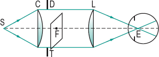
maxwellian view Method of observation in which a converging lens forms an image in the plane of the entrance pupil of the observer. If the observer’s eye is focused on the lens, the lens will appear as a disc filled with light of uniform intensity. This optical arrangement makes it possible to choose the point of incidence within the pupil, to minimize the effect of the optical aberrations of the eye and to avoid the effect of pupil size on the amount of light entering the eye (Fig. M6).
maxwellian view system, clinical Instrument designed to measure visual acuity by using a narrow beam or beams of light focused within the entrance pupil of the eye. The location of the beam or beams within the pupil can be controlled by the clinician. Such an instrument is valuable to assess acuity when part of the pupil is obstructed by a cataract or other opacity as the beam or beams of light can be directed to enter the eye through an area of the pupil where there is no opacity, thus providing an estimate of the visual acuity unaffected by optical image degradation. The results can contribute to the decision as to whether removal of a cataract will be beneficial. There are several types of these instruments. The Potential Acuity Meter (PAM) focuses a single beam of light in the pupil and a letter chart onto the retina. Others focus two beams of light in the pupil and a grating which can be produced on the retina by interference (if the two sources are coherent). This method is called laser interferometry. Examples of these are the Lotmar Visometer and the IRAS Randwal Interferometer, which are referred to as clinical interferometers. They tend to penetrate dense cataracts better than the PAM.
See coherent sources; entoptoscope, blue field; hyperacuity; interferometer.
Maxwell’s spot See spot, Maxwell’s.
McCollough effect See effect, McCollough.
Meares–Irlen syndrome See syndrome, Meares–Irlen.
media, ocular The transparent substances of the eye, i.e. the cornea, the aqueous humour, the crystalline lens and the vitreous humour.
medial Relating to the middle; nearer the median plane.
medial rectus muscle See muscle, medial rectus.
median line; plane See under the nouns.
medium, optical Any material, substance or space through which light can be transmitted.
medullated nerve fibres See fibres, myelinated nerve.
Meesmann’s dystrophy See dystrophy, Meesmann’s.
megalocornea A non-progressive enlargement of the cornea (more than 13 mm in diameter) without significant change in corneal thickness and normal clarity and function. Intraocular pressure is normal and high myopia and astigmatism often occur. It is usually transmitted as an X-linked recessive trait. Some systemic associations include Marfan’s syndrome, Apert’s syndrome, Down’s syndrome, Weil–Marchesani syndrome and osteogenesis imperfecta.
See cornea plana; keratoglobus; microcornea.
megalophthalmos A congenital condition in which the eye is abnormally large, particularly the structures of the anterior segment. The condition is associated with megalocornea, Marfan’s syndrome and Apert’s syndrome.
megalopsia See macropsia.
meibometry A method for quantifying the amount of lipids present in the tears. It may be used to facilitate the diagnosis of meibomian gland dysfunction.
See glands, meibomian.
meibomian cyst See chalazion.
meibomian gland dysfunction; glands
See glands, meibomian.
meibomianitis Inflammation of the meibomian glands. It is believed not to be a primary bacterial disease. It is characterized by the presence of a white, frothy secretion or ‘foam’ on the eyelid margin. The posterior lid margin is hyperaemic and the meibomian gland orifices are obstructed. Meibomianitis is often associated with blepharitis and conjunctivitis. Symptoms include mild itching of the lids and occasionally blurred vision due to the oily secretion spreading over the cornea. This condition may also result from hard contact lens wear. Management of this disease consists of tarsal massage and removal of the secretion with a moist cotton-tipped applicator and antibiotic medication (e.g. tetracycline, erythromycin). Syn. meibomitis. See blepharitis, marginal; blepharitis, posterior; glands, meibomian; hordeolum, internal.
meibomitis See
Stay updated, free articles. Join our Telegram channel

Full access? Get Clinical Tree


