C
CAB Cellulose acetate butyrate is a transparent thermoplastic material that is used in the manufacture of gas permeable contact lenses as it transmits some oxygen. It is a copolymer with varying percentages of cellulose, butyryl and acetyl. CAB lenses vary in their characteristics depending on the percentages of the three components. It is also used to make spectacle frames.
‘café au lait’ spots See disease, von Recklinghausen’s.
calcarine fissure See fissure, calcarine.
caliper A device used to measure distances between structures or surfaces. It usually comprises a scale at one end, while at the other end are two legs, which can be adjusted to the appropriate measurement. Example: lens thickness caliper.
caloric nystagmus See caloric testing.
caloric testing A neuro-ophthalmic technique in which cold and warm water is used to stimulate the vestibular system creating horizontal nystagmus (called caloric nystagmus or Barany’s nystagmus). Cold water placed in the ear induces a fastbeating vestibular nystagmus with the fast phase moving away from the stimulated ear, while warm water causes the fast phase to move in the direction of the stimulated ear. The mnemonic COWS (cold–opposite, warm–same) is used to describe this effect. By placing the subject at a 30-degree upright position, heated or cooled water stimulates the now vertical horizontal semicircular canals.
camera, fundus A camera attached to an indirect ophthalmoscope aimed at photographing the image of the fundus of the eye. This image is produced by the objective of the ophthalmoscope at the first focal point of the objective of the viewing microscope (and of the camera), which forms an image on the film. A flip mirror within the optical path of the viewing microscope allows the observer to view the image of the fundus and focus it, thus ensuring that the image being photographed is as clear as that being viewed. Fundus cameras usually require a dilated pupil of about 4 mm and their fields of view extend up to 45°. They provide an objective photographic record of any condition in the fundus. They can also be used to take photographs of the anterior segment of the eye.
See fundus, ocular; ophthalmoscope, indirect; ophthalmoscope, scanning laser.
camera obscura See camera, pinhole.
camera, pinhole A camera in which the lens is replaced by a pinhole (e.g. the camera obscura).
campimeter An instrument for the measurement of the visual field, especially the central region (usually within a radius of 30°).
See analyser, Friedmann visual field; chart, Amsler; perimeter; screen, tangent.
campimetry Measurement of the visual field with a campimeter.
Canada balsam A transparent resinous substance produced by the sap of the Canadian balsam fir and used to cement glass (doublet, beam-splitting prisms, the segment of a bifocal lens, etc). Its index of refraction is equal to about 1.54. It has nowadays been superseded by modern chemical adhesives.
canal A tubular channel which allows the passage of air, food, blood, excretions, secretions, or anatomical structures such as nerves or blood vessels.
Cloquet’s c. See canal, hyaloid.
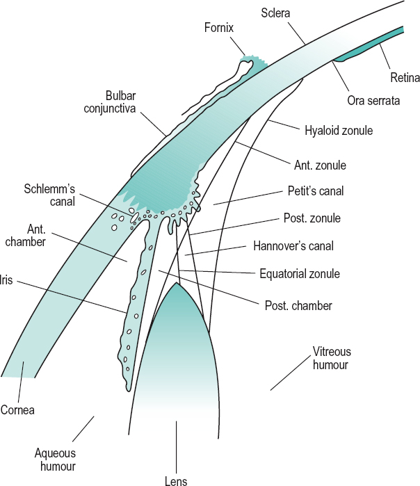
Hannover’s c . A space about the equator of the crystalline lens made up between the anterior and posterior parts of the zonule of Zinn and containing aqueous humour and zonular fibres (Fig. C1).
hyaloid c. A channel in the vitreous humour, running from the optic disc to the crystalline lens. In fetal life this canal contains the hyaloid artery, which nourishes the lens, but it usually disappears prior to birth. Syn. central canal; Cloquet’s canal; Stilling’s canal.
See hyaloid remnant.
infraorbital c. A channel beginning at the infraorbital groove in the floor of the orbit and ending at the infraorbital foramen of the maxillary bone opening onto the face below the inferior orbital margin. It is a channel for the infraorbital artery and the infraorbital nerve.
nasolacrimal c. See Table O4.
optic c. A canal leading from the middle cranial fossa to the apex of the orbit in the small wing of the sphenoid bone through which pass the optic nerve and the ophthalmic artery. Syn. optic foramen.
See Table O4.
c. of Petit A space between the posterior fibres of the zonule of Zinn and the anterior surface of the vitreous humour (Fig. C1).
Schlemm’s c. A circular venous sinus located in the corneoscleral junction, anterior to the scleral spur and receiving aqueous humour from the anterior chamber and discharging into the aqueous and the anterior ciliary veins (Fig. C1). Syn. scleral sinus; sinus circularis iridis; sinus venosus sclerae; venous circle of Leber.
See meshwork, trabecular; scleral spur; vein, aqueous.
Stilling’s c. See canal, hyaloid.
canaliculi See lacrimal apparatus.
canaliculitis Inflammation of a lacrimal canaliculus, most frequently the lower one. The patient presents with a red, irritated eye with ‘pouting’ of the punctum and a slight discharge, which can be expressed by compressing the canaliculus.
candela The candela is the luminous intensity in a given direction of a source emitting monochromatic radiation of frequency 540 × 1012 Hz and the radiant intensity of which in that direction is 1/683 watt per steradian. The candela so defined is the base unit applying to photopic quantities. Symbol: cd.
candela per square metre SI unit of luminance. Syn. nit. Symbol: cd/m2.
candelpower Designates a luminous intensity expressed in candelas.
canthal tendon See ligament, palpebral.
canthus The angle formed by the upper and lower eyelids at the nasal (inner canthus or medial canthus) or temporal (outer canthus or external angle) end. Plural: canthi. Syn. palpebral angle.
See caruncle, lacrimal; conjunctivitis, angular; epicanthus.
capsular fixation Process of inserting an intraocular lens implant into the capsular bag following cataract extraction.
See capsulectomy; phacoemulsification.
capsule, Bonnet’s See Tenon’s capsule.
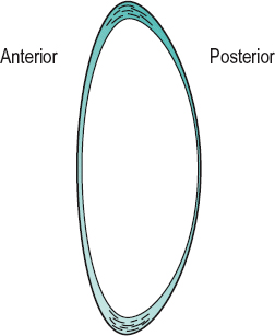
capsule, crystalline lens Transparent elastic capsule covering the crystalline lens. It is made up of collagen fibrils embedded in a glycosaminoglycan matrix. The thickness of the capsule varies; the anterior portion is thicker than the posterior and it is also thicker towards the periphery (or equator). This variation in thickness plays a role in moulding the lens substance, contributing to an increase in the curvature of the front surface, in particular, during accommodation. The capsule increases in thickness with age, and its modulus of elasticity decreases with age, which (besides flattening of the lens, and a hardening of the lens substance) contributes to presbyopia (Fig. C2). Under electron microscopy the capsule appears to have a lamellar structure that disappears with age. The capsule receives the insertion of the zonular fibres.
See fibres, lens; modulus of elasticity; shagreen of the crystalline lens; theory, Fincham’s; Zinn, zonule of.
capsule, Tenon’s See Tenon’s capsule.
capsulectomy The surgical removal of the capsule of the crystalline lens. In extracapsular cataract extraction only the anterior portion of the capsule is removed.
See capsulorhexis; capsulotomy.
capsulorhexis A form of capsulotomy in which the incision of the anterior capsule is made in a smooth circular pattern along the periphery of the lens to enable removal of opaque lens material during extracapsular cataract extraction. It is the preferred method of capsulotomy when inserting a rigid intraocular lens because the incision is less likely to tear than that made by the ‘can-opener’ technique.
capsulotomy Incision of the capsule of the crystalline lens. Capsulotomy of the anterior capsule is performed in extracapsular cataract extraction (anterior capsulotomy) to enable removal of opaque lens material. The cuts are made with a cystotome along the periphery of the lens in a circular, jagged configuration (called ‘can-opener’ capsulotomy). It is also performed on the posterior capsule (posterior capsulotomy) when it has become opaque following extracapsular extraction (manual or phacoemulsification) in order to regain the loss of visual acuity. The most common instrument used in this operation is the neodymium-yag laser.
See after-cataract; capsule, crystalline lens; capsulorhexis; cataract extraction, extracapsular.
carbachol See parasympathomimetic drug.
carbomer See tears, artificial.
carbon dioxide See hypercapnia.
carbonic anhydrase inhibitors Drugs which inhibit the carbonic anhydrase enzyme in the ciliary epithelium of the ciliary body. This enzyme is essential for the formation of aqueous humour; its reduction results in a decrease in intraocular pressure. Those in use are sulfonamide derivatives. They are administered systemically (e.g. acetazolamide) or topically in the treatment of glaucoma. Examples: acetazolamide, brinzolamide, dichlorphenamide, dorzolamide.
carboxymethylcellulose See tears, artificial.
carcinoma A malignant tumour of the epithelium, the tissue that lines the skin and internal organs of the body. It tends to invade surrounding tissues and to metastasize to distant regions of the body via the lymphatic vessels or the blood vessels. It is a form of cancer. Example: carcinoma of the skin.
See epithelioma; keratosis, seborrhoeic.
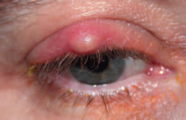
basal cell c. (BCC) A slow growing tumour derived from the basal cells of the epidermis of the skin. It is mainly located on the head and neck and most commonly on the eyelids, especially the lower eyelid. Old people who have had extensive sun exposure are primarily affected. It appears, initially, as a raised nodule with a pearly surface with small, dilated blood vessels on its surface and it may eventually become ulcerated (rodent ulcer) and invade other tissues but rarely metastasizes. Treatment includes surgical excision or cryotherapy.
sebaceous gland c. A malignant tumour arising from the meibomian glands or occasionally from the glands of Zeis. It frequently affects the upper eyelids of old people. Initially the tumour resembles a chalazion or a chronic blepharitis. However, this tumour is aggressive and may invade the orbit. It may metastasize. Treatment usually consists of thorough surgical excision.
See blepharitis; chalazion.
squamous cell c. A malignant skin cancer that affects the eyelids and conjunctiva. It is aggressive and may metastasize. It occurs most commonly in old people who have had extensive sun exposure. Treatment consists mainly of surgical excision.
Cardiff acuity test See test, Cardiff acuity.
cardinal planes; points See under the nouns.
cardinal positions of gaze See positions of gaze, cardinal.
cardinal rotation A rotation of the eye from the primary position to a secondary position about either the x-axis or the z-axis.
See axis, transverse; axis, vertical; position, primary; position, secondary.
carmellose See tears, artificial.
carrier See lens, lenticular.
carteolol hydrochloride See sympatholytic drugs.
caruncle, lacrimal A small pink fleshy structure situated in the inner canthus.
case-control study See study, case-control.
case history See history, case.
Cassegrain telescope See telescope.
cast See impression, eye.
cat’s eye syndrome See syndrome, cat’s eye.
catadioptric system See system, catadioptric.
cataphoria See kataphoria.
cataract Partial or complete loss of transparency of the crystalline lens substance or its capsule. Cataract may occur as a result of age, trauma, systemic diseases (e.g. diabetes), ocular diseases (e.g. anterior uveitis), high myopia, long-term steroid therapy, excessive exposure to infrared and ultraviolet light, heredity, maternal infections, Down’s syndrome, etc. The incidence of cataract increases with age, amounting to more than 50% in the population over 82 years. It is also more prevalent in Africa, Asia and South America than in Europe and North America. The main symptom is a gradual loss of vision, often described as ‘misty’. Some patients may also notice transient monocular diplopia, others fixed spots (not floaters) in the visual field and others better vision in dim illumination. Cataracts can easily be seen with the retinoscope, the ophthalmoscope and especially with the slit-lamp, although depending on the type, one instrument may be better than the other. At present the main treatment is surgical. Extraction is performed for one of three reasons: visual improvement, medical or cosmetic.
See after-cataract; arthritis, juvenile idiopathic; biometry of the eye; capsule, crystalline lens; disease, Wilson’s; entoptoscope, blue field; glare tester; hyperacuity; lens, crystalline; lens, intraocular; leukocoria; maxwellian view system, clinical; myopia, lenticular; phacoemulsification; syndrome, Down’s; vitreous, persistent hyperplastic primary.
age-related c. Cataract affecting older persons. It is the most common type of cataract and may take several forms: cortical, cuneiform, nuclear, mature or subcapsular. Syn. senile cataract.
anterior capsular c. A small central opacity located on the anterior lens capsule, either of congenital origin or due to a perforating ulcer of the cornea.
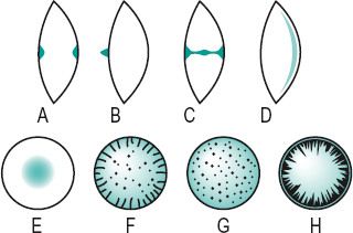
axial c. An opacity situated along the anteroposterior axis of the crystalline lens (Fig. C3).
bipolar c. An opacity involving both the anterior and the posterior poles of the lens (Fig. C3).
blue c. See cataract, blue-dot.
blue-dot c. A developmental anomaly of the crystalline lens characterized by numerous small opacities in the outer nucleus and cortex, which appear as translucent bluish dots. The condition is very common and does not usually affect acuity. Syn. blue cataract; diffuse cataract; punctate cataract.
brown; brunescent c. See cataract, nuclear.
capsular c. An opacity confined only to the capsule of the crystalline lens, anteriorly or posteriorly. It is usually congenital, although it may be acquired as a result of trauma or inflammation.
central c. See cataract, nuclear.
chalky c. A cataract characterized by the presence of lime salt deposits.
Christmas tree c. A rare type of diffuse age-related cataract in which the opacities appear as highly reflective crystals.
complicated c. A cataract caused by or accompanying another intraocular disease, such as glaucoma, cyclitis, anterior uveitis or a hereditary retinal disorder such as retinitis pigmentosa or Leber’s disease. Syn. secondary cataract.
See cataract, cuneiform; Leber’s hereditary optic atrophy; retinitis pigmentosa; syndrome, Down’s; syndrome, Fuchs’; syndrome, rubella.
congenital c. Cataract occurring as a result of faults in the early development of the lens. Some may be hereditary, usually autosomal dominant. The cause of others may be chromosomal abnormalities (e.g. Down’s syndrome, Turner’s syndrome), carbohydrate metabolic disorders (galactosaemia, galactokinase deficiency), rubella syndrome, etc. The condition requires urgent treatment to prevent the development of amblyopia.
See cataract, capsular; cataract, lamellar; cataract, nuclear; cataract, polar; cataract, sutural.
coronary c. A cataract characterized by a series of opacities having the shape of a crown or ring near the periphery of the lens (Fig. C3).
cortical c. Cataract affecting the cortex of the lens. The opacities often begin as spokes or isolated dots or clusters forming the cuneiform or subcapsular types of cataract, but eventually the opacity spreads through the entire cortex.
cuneiform c. Age-related cataract characterized by opacities distributed within the periphery of the cortex of the lens in a radial manner, like spokes on a wheel (Fig. C3).
See cataract, subcapsular.
cupuliform c. See cataract, sub-capsular.
diabetic c. Cataract associated with diabetes. In old eyes this type is similar to that of a non-diabetic person but in young eyes it is typically of the snowflake type.
diffuse c. See cataract, blue-dot; cataract, Christmas tree.
electric c. An opacity caused by an electric shock.
c. extraction, extracapsular (ECCE) Surgical procedure for the removal of a cataractous crystalline lens. The anterior capsule is excised, the lens nucleus is removed and the residual equatorial cortex is aspirated. The posterior capsule may be polished. An intraocular lens implant may then be inserted.
See after-cataract; biometry of the eye; capsulectomy; lens, intraocular; pearls, Elschnig’s; phacoemulsification; ring, Soemmering’s.
c. extraction, intracapsular (ICCE) Surgical procedure for the removal of a cataractous crystalline lens. The entire lens, together with its capsule, is removed. This procedure is rarely performed nowadays.
See ligament of Wieger.
fluid c. Hypermature cataract in which the lens substance has degenerated into milky fluid.
glassblower’s c. See cataract, heat-ray.
heat-ray c. Cataract due to excessive exposure to heat and infrared radiation. Syn. glassblower’s cataract; thermal cataract.
See exfoliation of the lens; infrared.
hypermature c. The last stage in the development of age-related cataract in which the lens substance has disintegrated.
See cataract, incipient; cataract, intumescent cortical; cataract, mature; glaucoma, phacolytic.
incipient c. The first stage in the development of age-related cataract characterized by streaks similar to the spokes of a wheel or with an increased density of the nucleus.
See cataract, intumescent cortical; cataract, mature; lens, crystalline; sight, second.
intumescent cortical c. A stage of development of a cataract in which the lens, especially the cortex, absorbs fluid and swells. It may lead to secondary angle-closure glaucoma. The cataract can progress to the hypermature stage in which case the fluid leaks out, resulting in shrinkage of the lens and wrinkling of the anterior capsule, leaving the harder nucleus free within the capsule.
See cataract, morgagnian.
lamellar c. A congenital cataract affecting one layer of the crystalline lens only. Syn. zonular cataract.
mature c. The middle stage in the development of age-related cataract characterized by a completely opaque lens and considerable loss of vision.
See cataract, hypermature; cataract, incipient.
morgagnian c. A hypermature age-related cataract in which the cortex has shrunk and liquefied and the nucleus floats within the lens capsule. Degraded lens proteins may leak into the aqueous humour and cause phacolytic glaucoma. Syn. cystic cataract; sedimentary cataract.
See cataract, intumescent cortical.
nuclear c. An opacity affecting the lens nucleus. It can be either congenital or age-related in origin. It frequently leads to an increase in myopia (or decrease in hyperopia). In some cases it reaches such a brown colour that it is called brunescent cataract (or brown cataract). Syn. central cataract (Fig. C3).
polar c. A congenital opacity found at either pole of the crystalline lens. Anterior polar cataract may be flat or project as a conical opacity (pyramidal cataract) into the anterior chamber (Fig. C3). Posterior types may be associated with persistent hyaloid remnant (Mittendorf’s dot).
punctate c. See cataract, blue-dot.
pyramidal c. See cataract, polar.
senile c. See cataract, age-related.
secondary c. 1. Syn. for complicated cataract. 2. Syn. for after-cataract.
snowflake c. A cataract characterized by greyish or whitish flakelike opacities. It is usually found in young diabetics or severe cases of diabetes (Fig. C3).
soft c. Cataract in which the lens nucleus is soft.
See lens, crystalline.
subcapsular c. An age-related opacity located beneath the anterior or posterior capsule. It may spread from the periphery of the cortex like spokes on a wheel (cuneiform cataract). This is the most common type of cortical cataract. The opacities may also be confined to the posterior layers of the cortex with a granular or lace-like appearance (cupuliform cataract). Subcapsular cataracts are often the result of radiation exposure, age, toxic damage (e.g. from corticosteroids), or secondary to eye diseases (e.g. uveitis, retinitis pigmentosa). (Fig. C3).
sunflower c. See chalcosis lentis.
c. surgery See capsulectomy; capsulorhexis; capsulotomy; cataract extraction, extracapsular; cataract extraction, intracapsular; phacoemulsification.
sutural c. A congenital cataract in which the opacities are found along the anterior and/or posterior lens sutures. The opacities may appear Y-shaped or flower-shaped. The condition is often associated with Fabry’s disease.
thermal c. See cataract, heat-ray.
traumatic c. Cataract following injury to the lens, its capsule, or to the eyeball itself. It is commonly unilateral. Penetrating trauma of the lens causes rapid opacification of the cortex or even most of the lens contents. Concussion of the lens may result in capsular, subcapsular or cortical opacities.
See ring, Vossius’.
zonular c. See cataract, lamellar.
catoptric image See image, catoptric.
catoptrics The branch of optics which deals with reflection and reflectors.
caustic The concentration of light in the caustic surface of a bundle of converging light rays which represents the focal image in an optical system uncorrected for spherical aberration. It appears as a hollow luminous cusp with its apex at the paraxial focus.
cavernous haemangioma; plexus; sinus See under the nouns.
cecocentral See centrocecal.
ceftazidime See antibiotic.
ceftriaxone See antibiotic.
cefuroxime See antibiotic.
cell 1. In biology, the basic, structural and functional units from which living organisms and tissues are built. A cell consists of a nucleus surrounded by all the cellular contents (cytoplasm) including various organelles (mitochondria, Golgi apparatus, lysosomes, ribosomes, etc.) and inclusions (glycogen, melanin, triglycerides, etc.) suspended in intracellular fluid (water, proteins, carbohydrates, lipids, inorganic and organic substances) all enclosed in a plasma membrane. There are many types of cells (blood cells, connective tissue cells, epithelial cells, muscle cells, nerve cells, secretory cells, etc.). Living cells are capable of reproduction (for body growth, wound healing, etc.) by mitotic activity. 2. In optics, a rim in a trial frame or in an optical instrument into which a lens can be placed.
A c. See cell, M.
acinar c. A type of cell found within the body of the lacrimal gland. This cell lines the lumens of glands in a lobular pattern and produces a serous secretion.
amacrine c. Retinal cell located in the inner nuclear layer connecting ganglion cells with bipolar cells. Some have an ascending axon synapsing with receptors.
B c. See cell, P.
basal c. See corneal epithelium.
binocular c. A cell in the visual cortex that responds to stimulation from both eyes. It may, however, show an ocular dominance for either eye. It responds more strongly when corresponding regions of each eye are stimulated by targets of similar size and orientation.
See column, cortical; hypercolumn.
bipolar c. Retinal cell located in the inner nuclear layer connecting the photoreceptors with amacrine and ganglion cells.
C c. A retinal ganglion cell with slow axonal conduction which sends information to the superior colliculus and to the centre involved in the control of pupillary diameter, rather than to the lateral geniculate body. There are very few such cells. Syn. Pγ cell; W cell (thus called in the cat).
Cajal’s c. See astrocytes.
clump c. Large, pigmented round cells found in the pupillary zone of the iris stroma. They are considered to be macrophages containing mainly melanin granules. The number of these cells increases with age.
colour-opponent c’s. Cells which respond by increasing response to light of some wavelengths and decreasing their response to others (usually complementary). If the light stimulus contains both sets of wavelengths the two responses tend to cancel each other. Two types of cells have been identified: red-green cells and blue-yellow cells. These cells are found mainly in the lateral geniculate bodies but also among retinal ganglion cells, and they form the blobs in the visual cortex. The responses of these cells support Hering’s theory of colour vision. Syn. opponent-process cell (although this term also includes a cell that increases its response to white light and decreases its response to dark).
See blobs; theory, Hering’s of colour vision.
complex c. A cell in the visual cortex whose receptive field consists of a large responsive area, approximately rectangular in shape, surrounded by an inhibitory region. The stimulus, which is usually a slit or a straight line, gives an optimum response if appropriately orientated but falling anywhere within the excitatory area. These cells tend to respond optimally to the movement of a specifically orientated slit. Many complex cells also respond better when the optimally orientated slit is moved in one direction rather than in the opposite direction. In general, complex cells show non-linear spatial summation properties.
See area, visual; cell, hypercomplex; cell, simple; field, receptive; summation.
cone c. Photoreceptor of the retina which connects with a bipolar cell and is involved in colour vision and high visual acuity and which functions in photopic vision. The outer segment of the cell is conical in shape, except in the fovea centralis where it is rodlike. In the outer segment (i.e. the part closest to the pigment epithelium) are contained hollow discs (or lamellae), the membranes of which are joined together and are also continuous with the boundary membrane of the cone cell. The visual pigments are contained in these discs. There are three types of cones, each containing a different pigment sensitive to a different part of the light spectrum. They are referred to as long-wave-sensitive (or L-cones), medium-wave-sensitive (or M-cones) and short-wave-sensitive (or S-cones). There are about six million cones in the retina, with the greatest concentration in the macular area (Fig. C4).
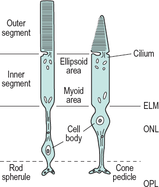
See cone pedicle; effect, Stiles–Crawford; ellipsoid; foveola; macula; pigment, visual; theory, duplicity; vision, photopic.
fixed c. See corneal corpuscle.
ganglion c. 1. Retinal cell that connects the bipolars and other cells in the inner plexiform layer with the lateral geniculate body. The axons of the ganglion cells constitute the optic nerve fibres. There are many types of ganglion cells. The two major types are: the magno (M or parasol) ganglion cells which project mainly to the magnocellular layers of the lateral geniculate bodies; and the parvo (P) ganglion cells which project to the parvocellular layers of the lateral geniculate bodies. Two types of P ganglion cells are noted: P1, which are midget cells and have small dendritic fields and P2, which have large dendritic fields. M and P cells comprise about 10% and 82% of the ganglion cells respectively. 2. One of a collection of nerve cell bodies found in a ganglion.
See cell, C; cell, M; cell, P; melanopsin.
glial c’s. Cells found throughout the nervous system. They provide support and nutrition for neurons, as well as being involved in the operation of the brain, especially the fluid surrounding the neurons and their synapses. They are also believed to be involved in the reuptake of neurotransmitters from within the synaptic cleft. There are three types of glial cells: astrocytes; microglia; oligodendroglia. Syn. glia; neuroglia.
goblet c. Cell of the conjunctival epithelium which secretes mucin.
See glands of Henle; mucin; xerophthalmia.
horizontal c. Retinal cell located in the inner nuclear layer which connects several cones and rods together.
hypercomplex c. A cell in the visual cortex that receives inputs from several simple and complex cells and therefore has an even more elaborate receptive field than a complex cell. It is most effectively stimulated by a stimulus of a specific size and of a specific orientation and which is moved in a specific direction.
See cell, complex; cell, simple.
Langerhans’ c’s. Dendritic cells located mainly in the epidermis, mucous membranes and lymph nodes. They have surface receptors for immunoglobulin (Fc), complement (C3) and surface HLA–DR (la) antigen. Langerhans’ cells are also found in the conjunctival epithelium and among the basal cells, mainly of the peripheral corneal epithelium. They have antigenic functions, stimulate T-lymphocytes, prostaglandin production and participate in cutaneous delayed hypersensitivity and corneal graft rejection. Extended wear of contact lenses tends to induce an increase of these cells. They are also found in histiocytic tumours.
Table C1
Main distinguishing features of the two principal types of ganglion cells of the retina
| properties | P cell (X cell) | M cell (Y cell) |
| size of cell body | small | large |
| dendritic spread | small | medium/large |
| receptive field size | small | medium/large |
| retinal distribution | 90% of these at the macula | 5% of these at the macula; about 13% overall |
| projection | LGN parvocellular layers | LGN magnocellular layers |
| type of response | sustained | transient |
| light sensitivity | low | high |
| wavelength response | selective (except P cells) | non-selective |
| spatial sensitivity | fine target detail | large target detail |
| temporal sensitivity | low target velocity | high target velocity |
M c. A retinal ganglion cell, mainly located in the periphery of the retina and which assists in movement perception. M cells tend to give transient responses to stimuli and to have non-linear spatial summation properties. This cell transmits information principally to the magno cells of the lateral geniculate bodies. Syn. A cell; Pαcell; Y cell (thus called in the cat).
magno c. See cell, ganglion; geniculate bodies, lateral.
Mueller’s c. Neuroglial cell in the retina with its nucleus in the inner nuclear layer and with fibres extending from the external to the internal limiting membrane. These cells support the neurons of the retina and possibly assist in their metabolism. Syn. Müller cell.
orientation-specific c. A cell that responds best to specifically orientated lines. This is the case for almost all cells in the visual cortex. Examples: complex cell; simple cell.
See cell, complex; cell, simple; field, receptive.
P c. A retinal ganglion cell, mainly located in the central region of the retina and which assists in high acuity and colour vision. P cells tend to give sustained responses to stimuli and to have linear spatial summation properties. This is the most common type of ganglion cells (about 82%). This cell transmits information principally to the parvo cells of the lateral geniculate bodies. Syn. B cell; Pβcell; X cell (thus called in the cat).
parasol c. See cell, ganglion.
parvo c. See cell, ganglion; geniculate bodies, lateral.
rod c. Photoreceptor cell of the retina which connects with a bipolar cell. It contains rhodopsin and is involved in scotopic vision. The molecules of rhodopsin are contained in about 1000 hollow discs (double lamellae or membranes), which are isolated from each other and from the boundary membrane of the rod cell. These discs are found in the outer segment (i.e. the part closest to the pigment epithelium) of the cell. There are about 100 million rod cells throughout the retina; only a small area, the foveola, is free of rods (Fig. C4).
See eccentricity; ellipsoid; foveola; rhodopsin; rod spherule; theory, duplicity; vision, scotopic.
Schwann c. A cell whose membrane spirals around the axon with layers of myelin in between each coil, as well as being a source of the myelin sheath. The cell provides insulation to the axon. It covers about one millimetre, so that hundreds may be needed to completely cover an axon. It also allows for an increase in the speed of the nervous impulse without an increase in axonal diameter. The gaps between the segments covered by the cells are called nodes of Ranvier.
simple c. A cell in the visual cortex whose receptive field consists of an excitatory and an inhibitory area separated by a straight line, or by a long narrow strip of one response flanked on both sides by larger regions of the opposite response. Responses occur only to a straight line or a narrow strip orientated approximately parallel to the boundary/ies between the two areas. In general, simple cells show linear spatial summation properties. They are presumably the first cells where the nervous impulses are processed as they enter the visual cortex.
See area, visual; cell, complex; field, receptive.
squamous c. See corneal epithelium.
W c. See cell, C.
wing c. See corneal epithelium.
X c. See cell, P.
Y c. See cell, M.
cellulose acetate butyrate See CAB.
cellulitis, preseptal Swelling or infection of the eyelid tissue in front of the orbital septum. There is redness, swelling and tenderness of the eyelid. The condition is treated with systemic antibiotics.
cellulitis, orbital Infection of the orbital contents caused by Staphylococcus aureus, Streptococcus or Haemophilus influenzae. It is often caused by the spread of infection from adjacent structures, especially the sinuses. The clinical signs are fever, pain, proptosis, redness, swelling of the lid and orbital tissue and restricted eye movements which may occasionally lead to diplopia and, as the condition worsens, visual acuity decreases. Initial management consists of parenteral antibiotics but surgery may become necessary.
See lamina papyracea.
central corneal optical zone See optical zone of cornea.
central fusion See fusion, sensory.
central retinal artery; vein See under the nouns.
central retinal artery occlusion See retinal arterial occlusion.
central retinal vein occlusion See retinal vein occlusion.
central serous retinopathy; vision; visual acuity
See under the nouns.
centration distance; near distance; point See under the nouns.
centre, boxing The point midway between the two horizontal and the two vertical sides of the rectangle enclosing the lens, in the boxing system. Syn. geometric centre of a cut lens.
centre, optical That point (real or virtual) on the optical axis of a lens which is, or appears to be, traversed by rays emerging parallel to their original direction. Applied to an ophthalmic lens, it is commonly regarded as coinciding with the vertex of either surface (British Standard).
centre of rotation of the eye When the eye rotates in its orbit, there is a point within the eyeball that is more or less fixed relative to the orbit. This is the centre of rotation of the eye. In reality, the centre of rotation is constantly shifting but by a small amount. It is considered, for convenience, that the centre of rotation of an emmetropic eye lies on the line of sight of the eye 13.5 mm behind the anterior pole of the cornea when the eye is in the straight ahead position (straightforward position), that is when the line of sight is perpendicular to both the base line and the frontal plane.
See axis, anteroposterior; line of sight.
centre, standard optical position A reference point specific to each spectacle lens shape. The standard optical position is on the vertical line passing through the boxed centre, and is at the boxed centre.
See centre, boxing; system, boxing.
centre, visual Centre of the brain concerned with vision.
See area, visual; fissure, calcarine.
centrocecal An area of the retina which includes the macula, the optic disc and the area in between. Note: also spelt centrocaecal. Syn. cecocentral.
cephalosporin See antibiotic.
cerium oxide A pink powder derived from the metallic element cerium. It is used to polish lenses and it is also added to ophthalmic glass to absorb ultraviolet radiations.
See polishing.
cetirizine See antihistamine.
cetrimide See antiseptic.
chalazion A chronic inflammatory lipogranuloma due to retention of the secretion (such as blocked ducts) of a meibomian gland in the tarsus of an eyelid. It is characterized by a gradual painless swelling of the gland without marked inflammatory signs and sometimes astigmatism which is induced by the cyst pressing on the cornea (Fig. C5). Small chalazia may disappear spontaneously but large ones usually have to be incised and curetted (i.e. removal of the pus with a scraper) through a tarsal incision. Resolution may also occur after local injection of a corticosteroid drug (e.g. dexamethasone or triamcinolone). Syn. meibomian cyst (although it is not a true cyst because its walls are made of granulomatous tissue and not lined with epithelium).
chalcosis lentis A cataract caused by excessive amount of copper in the eye. It appears as small yellowish-brown opacities in the subcapsular cortex of the lens and pupillary zone with petal-like spokes that extend towards the equator. It may be due to an intraocular foreign body containing copper, or from eyedrops that contain copper sulfate, or as part of Wilson’s disease. Management consists mainly of removal of the foreign body. Syn. sunflower cataract.
chamber In anatomy, a small cavity.
anterior c. (AC) Space within the eye filled with aqueous humour and bounded anteriorly by the cornea and posteriorly by the iris and the part of the anterior surface of the lens which appears through the pupil. Its average axial length is 3.2 mm.
See angle of the anterior chamber; flare, aqueous; gonioscope; method, van Herick, Shaffer and Schwartz; method, Smith’s; optics of the eye; test, shadow.
c’s. of the eye The anterior, posterior and vitreous chambers of the eye.
posterior c. Space within the eye filled with aqueous humour and bounded by the posterior surface of the iris, the ciliary processes, the zonule and the anterior surface of the lens.
vitreous c. Space within the eye filled with vitreous humour and bounded by the retina, ciliary body, canal of Petit and the postlenticular space of Berger.
Chandler’s syndrome See syndrome, Chandler’s.
channel A concept relating to the evidence that information about a particular feature of an image is transmitted and processed in the visual pathway approximately independently of information about other domains. The evidence was obtained from various experiments: matching, threshold elevation, after-effect, etc. Examples: the three channels of colour vision theory; the spatial frequency channels.
See after-effect, waterfall.
chaos, light See light, idioretinal.
Charles Bonnet syndrome See syndrome, Charles Bonnet.
chart 1. A tabular presentation of test targets for assessing vision. 2. A recording of clinical data relating to a patient’s case.
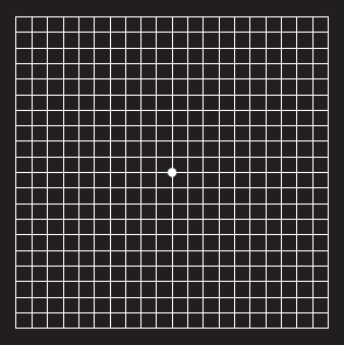
Amsler c. One of a set of charts used to detect abnormalities in the central visual field which are so slight that they are undetected by the usual methods of perimetry. There are various patterns, each on a different chart, 10 cm square. One commonly used chart consists of a white grid of 5 mm squares on a black background. Each pattern has a dot in the centre, which the patient fixates. When fixated at a distance of 30 cm the entire chart subtends an angle of 20°. If there is any visual impairment (usually as a result of macular disease) it is demonstrated by the absence or irregularities of the lines (Fig. C6). Syn. Amsler grid.
See macular degeneration, age-related; metamorphopsia.
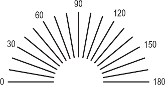
astigmatic fan c. A test pattern consisting of a semicircle of radiating black lines on a white background for determining the presence and the amount, as well as the axis of ocular astigmatism. If the chart resembles the ‘clock face’ type it is called an astigmatic dial or clock dial chart (Fig. C7).
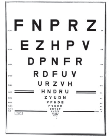
Bailey–Lovie c. A visual acuity chart with letter sizes ranging from 6/60 (20/200) to 6/3 (20/10) in 14 rows of 5 letters. Each row has letters which are approximately 4/5 the size of the next larger letters and the letters in each row have approximately the same legibility (within ±10%). It is most useful with low vision patients. This is the most commonly used type of log MAR charts (Fig. C8). There is also a Bailey–Lovie Word Reading Chart for near vision. It is composed of words rather than letters. The size progression of each line is logarithmic. The typeface used is the lower case Times Roman customarily used in newspapers and books, and the range of sizes varies between 80-point and 2-point print (or the Snellen equivalent at 40 cm of 6/144 or 20/480 to 6/3.6 or 20/12, respectively). There are 20 such charts, each with a different set of words.
See acuity, near visual.
clock dial c. See chart, astigmatic fan.
contrast sensitivity c. A chart designed to test contrast sensitivity. Such a test is useful with patients having low vision and in the early detection of diseases. Examples: Pelli–Robson chart; reduced contrast Bailey–Lovie near log MAR letter chart.
See acuity cards, Teller; test, Arden grating; Vistech.
illiterate E c. Chart for carrying out a subjective visual acuity test on a person who cannot read. It consists of a graduated series of the Snellen letter E orientated in various directions which the subject must recognize. This procedure is sometimes called the ‘E’ test or ‘E’ game.
See acuity cards, Teller; ‘E’ game; test, Cardiff acuity.
Landolt broken ring c. A visual acuity chart using a graduated series of Landolt rings in which the target thicknesses and gaps are equal to one-fifth of the outer diameter. The subject must indicate the orientation of the gap, which usually appears in one of four directions: right, left, up or down. This test is less subjective than the Snellen chart. Syn. Landolt C chart.
log MAR c. A visual acuity chart in which the rows of optotypes vary in a logarithmic progression. The multiplier of the geometric progression is usually equal to 1.2589 or 0.1 log unit. On one side of such a chart the rows of optotypes are usually labelled with the traditional Snellen notation. On the other side of each row visual acuity is labelled as the logarithm of the minimum angle of resolution (log MAR), which is the logarithm to the base 10 of the angular subtense of the stroke widths of the optotypes at a standard distance.
See chart, Bailey–Lovie; Glasgow acuity cards.
Pelli–Robson c. A contrast sensitivity chart consisting of eight lines of letters, all of the same size, subtending 3 degrees at a viewing distance of 1 m. On each line there are two groups, each containing three different letters; the letters in each group have the same contrast. The contrast of the different letters in each group decreases by a factor of 1/2 and the range of contrast varies between 100 and 0.6% in 16 steps. The subject is asked to read the letters starting with those of high contrast and continuing until two or three letters in one group are incorrectly named. The contrast threshold is represented by that of the previous group of letters. The chart gives the results in log contrast sensitivity. This test provides a measurement of contrast sensitivity at low to intermediate spatial frequencies depending upon the viewing distance. Syn. Pelli–Robson contrast sensitivity test.
Raubitschek c. A test target for determining the axis and the amount of astigmatism of the eye. It consists of two parabolic lines (known as wings) in an arrowhead pattern, parallel and closely spaced at one end, and each diverging from each other through a 90° angle at the other end. There are several methods of using this test. Syn. Raubitschek arrows; Raubitschek dial.
reduced contrast Bailey–Lovie near log MAR letter c. A set of 30 Bailey–Lovie charts designed to measure contrast sensitivity. Each chart has a different contrast, half of the charts contains the letters of the distance Bailey–Lovie chart and the other half contains the letters of the near Bailey–Lovie chart. All the charts have the same average reflectance and the contrast of the charts ranges from 0.95 to 0.0013%. The charts are presented to the subject in order of increasing contrast and the subject reads the letters from the largest to the smallest lines that they are able to. Threshold resolution in log min arc is determined for each of the 30 charts and a contrast sensitivity curve can thus be determined.
Snellen c. A visual acuity test using a graduated series of Snellen letters (or Snellen test types), in which the limbs and the spaces between them subtend an angle of one minute of arc at a specified distance. The letters are usually constructed so that they are 5 units high and 4 units wide, although some charts use letters that fit within a square subtending 5 minutes of arc at that distance.
See acuity, Snellen; Snellen fraction.
Table C2
Relationship between the Snellen fraction and the log MAR notation for distance visual acuity
| Snellen fraction | log MAR | |
| (m) | (ft) | |
| 6/150 | 20/500 | 1.4 |
| 6/120 | 20/400 | 1.3 |
| 6/95 | 20/320 | 1.2 |
| 6/75 | 20/250 | 1.1 |
| 6/60 | 20/200 | 1.0 |
| 6/48 | 20/160 | 0.9 |
| 6/38 | 20/125 | 0.8 |
| 6/30 | 20/100 | 0.7 |
| 6/24 | 20/80 | 0.6 |
| 6/19 | 20/63 | 0.5 |
| 6/15 | 20/50 | 0.4 |
| 6/12 | 20/40 | 0.3 |
| 6/9.5 | 20/32 | 0.2 |
| 6/7.5 | 20/25 | 0.1 |
| 6/6 | 20/20 | 0 |
| 6/4.75 | 20/16 | –0.1 |
| 6/3.75 | 20/12.5 | –0.2 |
| 6/3 | 20/10 | –0.3 |
test c. A board externally illuminated, an internally illuminated transparent sheet, a slide for projection or a computer based system which projects optotypes or other tests used in the subjective determination of refraction. Syn. letter chart.
See legibility; optotype.
Chavasse lens See lens, Chavasse.
check ligament See ligament, check.
checkerboard pattern See pattern, checkerboard.
cheiroscope An instrument used in the management of amblyopia, suppression and hand and eye coordination. It consists of presenting a line drawing to one eye (usually the dominant one), which is traced by a pencil or crayon in the field of view of the other eye. The two fields of view are separated by a septum and a small mirror is used to reflect the line drawing. Stereoscopes can easily be adapted into cheiroscopes.
chemical burn An injury caused, usually, by alkali (e.g. ammonia, caustic potash, lime, sodium hydroxide) or acid (e.g. hydrochloric, sulphuric). The type and severity of the injury depends on the properties of the chemical and upon which ocular tissue is involved. However, alkali burns are more severe than acid burns because they penetrate the tissues more rapidly and more deeply. In all cases, immediate copious irrigation is crucial, followed by a topical anaesthetic to relieve pain. Irrigation is continued until repeated measurements of ocular pH reach and retain a normal value. Treatment includes cycloplegics, antibiotics, steroids, ascorbate (only in alkali burns) to restore collagen synthesis, and glaucoma medication may be needed to prevent an increase of intraocular pressure. In some cases, surgery may also be required.
Table C3
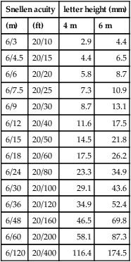
Relationship between Snellen visual acuity and letter height at two viewing distances (the letter corresponding to an acuity of 6/6 subtends 5’ and the gap in the letter 1’)
| Snellen acuity | letter height (mm) | ||
| (m) | (ft) | 4 m | 6 m |
| 6/3 | 20/10 | 2.9 | 4.4 |
| 6/4.5 | 20/15 | 4.4 | 6.5 |
| 6/6 | 20/20 | 5.8 | 8.7 |
| 6/7.5 | 20/25 | 7.3 | 10.9 |
| 6/9 | 20/30 | 8.7 | 13.1 |
| 6/12 | 20/40 | 11.6 | 17.5 |
| 6/15 | 20/50 | 14.5 | 21.8 |
| 6/18 | 20/60 | 17.5 | 26.2 |
| 6/24 | 20/80 | 23.3 | 34.9 |
| 6/30 | 20/100 | 29.1 | 43.6 |
| 6/36 | 20/120 | 34.9 | 52.4 |
| 6/48 | 20/160 | 46.5 | 69.8 |
| 6/60 | 20/200 | 58.1 | 87.3 |
| 6/120 | 20/400 | 116.4 | 174.5 |
chemodenervation A technique in which a pharmacologic compound (e.g. atropine, botulinum toxin) is used to paralyse a muscle or group of muscles. This technique is most often used in the treatment of certain forms of strabismus as well as blepharospasm.
chemosis Severe oedema of the conjunctiva.
See ophthalmopathy, thyroid.
cherry-red spot See spot, cherry-red.
Cheshire cat effect See effect, Cheshire cat.
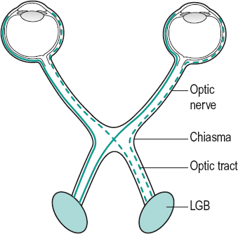
chiasma, optic A structure located above the pituitary gland and formed by the junction and partial decussation (crossing-over) of the optic nerves. The fibres from the nasal half of the retina of the left eye cross over to join the fibres from the temporal half of the right retina to make up the right optic tract and vice versa. About 53% of the axons of the optic nerves cross to the opposite tract (Fig. C9). A lesion of the chiasma produces a typical field defect (heteronymous hemianopia). Note: also spelt chiasm.
See circle of Willis; decussation; pathway, visual; stereo-blindness; tracts, optic.
chiastopic fusion See fusion, chiastopic.
Chievitz, transient layer of See layer of Chievitz, transient.
chlamydial infection See conjunctivitis, adult inclusion; keratitis, epithelial; ophthalmia neonatorum; trachoma.
chlorambucil See
Stay updated, free articles. Join our Telegram channel

Full access? Get Clinical Tree


