Lids and Adnexa
Alex V. Levin
Thomas W. Wilson
Robert Pashby
Dan DeAngelis
The lids and adnexa are composed of the anterior and posterior lamella. The anterior lamella is composed of the skin, subcutaneous tissue (which is the thinnest in the body), cilia, and orbicularis muscle. The posterior lamella consists of the orbital septum, tarsus (including meibomian glands), and conjunctiva. Embryologically, the upper eyelids are derived from the frontonasal process and the lower eyelid is derived from the maxillary process of the face. The eyelids fuse at approximately 7 weeks’ gestation and typically open by the sixth month of gestation. The events that lead to eyelid separation in utero are thought to be related to either keratinization of the eyelid margin or secretions by the primitive meibomian glands. Proper development of the eyelids and adnexa is dependent on normal fusion of the eyelids. Inadequate fusion of the eyelids can result in eyelid colobomas and deficiencies in the adnexal structures, namely the cilia and tarsus. Similarly, abnormal separation of the eyelids can result in persistent bands to the globe or the opposing lid.
Early in the development of the adnexa, the meibomian glands are pluripotential structures that most commonly develop into glandular elements. However, they also have the potential to develop into follicular structures. If this occurs, then cilia are seen emerging from the posterior half of the eyelid margin, in the meibomian orifices. Thus, the patient will have an extra accessory row of anatomically normal eyelashes, which can cause significant ocular irritation.
The normal eyelid crease is formed by the projection of the levator fibers anteriorly, as they interdigitate among the fibers of the orbicularis oculi muscle. Congenital ptosis is caused by a dystrophic change in the levator muscle, resulting in a muscle that is variably functional. As such, the fibrosis in the muscle fibers results in an absent lid crease and in lid retraction on downgaze, both prominent features of congenital ptosis.
Lid abnormalities may occur in isolation or as part of systemic syndromes and disease processes. The underlying globe may also be abnormal. Lid and adnexal malformations may also be unilateral or bilateral. An understanding of these congenital and acquired lid disorders may assist the physician in understanding coexisting ocular and systemic abnormalities.
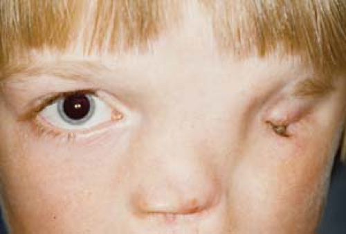 Figure 2.1 Cryptophthalmia Cryptophthalmia is complete or partial fusion of the eyelids usually with aberrant or no eyelashes. The underlying eye is typically microphthalmic and often fused to the overlying eyelid skin. Attempts to repair the skin defect often involve corneal transplantation with poor visual prognosis. The nasolacrimal system is usually absent or malformed. Cryptophthalmos can be either unilateral or bilateral. Autosomal recessive, and less commonly autosomal dominant, inheritance has been reported. Most cases are sporadic. Approximately one fourth of cases are isolated and the rest are associated with other malformations. Fraser syndrome is the association of cryptophthalmos (not obligatory), syndactyly, and abnormalities of the urogenital system. Some cases are due to mutations in the FRAS1 gene at 4q21. |
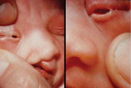 Figure 2.2 Ankyloblepharon Ankyloblepharon is complete or partial fusion of the eyelid margins. Unlike cryptophthalmus, the globe is usually normal and proper lid structures are recognizable. Most often the connections are thin bands as shown in these images. The skin adjoining the lids is called filiform adnatum. Although the strands look thin, one should not separate the lids by manual traction. Treatment is by surgical separation of the eyelid margins. Rarely, ankyloblepharon can be part of systemic conditions such as Hay-Wells syndrome. More often it is a sporadic nongenetic anomaly. |
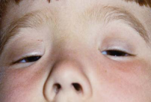 Figure 2.3 Blepharophimosis Blepharophimosis syndrome (BPES) is a combination of congenital ptosis, epicanthus inversus, and short horizontal palpebral fissure length with telecanthus (increased distance between the medial canthi). Ectropion may also be seen. As shown here, the patient may use a chin lift in the straight-ahead viewing position. There is an autosomal dominant inheritance. Type 1 BPES is also characterized by female infertility and is due to abnormalities in the FOXL2 gene at 3q22. Type 2 BPES is due to abnormalities in the same locus but with no systemic abnormalities. Another locus for isolated BPES is proposed on chromosome 7q. Treatment options include a resection of the medial canthal skin (Roveda procedure) and ptosis surgery. |
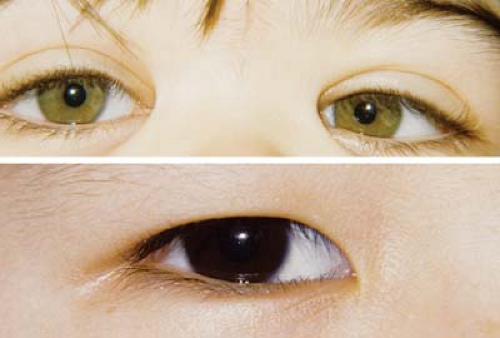 Figure 2.4 Epicanthus There are four types of epicanthal folds: supraciliaris (upper photo), palpebralis (not pictured), tarsalis (lower photo), and inversus (see Fig. 2.3). Although epicanthus inversus is more common in blepharophimosis, epicanthus is otherwise generally a nonspecific minor malformation often seen in normal individuals. Epicanthus is more prevalent in certain ethnic groups. |
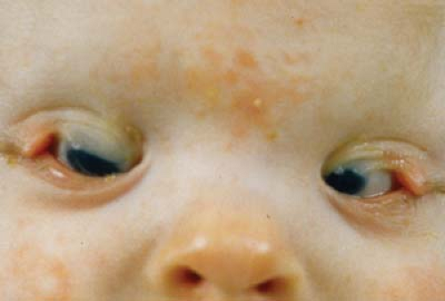 Figure 2.5 Coloboma Eyelid coloboma is a defect in the upper or lower eyelid margin. The most common location is in the nasal half of the upper lid and may be isolated or seen in association with Goldenhar syndrome. Characteristic lower eyelid colobomas are associated with Treacher Collins syndrome or mandibulofacial dysostosis (Chapter 14: Craniofacial, Figs. 14.14, 14.15 and 14.16), where the medial aspect of the lid downslopes gradually, followed by a sharp uprise to the normal lateral margin. When the medial aspect of the lid is involved with coloboma, the lacrimal puncta and canaliculus may be absent and the caruncle abnormal. As seen in this image, lid coloboma may also be associated with a form of symblepharon where conjunctiva and/or lid tissue may be attached to the globe with or without scarring of the cornea. |
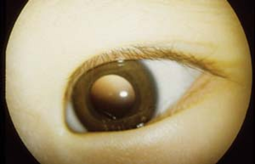 Figure 2.6 Epiblepharon Epiblepharon is caused by a horizontal fold of skin under the medial lower eyelid, which rotates the eyelashes toward the globe. It is more common in some Asian children, but may be seen in any ethnic group. Epiblepharon can often resolve with midfacial growth. Surgery is indicated for corneal irritation and scarring due to trichiasis. A horizontal strip of skin and orbicularis corresponding to the fold is removed in order to reorient the eyelashes. It is important not to include the epicanthal folds with the resection. This entity is not considered a major malformation and is often seen in the otherwise normal general population. |
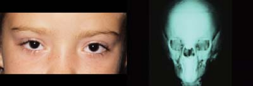 Figure 2.7 Telecanthus and Hypertelorism
Stay updated, free articles. Join our Telegram channel
Full access? Get Clinical Tree
 Get Clinical Tree app for offline access

|