Laser Devices in Measuring Visual Acuity
Daniel G. Green
Visual acuity, the ability to distinguish fineness of detail, is ordinarily tested by presenting a patient with a series of objects which vary in size or viewing distance. To resolve the test object, the patient’s eye must first form an image of the object on the photoreceptors. It is this retinal image which the eye senses. The various optical defects of the ocular media tend to degrade the retinal image. Optical aberrations cause a point source to fail to be brought to focus at a unique point. Scattering causes light to spread diffusely over large areas of the retina.
Fortunately, placing an object to be viewed before the patient is not the only means one has for producing a regular pattern on the retina. A much less commonly used procedure bypasses the effects of the eye’s optics. A pattern can be formed using the light from a laser. These patterns are produced on the retina by the interaction of the waves from this highly monochromatic source of light. Regular striped patterns (interference fringes) are formed by the superposition of waves directly on the surface of the retina. Since these are not images in the usual sense, they are not affected by ordinary optical defects. Defects of focus or imperfections in the refracting system of the eye do not blur them. Whether an observer sees these fringes depends only on the ability of his retina to conduct signals from the photoreceptors into the nervous system. Therefore, by using interference fringes it is possible to separate the retinal and neurologic factors from the optical factors limiting visual resolution.
This ability to measure visual acuity independent of the ocular refractive media has been put to good use in the study of the normal visual apparatus (1,2,3). However, one of the most exciting applications has been in evaluating potential macular function in the presence of opacities of the ocular media (4,5,6,7,8,9). For example, in the patient who has a cataractous lens and concomitant decrease in vision, the ophthalmologist is faced with the problem of deciding whether the loss in vision can be explained completely by lens changes. The problem may be compounded by past or present evidence of degenerative retinal disease or by the fact that the physician is unable to obtain a satisfactory view of the fundus. The various tests used, eg, discrimination between two lights, projection, and color perception, are, at best, indicators only of gross retinal function and may be of limited value in evaluating potential macular function. In many instances, by using the phenomena of constructive and destructive interference of coherent light, it is possible to produce graded patterns on the retinas of these patients. The fineness of the interference bands that can be seen by the patient provides the physician with a better idea of how much improvement in visual acuity may be offered his patient by cataract surgery.
PRINCIPLE
An essential characteristic of light is that it is a periodic disturbance which propagates through space. The wave motion travels in space with velocity c and wavelength λ. The periodic nature of this wave motion cannot be directly appreciated by the eye, as the number of vibrations per second is approximately 5 × 10¹⁴; however, this is not to say that the wave properties of light cannot be made visible. In 1807, Young performed an experiment which conclusively showed the existence of light waves. The essential idea was to make the wave motion apparent by interacting the wave motions from two sources of light. Fig 1 shows an analogous effect. Two regularly striped patterns of slightly different frequencies are overlapped. When viewed at a distance of several feet, the periodic nature of the separate patterns is no longer visible and they appear uniformly gray. This, however, is not the case where they overlap. Here, regular patterns called Moiré fringes, which are produced by the combination, are apparent even though the regularity of the individual patterns is not. Inspection of the fringes at closer range reveals that what appears as a light band is in fact a position where dark and light portions of the individual patterns are in close register; they are said to be “in-phase.” A dark band results from dark stripes in one pattern overlaying light stripes in the other; the patterns are “out-of-phase” with each other.
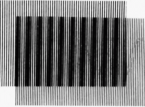 Fig. 1. Overlapping patterns (Moiré fringes) which differ slightly in the number of dark bars per unit of distance. If one views these from far enough away, the individual bars will not be resolved and patterns may still be seen in the region of overlap. |
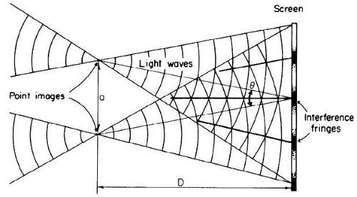 Fig. 2. Spherical wavefronts from two coherent sources interact to produce interference fringes. |
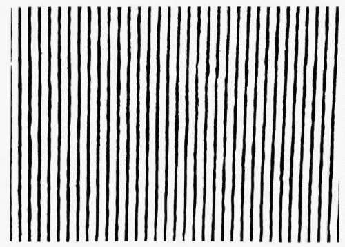 Fig. 3. Interference pattern resulting from the interactions of two coherent sources such as those illustrated in Figure 2. |
Likewise a wave in the negative x direction is given by
The sum is
Stay updated, free articles. Join our Telegram channel

Full access? Get Clinical Tree


