Laryngeal Stenosis


Laryngeal Stenosis


Michael J. Rutter

Aliza P. Cohen

Alessandro de Alarcón


Airway management of the infant, child, or adolescent may transform from the stable and routine to abrupt and life-threatening loss of the airway. This may occur either as a result of the underlying pathology or from the management of the pathology. Given the importance of a potentially emergent situation, the clinician must have a sound understanding of the causes, clinical presentations, and management of laryngotracheal stenosis. Our chapter therefore focuses on this content.


AGE-RELATED ANATOMIC DIFFERENCES IN THE LARYNX

Although the three portions of the larynx (supraglottis, glottis, and subglottis) are the same in children and adults, there are a number of age-related differences that play a role in the development of laryngotracheal stenosis.


Laryngeal Size

The infant larynx is approximately one-third the size of the adult larynx; however, it is proportionally larger than the adult larynx relative to the rest of the tracheobronchial tree. The infant vocal fold also differs in length from the adult vocal fold; it is approximately 7 to 8 mm long, whereas the adult vocal fold is 14 to 23 mm long. In the infant, half of the length of the true vocal fold is composed of the vocal process of the arytenoid. In the adult, the vocal process occupies only one-fourth to one-third of the total length of the true vocal fold. The subglottis is the narrowest part of the airway because of the complete ring structure of the cricoid cartilage. In the infant, the subglottis is approximately 4.5 to 7 mm in diameter. A child with a subglottic diameter of 4.0 mm or less has subglottic stenosis (1).


Laryngeal Location

The position of the larynx in relation to other structures of the neck is different in infants and adults. In the infant, the superior border of the larynx is located as high as the first cervical vertebra, with the cricoid cartilage positioned at approximately the level of the fourth cervical vertebra. This results in the hyoid overriding the superior larynx in an infant, with the thyroid notch usually being impalpable as a consequence. Because of the superior positioning of the larynx, the epiglottis approximates the dorsal surface of the soft palate. This contributes to the obligate nasal breathing seen in the first months of life. As the child grows into adulthood, the larynx gradually descends, and the cricoid cartilage eventually rests at the level of the sixth cervical vertebra. The infant epiglottis is the structure that demonstrates the most dramatic change in configuration. At birth, the epiglottis, which is shaped like the Greek letter omega (Ω), is narrower and softer than the epiglottis in older children and adults. It has a less stable base, and there is a more acute angle between the epiglottis and glottis, allowing the epiglottis to fall into the laryngeal inlet. As the child grows, cartilaginous support of the epiglottis becomes more rigid, and the angle of the thyroid cartilage changes from 110 to 120 degrees to an angle of 90 degrees in the adolescent male. In the adult female, this angulation remains more obtuse, as in childhood. Because cartilage, muscle, and submucosal tissues are more pliable and less fibrous in the infant than in the adult and because the airway is so narrow at the subglottis, any process that produces edema can cause significant airway obstruction. Circumferential mucosal edema of 1 mm within the larynx of an infant narrows the subglottic space by more than 60% (2). Within the fixed ring of the cricoid cartilage, edema will cause a marked diminution in potential
airflow. This diminution is best explained by Poiseuille’s Law, which relates resistance to airflow (Q) across an area of narrowing to the inverse of the radius of the lumen to the fourth power (Q ≈ 1/r4). Thus, 1 mm of edema in an infant results in a 75% decrease in luminal diameter and a 16-fold increase in resistance through the airway. This same degree of narrowing in the adult reduces the diameter of the airway by only 30% and doubles airway resistance.


LARYNGEAL STENOSIS

Laryngeal stenosis may occur in the supraglottis, glottis, or subglottis. Clinically, subglottic stenosis is by far the most common problem requiring intervention, followed by glottic stenosis. Supraglottic stenosis is rare in children and is usually a consequence of thermal or chemical injury or iatrogenic injury following previous airway reconstructive surgery. Evaluation of congenital stenosis of the larynx can often be determined by the signs and symptoms of obstruction, which differ depending on the specific anatomic level of laryngeal narrowing. These signs and symptoms help the clinician in the initial assessment of the patient (Table 90.1).

Subglottic laryngeal obstruction produces a hoarse and husky voice with pathognomonic biphasic stridor. Patients may also have a barking cough. Feeding is normal unless there is a severe airway obstruction.

Signs and symptoms of glottic obstruction include a hoarse, aphonic voice with inspiratory stridor that may become biphasic as the severity of the obstruction increases. As in patients with subglottic obstruction, feeding is normal unless the airway obstruction is severe. Patients usually have no cough. Patients with posterior glottic stenosis usually present with a normal voice, and may mimic bilateral vocal fold paralysis in this regard.

Supraglottic laryngeal obstruction produces a muffled or throaty voice with an inspiratory fluttering stridor. Patients may present with significant feeding problems but no cough. The airways of children with supraglottic stenosis are among the most challenging airways to manage, as the stenosis is often further complicated by dynamic supraglottic collapse. An individualized approach is essential in these patients.


TABLE 90.1 SIGNS AND SYMPTOMS OF LARYNGEAL STENOSIS


Laryngeal Region


Signs and Symptoms


Subglottis


Hoarse or normal voice, biphasic stridor, normal feeding (except with severe obstruction), barking cough


Glottis


Hoarse voice or aphonia, inspiratory (early) or biphasic (late) stridor, normal feeding (with or without severe obstruction), no cough


Supraglottis


Muffled voice, fluttering inspiratory stridor, severe feeding problems, no cough


Adapted from Reichert TJ. Diseases of the Larynx in Infants and Children. Washington, DC: American Academy of Otolaryngology-Head and Neck Surgery, 1987.


Congenital Laryngeal Stenosis


Subglottic Stenosis

Subglottic stenosis in the neonate is defined as a lumen 4.0 mm in diameter or less at the level of the cricoid. A relevant reference point is the outer diameter of an endotracheal tube. For example, a 2.5 endotracheal tube has an outer diameter of 3.6 mm, and a 3.0 endotracheal tube has an outer diameter of 4.2 mm.

It is important to differentiate congenital from acquired subglottic stenosis. Acquired subglottic stenosis is caused by intubation or other forms of laryngeal trauma; this is often an iatrogenic injury caused by a complication of medical therapy. Acquired subglottic stenosis is generally more severe, requiring aggressive, long-term management (discussed later in this chapter). In the absence of trauma, an abnormality of the cartilage or subglottic tissues is usually considered to be congenital. The cause of congenital subglottic stenosis is thought to be a failure of the laryngeal lumen to recanalize (3). This abnormality lies on a continuum of embryologic failures that includes laryngeal atresia, stenosis, and webs. In its mildest form, congenital subglottic stenosis manifests with a normal-appearing cricoid with a smaller-than-average diameter, usually with an elliptical shape.

A mild case of subglottic stenosis may produce a clinical picture of recurrent upper respiratory infections, often diagnosed as croup, in which minimal subglottic swelling precipitates airway obstruction. The greatest obstruction is usually 2 to 3 mm below the true vocal folds in a young child.

More severe cases of congenital subglottic stenosis may present with acute compromise of the airway at delivery. If endotracheal intubation is successful, the patient may require intervention before extubation. In more severe cases, when intubation cannot be achieved, a tracheotomy may be lifesaving at the time of delivery. Interestingly, however, infants with congenital subglottic stenosis may have surprisingly few symptoms, and even children with grade III subglottic stenosis may not present for weeks or months.

Congenital subglottic stenosis is often associated with other congenital head and neck lesions and syndromes (e.g., a small larynx in Down syndrome). After the initial management of congenital subglottic stenosis, the larynx will grow with the patient and may not require further surgical intervention.

Congenital subglottic stenosis can be divided histopathologically into membranous and cartilaginous types (1,2) (Table 90.2). The membranous type is usually
circumferential and presents as a fibrous soft tissue thickening in the subglottis caused by increased fibrous connective tissue or hyperplastic mucous glands. It usually includes the area 2 to 3 mm below the true vocal folds, but it may extend upward to include the true cords. The cartilaginous type commonly presents as a thickening or deformity of the cricoid cartilage that creates a shelf-like plate of cartilage on the inner surface of the cricoid ring, extending posteriorly as a solid sheet and leaving only a small posterior opening (1). Stenosis may also be caused by a trapped first tracheal ring.


TABLE 90.2 HISTOPATHOLOGIC CLASSIFICATION OF SUBGLOTTIC STENOSIS


Membranous stenosis


Granulation tissue


Submucosal gland hyperplasia


Submucosal fibrosis


Cartilaginous stenosis


Cricoid cartilage deformity


Normal shape that is small for infant’s size


Abnormal shape


Large anterior lamina


Large posterior lamina


Generalized thickening


Elliptical shape


Submucosal cleft


Other congenital cricoid stenosis


Trapped first tracheal ring


Combined stenosis


From Bluestone CD, Stool SE, eds. Pediatric Otolaryngology, 2nd ed. Philadelphia, PA: WB Saunders, 1990:1098, with permission.


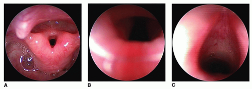
Although the histopathologic picture may vary, the diagnosis of subglottic stenosis is based on endoscopic assessment. The stenosis due to a scar, granulation tissue, submucosal thickening, or a congenitally abnormal cricoid can be differentiated from subglottic stenosis with a normal cricoid, but endoscopic measurement with endotracheal tubes or bronchoscopes is required for an accurate evaluation. Many laryngeal stenoses represent a combination of a congenitally small larynx further traumatized by endolaryngeal intubation or instrumentation.

Radiographic evaluation of a patient’s unintubated airway may give the clinician clues about the site and length of the stenosis. Useful imaging modalities include inspiratory and expiratory lateral soft tissue neck films; fluoroscopy to demonstrate the dynamics of the trachea and larynx; and a chest radiograph. The single most important investigation, however, remains high-kilovoltage airway films. These films are helpful in identifying the classic steepling seen with subglottic stenosis and also in providing a warning of tracheal stenosis; this is usually caused by complete tracheal rings, which may place the child in a life-threatening situation during rigid endoscopy.


TABLE 90.3 GRADING SCALE FOR LARYNGEAL STENOSIS


Grade


Laryngeal Lumen Obstruction


I


<50%


II


51%-70%


III


71%-99%


IV


Complete obstruction


Endoscopy is necessary for the diagnosis of laryngeal stenosis. Flexible fiberoptic endoscopy provides information on dynamic vocal fold function. Rigid endoscopy with Hopkins rod lens telescopes provides the best possible examination. Precise measurement of the endolarynx and staging of the laryngeal stenosis can be carried out. The most commonly used system for staging subglottic stenosis is the Myer-Cotton grading scale (Table 90.3). Most grade I and many grade II lesions do not require surgical intervention; however, grade III and grade IV lesions generally necessitate a surgical approach.


Laryngeal Webs

Supraglottic webs represent fewer than 2% of congenital laryngeal webs (Fig. 90.1). These webs are diaphragmatic outgrowths that usually arise anteriorly. If partial, they provide air passage posteriorly. The web is typically thickened anteriorly and thins out toward the posterior edge. Symptoms, including dyspnea and voice change, depend on the size and position of the web. Ten percent of children with supraglottic webs have associated congenital anomalies.

More than 90% of laryngeal webs are anterior glottic webs, representing a failure to completely recanalize the larynx. Patients usually present with an abnormal cry or respiratory distress at birth. Virtually all glottic webs are anterior, with varying degrees of compromise of the glottic airway. Although some webs are gossamer thin, most anterior glottic webs are thick and usually associated with a subglottic “sail” compromising the subglottic lumen. As such, they may be considered a form of partial laryngeal atresia. Thin webs may escape detection because neonatal intubation for airway distress may lyse the web. Thick webs require open reconstruction with either reconstruction of the anterior commissure or placement of a laryngeal keel. Approximately 40% of patients with thick membranous webs require tracheotomy placement. There is a strong association between anterior glottic webs and velocardiofacial syndrome (4).

A web can also form in the subglottic region, mimicking cricoid cartilage deformities or subglottic stenosis. Subglottic webs account for approximately 7% of laryngeal webs and are anteriorly based, with a small posterior
opening that may be only the size of a pinpoint. The superior surface is covered with squamous epithelium, and the inferior surface is mucous membrane. A slight female predominance has been reported. Symptoms include aphonia, respiratory distress, and biphasic stridor. The diagnosis must be made by flexible fiberoptic or rigid endoscopy.


Figure 90.1 Endoscopic view of a patient with a congenital supraglottic web A: 0-degree telescope. B: 30-degree telescopic view. C: 30-degree telescopic view showing normal vocal folds beyond the web.


Atresia

Laryngeal atresia (supraglottic, glottic, subglottic) represents complete failure of the lumen of the larynx to recanalize, and is therefore the most severe form of laryngeal stenosis. This condition is associated with a high frequency of other congenital anomalies, including esophageal atresia, tracheoesophageal fistula (TEF), urinary tract anomalies, and limb defects, particularly those that involve the radius. If a TEF exists, ventilation may occur through this communication. Tracheotomy is essential if the patient is to survive. Many children are not properly diagnosed in time for the appropriate intervention, and the diagnosis may be made postmortem. In the absence of a TEF, the lungs and major airways expand due to the pressure of fluid secreted by the lungs, leading to eversion of the diaphragms, development of a prune belly, and fetal hydrops; this constellation is known as congenital high airway obstructive syndrome.

Complete laryngeal atresia is challenging to repair due to the associated small cricoid. Reconstruction involves anterior cricoid resection, posterior cricoid expansion, web division, and prolonged stenting. This is usually best left until the child is older than 4 years of age. It is unlikely that a single operation will achieve decannulation.


Acquired Laryngeal Stenosis


Postintubation Stenosis

In 1965, McDonald and Stocks (5) advocated longterm nasotracheal intubation in the management of the unstable neonatal airway. Although this revolutionized neonatal care, especially for the premature infant, there was a corresponding increase in the incidence of acquired subglottic stenosis in neonates with prolonged stays in neonatal intensive care units. The incidence of combined congenital and acquired subglottic stenosis also rose as infants with congenital subglottic stenosis were intubated for airway compromise, often with inappropriately large endotracheal tubes. Acquired subglottic stenosis resulting from prolonged neonatal intubation is now more common than congenital stenosis in the pediatric age group.

The pathogenesis of acquired subglottic stenosis is still not understood, although several theories have been proposed. Autopsy studies demonstrate that there is a period of ulceration and necrosis of cricoid mucosa in the first hours and days of intubation (5). Quiney and Gould (6) reported healing and reepithelialization in the cricoid region, even while the endotracheal tube remained in place. However, 1% to 8% of neonates develop stenosis after prolonged intubation (7). The incidence has steadily dropped over the last three decades despite the longer periods of intubation that have become commonplace in increasingly smaller infants. Although several factors are involved, the two most significant factors have been the abandoning of red rubber endotracheal tubes and the realization that the ideal endotracheal tube size is not the largest that will fit but rather the smallest that can permit adequate ventilation. Ideally, an endotracheal tube should leak air around it with subglottic pressures below 20 to 25 cm of water.

Flexible fiberoptic laryngoscopy should be a routine part of the initial evaluation of the larynx. Vocal fold paralysis may prohibit decannulation, even after successful surgical reconstruction of a compromised airway. Rigid endoscopy using Hopkins telescopes is necessary for diagnosis and photo documentation. Correct sizing of the airway should be accomplished with appropriate-sized endotracheal tubes or rigid bronchoscopes. Radiographic assessment can provide information about the site and length of the stenosis that will assist in management decisions. In patients
with acquired stenosis, a prolonged period of observation after tracheotomy is inappropriate because of the high risk of death should the tracheotomy tube become obstructed. Therefore, surgical reconstruction is often undertaken to create a safer airway.


Post-Tracheotomy Stenosis

During a tracheotomy, additional injury may result in laryngotracheal stenosis or suprastomal collapse. Secondary infection, chondritis, or pressure exerted by the curvature of the tracheotomy may result in stenosis or suprastomal collapse above the tracheotomy site. Stenosis may occur because of a high tracheotomy, which is performed through the thyroid, cricoid, or first tracheal cartilage; a high tracheotomy also includes cricothyroidotomy. Although this procedure is well known to otolaryngologists and emergency room physicians as a lifesaving intervention, it may result in a complication rate as high as 32% (8).


Post-Reconstruction Restenosis

Subglottic stenosis may also result after failed laryngotracheoplasty. This is more likely to occur if the original procedure was not aggressive enough or there is concomitant bacterial infection, development of granulation tissue, malalignment or improper stent sizing, or histopathologic changes. Indeed, any form of airway surgery, endoscopic or open, has an attendant risk of resultant airway restenosis. There is greater risk of restenosis following airway reconstruction if the larynx is actively inflamed. This is most commonly seen in patients with gastroesophageal reflux disease (GERD), but may also be seen in patients with eosinophilic esophagitis (EE). Optimally, both of these conditions should be treated before reconstructive surgery is performed. Occasionally, an active larynx is seen in a patient who does not have either GERD or EE. In this setting, spontaneous resolution with time is expected, and patience prior to airway surgery is recommended.


Infectious or Inflammatory Stenosis

Laryngeal stenosis caused by infectious or inflammatory disorders is usually the result of end-stage processes of untreated disorders.


Granulomatous Disease

Tuberculosis of the larynx is the most common granulomatous disease of the larynx, and it is usually associated with pulmonary disease. The most common sites for laryngeal tuberculosis are the interarytenoid space, arytenoid cartilages, posterior surface of the true vocal folds, and laryngeal surface of the epiglottis. The patient may present in the early stages of disease with diffuse edema and erythema of the cords, which may mimic an early-stage glottic carcinoma. However, disease progression manifests with nodular lesions and ulceration of the epithelium, which can lead to perichondritis and chondritis. Due to interarytenoid muscular involvement or cricoarytenoid joint fixation, patients may also present with symptoms that mimic vocal fold paralysis. Diagnosis is made by demonstration of Mycobacterium tuberculosis. Successful treatment usually leads to complete healing of the larynx. If the disease is not treated, chondritis and necrosis will destroy the larynx with extensive scarring and stenosis.

Sarcoidosis, rhinoscleroma, and Wegener granulomatosis are all rare disorders in children. Wegener granulomatosis may respond well to steroids, cyclophosphamide, and possibly, co-trimoxazole. It has a predilection for the subglottis and, once quiescent, may require airway reconstruction with cricotracheal resection being more efficacious than standard laryngotracheal reconstruction utilizing expansion cartilage grafting techniques.


Trauma

Because laryngeal trauma is discussed in Chapter 77, our comments here are brief. If trauma is mismanaged or unrecognized, it may lead to laryngeal stenosis. Sources of internal trauma include foreign bodies and instrumentation during endoscopic procedures. These forms of trauma usually lead to glottic and subglottic scarring and resultant stenosis. External forms of laryngeal trauma, such as motor vehicle accidents, sports-related injuries, and assaults, including blunt and penetrating trauma, can also produce laryngotracheal stenosis.

Anterior blunt trauma, as sustained in motor vehicle accidents or sports-related injuries, usually leads to posterior supraglottic and glottic stenosis. An external force at the hypopharyngeal level may cause scar formation between the epiglottis and posterior pharyngeal wall. Fracture of the hyoid bone displaces soft tissues posteriorly, narrowing the laryngeal inlet. These injuries may also cause web formation on the posterior hypopharyngeal wall and stenosis in the postcricoid area. Blunt and penetrating trauma may lead to laceration or hematoma formation in the glottis, which will produce laryngeal stenosis if not treated appropriately.


Systemic Diseases

Laryngeal stenosis due to fixation of the cricoarytenoid joint may be caused by rheumatoid arthritis or juvenile rheumatoid arthritis (Still disease). About 25% of patients with rheumatoid arthritis present with limitation of the motion of the cricoarytenoid joint. Systemic lupus erythematosus and gout may also cause arthritis of this joint, leading to narrowing of the laryngeal inlet. Hoarseness, stridor, dyspnea, and pain are the signs and symptoms of arthritic involvement. Erythematous edema of the arytenoid with the vocal fold fixed in the paramedian or intermediate position may be found during examination. Diagnosis must include direct laryngoscopy with palpation of the arytenoid to differentiate this from vocal fold paralysis. Appropriate laboratory studies
include erythrocyte sedimentation rate, C-reactive protein, antinuclear antibodies, and rheumatoid factor.

May 24, 2016 | Posted by in OTOLARYNGOLOGY | Comments Off on Laryngeal Stenosis

Full access? Get Clinical Tree

Get Clinical Tree app for offline access