L
Labrador keratopathy See keratopathy, actinic.
lacquer cracks See spot, Fuchs’.
lacrimal Relating to tears.
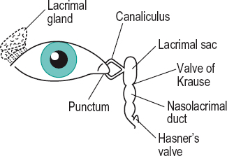
lacrimal apparatus The system involved in the production and conduction of tears. It consists of the lacrimal gland and accessory lacrimal glands (glands of Krause and Wolfring); the eyelid margins; and the two puncta lacrimale. Each punctum is a small round or oval aperture situated on a slight elevation at the inner end of the upper and lower lid margin (lacrimal papilla) and forms the entrance to the canaliculi. Each canaliculus consists of a vertical portion of about 2 mm long and then bends inward for some 8 mm, the upper one being slightly shorter. The canaliculi pierce the lacrimal fascia (i.e. the periorbita covering the lacrimal sac or tear sac) and unite (forming the common canaliculus) to enter a small diverticulum of the sac called the sinus of Maier. The lacrimal sac is closed above and open below where it is continuous with the nasolacrimal duct which extends over some 1.5 cm in length to Hasner’s valve (or Bianchi’s valve or plica lacrimalis) (folds of mucous membrane) at the inferior meatus of the nose. The inferior opening of the duct is called the ostium lacrimale (Fig. L1).
See dacryocystitis; epiphora; fistula, lacrimal; fossa for the lacrimal sac; syndrome, Sjögren’s; tear duct; test, dye dilution; test, Jones II; valve of Krause.
lacrimal artery See artery, lacrimal.
lacrimal bone See orbit.
lacrimal canaliculi See lacrimal apparatus.
lacrimal caruncle See caruncle, lacrimal.
lacrimal crest, anterior The anterior margin of the fossa for the lacrimal sac located on the frontal process of the maxilla.
See fossa for the lacrimal sac.
lacrimal crest, posterior The posterior margin of the fossa for the lacrimal sac situated on the lacrimal bone.
lacrimal duct See tear duct.
lacrimal fascia See lacrimal apparatus.
lacrimal fluid See tears.
lacrimal gland See gland, lacrimal.
lacrimal lake Accumulation of tears in the angle between the eyelids and the inner canthus prior to draining into the lacrimal puncta.
See epiphora; tear meniscus.
lacrimal layer See film, precorneal.
lacrimal lens See lens, liquid.
lacrimal nerve See nerve, ophthalmic.
lacrimal papilla A small elevation at the inner canthus of each eyelid containing a puncta lacrimale.
lacrimal prism See tear meniscus.
lacrimal punctum A small, round or oval, opening of the lacrimal canaliculus (duct) on the margin of each eyelid near the inner canthus and situated in the middle of a small elevation the lacrimal papilla. A lacrimal punctum is normally visible only if the lid is everted. Plural: lacrimal puncta. Syn. lacrimal point.
lacrimal reflex See reflex, lacrimal.
lacrimal sac See lacrimal apparatus.
lacrimal tubercle A small bump on the frontal process of the maxilla situated near the lower orbital and the anterior lacrimal crest, to which the medial palpebral ligament attaches. Syn. papilla lacrimalis.
See ligament, palpebral; lacrimal crest, anterior.
lacrimation 1 . Secretion and flow of tears. 2. Synonym for weeping.
See epiphora; reflex, lacrimal; tear secretion.
lacrimation, paradoxic See tears, crocodile.
laevoclination Rotation of the upper pole of an eye towards the subject’s left. Syn. laevocycloduction; laevotorsion.
laevocycloduction See laevoclination.
laevocycloversion Rotation of the upper poles of the vertical meridians of both eyes towards the subject’s left.
See dextrocycloversion.
laevodeorsumversion Movement of the eyes down and to the left.
laevoduction Rotation of one eye to the left. Note: also spelt levoduction.
See duction.
laevophoria A tendency of the visual axes of both eyes to deviate to the left, in the absence of a stimulus to fusion.
See dextrophoria; heterophoria.
laevosursumversion Movement of the eyes up and to the left.
laevotorsion See laevoclination; torsion.
laevoversion Movement of both eyes to the left. See version.
lag of accommodation See accommodation, lag of.
lagophthalmos Failure of the upper eyelid to close the eye completely. During sleep, about one-third of the population has a slight lagophthalmos.
See ectropion; keratopathy, exposure.
Lagrange’s law See law, Lagrange’s.
lambert Unit of luminance equal to 3183 candelas per m2. Symbol: L.
Lambert’s cosine law See diffusion.
lamellar keratoplasty See keratoplasty.
lamina Thin sheet or layer. Example: the lamina vitrea of Bruch’s membrane.
lamina cribrosa See cribriform plate.
lamina elastica See membrane, Bruch’s.
lamina elastica anterior See, layer, Bowman’s.
lamina elastica posterior See membrane, Descemet’s.
lamina fusca See choroid.
lamina papyracea Synonym for the orbital plate of the ethmoid bone, which forms part of the medial wall of the orbit. It is thus named because it is as thin as paper and this may contribute to an infection of an ethmoidal sinus spreading into the orbit and resulting in orbital cellulitis.
See cellulitis, orbital.
lamina vitrae See membrane, Bruch’s.
laminated lens See lens, laminated.
lamp Any device that produces light or heat. Burton l. Ultraviolet lamp, including some short wavelengths from the visible spectrum (e.g. Wood’s light), mounted with a magnifying lens in a rectangular frame. It is used primarily in the evaluation of the fit of a hard contact lens, in conjunction with the instillation of fluorescein into the eye.
See staining.
filament l. A lamp in which light is produced by electrically heating a filament, usually of tungsten. The filament is contained in a bulb in which there is either a vacuum or an inert gas. The emitted spectrum is continuous.
See spectrum, continuous.
fluorescent l. Discharge lamp in which most of the light is emitted by a layer of fluorescent material excited by the ultraviolet radiation from the discharge (CIE).
See fluorescence.
halogen l. A tungsten filament lamp in which the glass envelope is made of quartz and is filled with gaseous halogens. This permits a higher filament temperature and consequently provides a higher luminance and a higher colour temperature as well as a longer operating life than a conventional filament lamp of the same input power. Halogen lamps are used in some ophthalmoscopes and retinoscopes and as very bright sources for people with low vision. Syn. tungsten-halogen lamp.
incandescent electric l. Lamp in which light is produced by means of a body (filament of carbon or metal) heated to incandescence by the passage of an electric current (CIE).
See incandescence; luminescence.
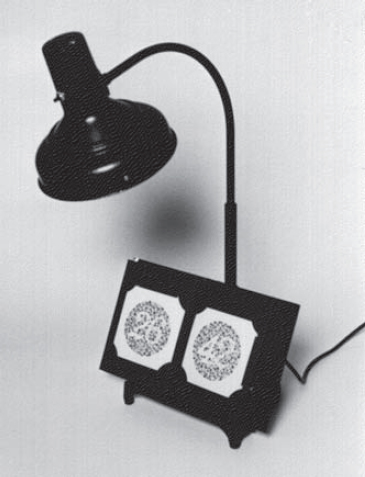
Macbeth l. A lamp used in testing colour vision. It contains a powerful tungsten filament bulb with a blue filter of specific absorption properties such that it produces a source of a colour temperature of about 6800 K, thus approximating the spectral characteristics of natural sunlight. The lamp is also fitted with a stand to hold the colour vision booklet (Fig. L2). Syn. Macbeth illuminant C.
See illuminants, CIE standard; plates, pseudoisochromatic; test, Farnsworth. tungsten-halogen l. See lamp, halogen.
Landolt ring A test object used for measuring visual acuity consisting of an incomplete ring resembling the letter C. The width of the break and of the ring are each one-fifth of its overall diameter. The subject must indicate where the break is located, the break being positioned in any direction. The minimum angle of resolution corresponds to the angular subtense of the just noticeable break at the eye (Fig. L3). Syn. Landolt broken ring; Landolt C; Landolt test type.
See chart, Landolt broken ring.

Lang stereotest See stereotest, Lang.
Langerhans’ cells See cells, Langerhans’.
lantern test See Edridge–Green lantern; test, lantern.
Lanthony desaturated D-15 test See test, Farnsworth.
Lanthony tritan album See plates, pseudoisochromatic.
LASEK A surgical procedure on the cornea aimed at correcting ametropia. An alcohol solution is applied to the cornea (usually for less than 30 seconds) to loosen the epithelium. A trephine is used to make an incision in the epithelium leaving a hinge of 2–3 clock hours of intact margin. The loosened edges of the epithelium are lifted along the trephine mark and the epithelium is folded or rolled back exposing Bowman’s layer. Excimer laser ablation is then performed on the anterior stromal surface and Bowman’s layer is ablated away over the treatment area. The epithelial flap is repositioned and a bandage soft contact lens is worn for 3–4 days to minimize discomfort and to protect the epithelium. The method is less invasive than LASIK and appears to give rise to fewer complications. LASEK is an acronym made from the following italic letters ‘laser assisted epithelial keratomileusis’.
See keratectomy, photorefractive; pachometer.
laser An intense luminous source of coherent and monochromatic light. The term is an acronym for light amplification by stimulated emission of radiation. Lasers are used in the treatment of a variety of ocular conditions, especially of the cornea, the retina (e.g. detached retina, diabetic retinopathy), glaucoma and refractive errors.
See cyclodiode; iridotomy; keratectomy, photorefractive; LASEK; LASIK; ophthalmoscope, scanning laser; photocoagulation; trabeculoplasty.
argon l. A laser with ionized argon gas as the active medium, which emits a bluegreen light beam with a wavelength of 514 nm. It may be used to perform iridectomy, iridoplasty, iridotomy, photocoagulation or trabeculoplasty.
excimer l. A gas laser that emits pulses of light in the ultraviolet region (at 193 nm). All the energy is absorbed by the superficial layers (e.g. the corneal epithelium), which are then exploded away or ablated without any change to the underlying or adjacent tissue or material.
See keratectomy, photorefractive; LASEK; LASIK.
l. interferometry See maxwellian view system, clinical.
l. iridotomy See iridotomy.
krypton l. A laser with krypton gas ionized by electric current as the active medium, which emits a light beam in the yellow-red region of the visible spectrum (521 nm, 568 nm or 647 nm). It may be used to perform photocoagulation or trabeculoplasty.
neodymium-yag l . (Nd-Yag) A solid-state laser whose active medium is a crystal of yttrium, aluminium and garnet doped with neodymium ions. It emits an infrared light beam with a wavelength of 1064 nm. It is typically used with a slit-lamp and in conjunction with a helium-neon laser which produces a red beam of light (633 nm) to allow focusing. It may be used to perform capsulotomy, iridotomy or trabecular surgery. Yag is an acronym for yttrium–aluminium–garnet.
l. refraction See refraction, laser.
l. refractive keratoplasty See keratectomy, photorefractive.
l. trabeculoplasty See trabeculoplasty, laser.
LASIK A surgical procedure on the cornea aimed at correcting ametropia. A suction ring is applied to the globe and an increase in intraocular pressure to approximately 65 mmHg is induced for a maximum of two minutes. During that time an automated microkeratome advances across the cornea creating a corneal flap of about 8.5 mm in diameter, which contains the epithelium, Bowman’s layer and a portion of the anterior stroma. The vacuum is then switched off and the suction ring removed. The corneal flap, which is hinged on one side of the cornea, is turned round onto the conjunctiva and the exposed stroma is ablated with the excimer laser. On completion of the laser ablation, the corneal flap is repositioned and left to adhere without sutures. There are some complications associated with this procedure, but it gives rise to less postoperative pain and more rapid visual rehabilitation than other similar surgical procedures (Fig. L4). LASIK is an acronym made from the following italic letters ‘laser in situ keratomileusis’ or ‘laser assisted intrastromal keratoplasty’.
See ectasia, corneal; epikeratoplasty; Intacs; keratome; keratomileusis; keratophakia; keratectomy, photorefractive.
latanoprost See prostaglandin analogues.
latent hyperopia See hyperopia, latent.
lateral geniculate body See geniculate bodies, lateral.
lateral inhibition See inhibition, lateral.
lateral rectus muscle See muscle, lateral rectus; muscles, extraocular.
lateral rectus paralysis See paralysis of the sixth nerve.
lathe-cut contact lens See lens, lathe-cut contact.
lattice degeneration of the retina See retina, lattice degeneration of the
lattice dystrophy See dystrophy, lattice.
lattice theory See theory, Maurice’s.
Laurence–Moon–Bardet–Biedl syndrome
See syndrome, Laurence–Moon–Bardet– Biedl.
law In science, a statement of facts or principles which is considered invariable under the given conditions having been tested and tried.
Abney’s l. The total luminance of an area is equal to the sum of the luminances that compose it.
Alexander’s l. An increase in the intensity of a jerk nystagmus when the eyes move in the direction of the fast phase.
all or none l. The response in a nerve fibre to any stimulus strong enough to produce a response is always of the same amplitude. However, different nerve fibres have action potentials with different amplitudes. An increase in the intensity of the stimulus yields only an increase in the frequency of nerve impulses (or action potentials). Syn. all or nothing law.
Aubert–Förster l. See phenomenon, Aubert–Förster.
Bloch’s l. The luminance L of a stimulus required to produce a threshold response is inversely proportional to the duration of exposure t of the stimulus, i.e.

where C is a constant. This law is only valid for exposure t below about 0.1s.
Bunsen–Roscoe l. In photochemistry, the product of the intensity of the light stimulus and the duration of exposure is a constant. Syn. law of reciprocity.
cosine l. See diffusion.
Descartes’ l. See law of refraction.
Donders’ l. For any determinate position of the line of fixation with respect to the head there corresponds a definite and invariable angle of torsion.
Draper’s l. An effect is produced in a medium only by that portion of the spectrum which is absorbed by the medium. The effect may be thermal, chemical or the production of fluorescence. Syn. Grotthus’ law.
Emmert’s l. The apparent size of a projected after-image varies in proportion to the distance of the surface on which it is projected. The law can be expressed by the following relationship h/H = d/D, where h is the linear size of the object, H the apparent size of the projected after-image, d the object’s distance from the observer and D the distance between the observer and the surface on which the after-image is projected. It follows from the above expression that H = hD/d, i.e. the greater the distance of the projected image the larger its apparent size.
l. of equal innervation See law of equal innervation, Hering’s.
Fechner’s l. The intensity of a sensation S varies as the logarithm of the intensity I of the stimulus, i.e.

where k is a constant. However, in some conditions this law is not valid and Stevens’ law (or power law) is more appropriate. This stipulates that the intensity of a sensation S varies as the intensity of the stimulus I to the power of x, i.e.

where x is a constant which depends on the stimulus.
See magnitude estimation.
Fermat’s l. The path taken by a light ray in going from one point to another is that route which takes the least time. Syn. Fermat’s principle.
Ferry–Porter l . The critical flicker frequency F is directly proportional to the logarithm of the luminance L of the stimulus, i.e.
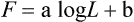
where a and b are constants. See frequency, critical fusion,
Granit–Harper l. The critical fusion frequency increases with the logarithm of the retinal area stimulated.
Grassmann’s l’s. Laws of colour mixture. 1. The first law states that any colour C of the visible spectrum can be matched in appearance by a mixture of three primary colours, such as red R, green G and blue B, provided that none of these can be matched by a mixture of the other two, i.e.
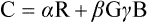
where α, β and γ are the relative proportions of the chosen primaries.
2. Additive property: if a colour is added in an identical manner to two equivalent mixtures (or single colours) the two new mixtures will appear identical, i.e. if A + B = C + D, then A + B + X = C + D + X, or if A = B, then A + X = B + X.
3. Scalar property: if the brightness of each of two equivalent mixtures is increased or decreased by the same factor the two new mixtures will appear identical, i.e. if A + B = C + D, then k (A + B) = k (C + D).
4. Associative property: if a colour is substituted in one of the mixtures by an equivalent colour the two new mixtures will appear identical, i.e. if A + B = C + D and X = B, then A + X = C + D.
Grotthus’ l. See law, Draper’s.
Helmholtz’s l. of magnification See law, Lagrange’s.
Hering’s l. of equal innervation
Innervation to the extraocular muscles is equal to both eyes. Thus, all movements of the two eyes are equal and symmetrical. Syn. Hering’s law; law of equal innervation.
See muscles, yoke.
l. of identical visual directions An object stimulating corresponding retinal points is localized in the same apparent monocular direction in each eye. Syn. law of oculocentric visual direction.
See line of direction; retinal corresponding points.
inverse square l. of illumination The illuminance E of a surface by a point source is directly proportional to the luminous intensity I of a point source and to the cosine of the angle θ of incidence and inversely proportional to the square of the distance d between the surface and the source, i.e.
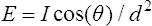
Syn. law of illumination.
See illuminance.
Imbert–Fick l . Applied to applanation tonometry, this law states that the intraocular pressure P (in mmHg) is equal to the tonometer weight W (in g) divided by the applanated area A (in mm2), hence,

This law is correct only for infinitely thin, dry, elastic, spherical membranes.
Kirschmann’s l. The greatest contrast in colour is seen when the luminosity difference is small.
Knapp’s l. A correcting lens placed at the anterior focal plane of an axially ametropic eye forms an image equal in size to that formed in a standard emmetropic eye. Knapp’s law applies to the relative spectacle magnification but not to the spectacle magnification. Syn. Knapp’s rule.
Kollner’s l. See rule, Kollner’s.
Lagrange’s l. In paraxial optics, the product of the index of refraction of image space n’, the image size h’ and the half-angle of the refracted cone in image space u’ is equal to the product of the index of refraction of object space n, the object size h and the half-angle of the incident cone in object space u, i.e.
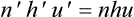
Syn. Helmholtz’s law of magnification; Lagrange’s relation; Smith–Helmholtz law. See sign convention.
Lambert’s l. See diffusion.
Listing’s l. When an eye moves to any position from the primary position, it may be considered to have made a single rotation about an axis that is perpendicular to both the initial and final lines of fixation at their point of intersection.
l. of oculocentric visual direction See law of identical visual directions.
Planck’s l. Law giving the energy distribution of a black body as a function of wavelength, for a specified temperature.
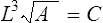
Prentice’s l. The prismatic effect P in prism dioptres at a point on a lens is equal to the product of the distance c in centimetres of the point from the optical centre of the lens, and the dioptric power F of the lens, i.e.

Syn. Prentice’s rule.
See effect, differential prismatic; power, prism; prism, induced.
l. of reciprocity See law, Bunsen–Roscoe.
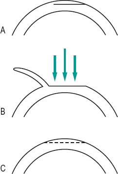
l. of reflection The incident and reflected rays and the normal to the surface at the point of incidence lie in the same plane and the angle of incidence is equal to the angle of reflection (Fig. L5).
l. of refraction The incident and refracted rays and the normal to the surface at the point of incidence lie in the same plane and the ratio of the sine of the angle of incidence i to the sine of the angle of refraction i’ is a constant for any two media, i.e.
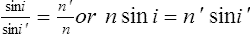
where n and n’ are the refractive indices of the first and second medium, respectively. This constant (n’/n) is called the relative index of refraction for the two media. Syn. Descartes’ law; Snell’s law.
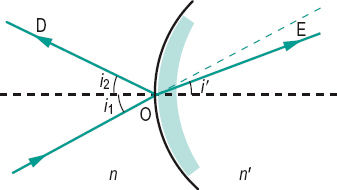
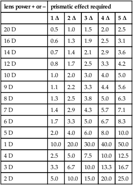
Table L1
Approximate amount of spectacle lens decentration (in mm) of its optical centre away from the pupillary centre of the eye to produce five prismatic effects (in prism dioptres) for distance vision. The results ignore the effect of spherical aberration
| lens power + or – | prismatic effect required | ||||
| 1 Δ | 2 Δ | 3 Δ | 4 Δ | 5 Δ | |
| 20 D | 0.5 | 1.0 | 1.5 | 2.0 | 2.5 |
| 16 D | 0.6 | 1.3 | 1.9 | 2.5 | 3.1 |
| 14 D | 0.7 | 1.4 | 2.1 | 2.9 | 3.6 |
| 12 D | 0.8 | 1.7 | 2.5 | 3.3 | 4.2 |
| 10 D | 1.0 | 2.0 | 3.0 | 4.0 | 5.0 |
| 9 D | 1.1 | 2.2 | 3.3 | 4.4 | 5.6 |
| 8 D | 1.3 | 2.5 | 3.8 | 5.0 | 6.3 |
| 7 D | 1.4 | 2.9 | 4.3 | 5.7 | 7.1 |
| 6 D | 1.7 | 3.3 | 5.0 | 6.7 | 8.3 |
| 5 D | 2.0 | 4.0 | 6.0 | 8.0 | 10.0 |
| 1 D | 10.0 | 20.0 | 30.0 | 40.0 | 50.0 |
| 4 D | 2.5 | 5.0 | 7.5 | 10.0 | 12.5 |
| 3 D | 3.3 | 6.7 | 10.0 | 13.3 | 16.7 |
| 2 D | 5.0 | 10.0 | 15.0 | 20.0 | 25.0 |
See index of refraction; sign convention.
Ricco’s l. The product of the absolute threshold of luminance L and the image area A is a constant, i.e.

This law is valid for small images subtending an angle of a few minutes of arc in the fovea and to one degree in the near macular region. For larger images in the macular area, Piéron’s law applies; it states that the product of the luminance L of the image at threshold and the cube root of the retinal area A stimulated is a constant, i.e.
In the peripheral retina, Piper’s law becomes valid. This law states that the product of the luminance of the stimulus L and the square root of the area A is a constant, i.e.

In the far periphery of the retina, L tends to become independent of A.
Sherrington’s l. of reciprocal innervation The contraction of a muscle is accompanied by simultaneous and proportional relaxation of its antagonist. For example, if the superior oblique muscle contracts, its antagonist, the inferior oblique muscle, relaxes. The validity of this law has been established by electromyography.
Smith–Helmholtz l. See law, Lagrange’s.
Snell’s l. See law of refraction.
Stevens’ l. See law, Fechner’s.
Talbot’s l. See law, Talbot–Plateau.
Talbot–Plateau l. The brightness of a light source presented at short intervals above the critical fusion frequency is equal to that which would be produced by a constant light source of an intensity equal to the mean value of the intermittent stimuli. Syn. Talbot’s law.
Weber’s l. The just noticeable difference (or difference threshold) in intensity of a stimulus ΔI varies as a constant ratio of the initial intensity of the stimulus I, i.e.

where k = {ΔI/l} is a constant called Weber’s fraction (Weber’s constant).
Example: if the initial stimulus was a light source of 1000 cd/m2 and k = 0.01 (or 1%), ΔI = 0.01 × 1000 = 10 cd/m2. Syn. Weber–Fechner law.
See threshold, differential.
Weber–Fechner l. See law, Weber’s.
layer A sheet of one thickness lying over or under another and distinguished from it by a difference in composition or colour.
Bowman’s l. Thin layer of the cornea (about 12 μm) located between the anterior stratified epithelium and the stroma. This layer is acellular; it is a modified superficial stromal layer found only in primates. It is composed of a randomly orientated array of fine collagen fibrils, primarily of collagen types I, III and V. Syn. anterior limiting layer; Bowman’s membrane; lamina elastic anterior.
l. of Chievitz, transient A temporary layer found in the developing embryonic retina lying between the inner neuroblastic layer (which will form the ganglion, amacrine and Mueller cells) and the outer neuroblastic layer (which will form the bipolar, horizontal and photoreceptor cells). It contains the inner processes of Mueller’s fibres.
Haller’s l. An outer layer of the choroid lying between Sattler’s layer and the suprachoroid. It contains connective tissue and large vessels, mainly veins.
l. of Henle, fibre Located in the macular region, it is formed by the cone and rod fibres that run parallel to the retinal surface within the outer molecular layer of the retina.
See Haidinger’s brushes; macular star.
retinal l’s. See retina.
Sattler’s l. An inner layer of the choroid lying between the choriocapillaris and Haller’s layer. It contains small blood vessels.
leaf room See room, leaf.
Leber’s congenital amaurosis A hereditary, bilateral blindness present at birth or in early childhood. It is caused by mutations in the gene encoding the retinal guanylate cyclase (GUCY2D) on chromosome 17. Initially, the ocular fundus appears normal, although the ERG is markedly reduced. A salt and pepper fundus and optic atrophy appear later. The condition is often accompanied by nystagmus and photophobia.
Leber’s disease See Leber’s hereditary optic atrophy.
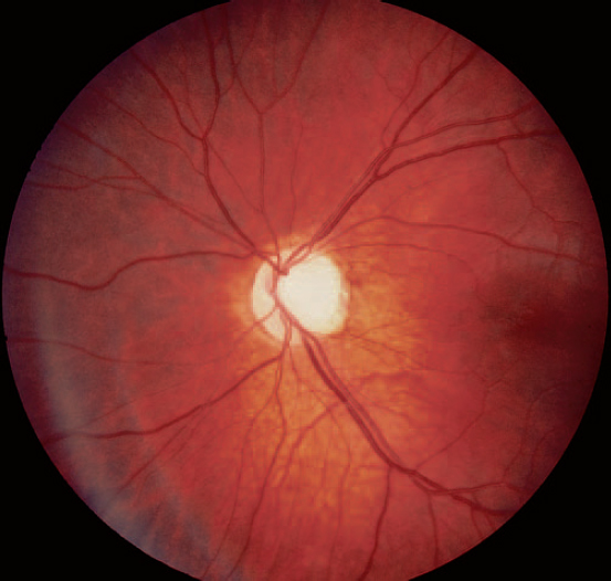
Leber’s hereditary optic atrophy A mitochondrial inherited bilateral condition, which appears suddenly in healthy people, primarily males, of about the age of 20 and results in a marked loss of vision and ultimately optic atrophy. A very small percentage of people recover some visual acuity in one or both eyes after the disease has run its course (Fig. L6). Syn. Leber’s disease; Leber’s hereditary optic neuropathy.
See atrophy, optic; inheritance; mitochondrion.
Leber’s miliary aneurysms See disease, Coats’.
legibility Term referring to the difference in the ease of difficulty with which optotypes can be read. Some letters are easier to recognize (e.g. L, T, U, V, Z and C) than others (e.g. S, G, H, F, R and B). Test charts either mix these letters or use letters of similar difficulty; the latter facilitates standardization of charts.
length, equivalent focal In an optical system composed of more than one lens, it is the linear distance separating the principal focus from the corresponding principal point. It is usually the most important quantity in the specification of an optical system as in objectives, eyepieces, etc.
See focus, principal; power, equivalent.
length of the eye, axial The distance between the anterior and posterior poles of the eye. In vivo, it is measured either by ultrasonography or by partial coherence interferometry (PCI). These measurements represent the distance between the anterior pole and Bruch’s membrane. (In young eyes in which there is a refractive index difference at the retina-vitreous interface ultrasonography measures the distance between the anterior pole and the anterior surface of the retina.) The axial length of the eye at birth is approximately 17 mm and reaches approximately 24 mm in adulthood. It is typically longer than 24 mm in myopes and shorter than 24 mm in hyperopes. Each mm of change in axial length of the eye corresponds to approximately 2.5 D
See biometry of the eye; ultrasonography.
length, focal The linear distance separating the principal focal point (or focus) of an optical system from a point of reference (e.g. vertex, principal point, nodal point). The first (anterior) focal length is the distance from the lens (or first principal point) to the first principal focus. The second (posterior) focal length is the distance from the lens (or second principal point) to the second principal focus. Symbol: f.
In a spherical mirror the focal length f, i.e. the distance between the focal point and the pole of the mirror, is equal to half its radius of curvature r

Syn. focal distance. See focus, principal; mirror; Points, cardinal; Points, principal; power, equivalent; power, refractive; sign convention.
lens A piece of transparent glass, crystal, plastic or similar substance (e.g. liquid) having two opposite regular surfaces which can be plane or curved and which alter the vergence of pencils of light transmitted through it. There are many types of lenses, which are described below.
See coquille; glass; moulding; surfacing.
absorptive l. A lens that absorbs a proportion of the incident radiation. Some lenses absorb mostly in the infrared region of the spectrum, others absorb mostly in the ultraviolet region and others absorb more or less equally throughout the visible spectrum.
See CR-39; filter; lens, coated; pterygium.
achromatic l. A compound lens designed to reduce or eliminate chromatic aberration. The most common type is called a doublet. Syn. achromat.
See lens, apochromatic; lens, hyperchromatic.
achromatizing l.
Stay updated, free articles. Join our Telegram channel

Full access? Get Clinical Tree


