Keratouveitis
Eva C. Kim
Rocio Murphy
Stephen D. McLeod
Keratouveitis refers to a clinical picture of active corneal disease associated with anterior chamber inflammation. In certain cases, an inflammatory process such as active infection with herpes simplex virus (HSV) or herpes zoster virus (HZV) involves multiple anterior segment tissues, including the cornea, but in other situations the anterior chamber inflammation appears to be secondary only to corneal disease. This “reflex” anterior chamber inflammation is commonly seen in association with dendritic HSV keratitis, bacterial or fungal infectious keratitis, and corneal epithelial disease in the setting of diabetes or aphakic bullous keratopathy or pseudophakic bullous keratopathy. Herpetic uveitis in the absence of corneal disease is considered to be relatively uncommon, and when it is seen, it is likely to be attributed to varicella-zoster virus (VZV) rather than HSV. A rare idiopathic, dominantly inherited familial form of recurrent keratouveitis has also been described in two Swedish families with autosomal dominantly inherited Osler-Rendu-Weber disease.1 Among the differential diagnoses of infectious keratouveitis are HSV, HZV, bacterial, fungal, acanthamoebal, and, rarely, vaccinia infection.2 Scleritis and peripheral ulcerative keratitis are important noninfectious causes of keratouveitis. A rare cause of keratouveitis is endogenous Listeria monocytogenes endophthalmitis, which can occur in immunocompetent patients.3
Herpes Simplex Keratouveitis
Clinical Features
Ocular HSV infection is a leading cause of corneal blindness in the developed world due to its recurrent nature. Loss of vision is due to the corneal scarring that results from inflammation and structural corneal damage that accompanies repeated episodes of HSV stromal inflammation and iritis. In a study looking at ocular HSV patients in Rochester, Minnesota, between 1950 and 1982, the estimated prevalence was roughly 150 per 100,000 population.4 An estimated 48,000 episodes of active HSV eye disease occur annually in the United States, 20,000 of which are new. Incidence and prevalence are influenced by factors that affect the degree of exposure to these sources of infection, such as crowding, poor hygiene, and age.5
Experimental infection can be induced in animals, but humans are the only natural reservoirs of HSV, and infection is ubiquitous among us. It is estimated that 50% of U.S. citizens are infected with HSV type 1 (HSV-1), which is less than in prior decades. By age 5, 20% of middle-class children seroconvert, and by age 20, the prevalence of HSV antibodies increases to about 50%. Seventy to 80% of adolescents in lower socioeconomic classes have serologic evidence of HSV.6 This proportion rises to 97% by 60 years of age.7 HSV can be detected by polymerase chain reaction (PCR) in the trigeminal ganglia of 18.2% of cadavers of people age 20 years and younger, increasing to roughly 100% in cadavers of people older than 60 years.8 Transmission of the virus is by direct contact, with an incubation period of 3 to 6 days. The initial infection is subclinical in 85% to 99% of cases, and all infected individuals will continue to carry latent virus. The oral mucosa is most commonly infected by contaminated saliva droplets, and the virus can be transmitted to the eye via the trigeminal nerve. Ocular HSV type 2 (HSV-2) infection is likely spread directly by oculogenital contact or by contaminated fingers, but hematogenous spread has also been demonstrated. Atopy, HIV infection, and other forms of immunosuppression predispose to prolonged or bilateral disease. Unlike cytomegalovirus and VZV infections, ocular HSV does not appear with greater frequency or severity among patients who are HIV positive.5
The clinical manifestations of HSV ocular disease can be categorized as congenital or neonatal infection, primary infection, or recurrent disease. HSV-1 and -2 can be acquired in utero (4%) transplacentally or by ascending infection, as well as by exposure to genital lesions during delivery (86%) or by postnatal infection by attendants (10%).9,10 Congenital ocular HSV disease includes microphthalmos, cataracts, optic atrophy, and retinal scarring. Neonatal ocular HSV disease is often bilateral and includes skin vesicles, conjunctivitis, epithelial ulceration or stromal keratitis, cataract, retrolental masses, iridocyclitis and synechiae, iris atrophy, chorioretinitis, and optic neuritis and optic atrophy.11,12
In primary disease, the oral mucosa is much more commonly involved than the eye, and the disease is often subclinical or mild. Periocular disease is characterized by a vesicular or ulcerative dermatitis and blepharitis. Conjunctival surface disease can be characterized by a unilateral, acute follicular conjunctivitis that can involve pseudomembranes or demonstrate dendrites. Up to 21% of acute conjunctivitis may be caused by HSV.13 In a study of 108 patients examined at Moorfields Eye Hospital in London between 1973 and 1980, moderate or severe conjunctivitis was present in 84%, dendritic ulcers in 15%, and disciform keratitis in 2%.14 Keratitis tends to lag conjunctivitis and lid disease by 1 to 2 weeks. These same 108 patients were followed for 2 to 15 years with 32% experiencing a recurrence.15 Patients tend to have recurrence of the same type of disease they had previously.
Recurrent disease is a risk for significant ocular morbidity with reduced visual acuity and can affect all layers of the cornea, with roughly 20% of recurrences presenting as stromal disease.4 Recurrences may be caused by reactivation of endogenous HSV-1 or, rarely, by reinfection with exogenous HSV-1.16 In the Herpetic Eye Disease Study (HEDS), the risk of a patient developing an ocular HSV recurrence without regard for the type of recurrence was associated with the number of previous episodes of ocular HSV disease that the patient experienced.15 In a recent report from this group, of 346 patients from the placebo group of the Acyclovir Prevention Trial who had experienced an episode of HSV eye disease in the previous year, 18% developed epithelial keratitis and 18% developed stromal keratitis during 18 months of follow-up.17 Statistically, previous and multiple episodes of stromal keratitis significantly increased the probability of subsequent stromal keratitis.17
In HSV epithelial disease, the characteristic lesions are dendrites: thin, meandering, arborizing epithelial ulcerations, with terminal bulbs at the ends of fine branches. With PCR, HSV DNA can been detected within dendritic lesions and in tear fluid during stromal keratitis.18
Stromal disease is immune mediated; however, without systemic antiviral medication such as acyclovir, 28% and 61% of eyes with a history of stromal keratitis develop a recurrence within 6 months and 2 years, respectively.19,20 The commonly described patterns of stromal herpetic disease include disciform edema, necrotizing stromal keratitis, and immune ring formation. In disciform edema, the dominant feature is circular stromal edema with accompanying folds in Descemet’s membrane and keratic precipitates accumulating underneath the area of edema (Fig. 39.1). The edema occurs and resolves without neovascularization or necrosis, but may result in a permanent stromal haze. Disciform keratitis is usually accompanied by a mild to moderate uveitis. A type of HSV-related corneal edema has been observed in which a line of endothelial dysfunction that is reminiscent of corneal allograft rejection develops and progresses from the periphery centrally, accompanied by anterior chamber inflammation and overlying corneal edema.21,22 This endotheliitis may be accompanied by trabeculitis, which leads to elevated intraocular pressure.
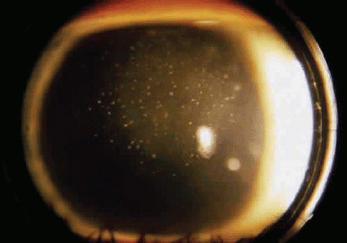 Figure 39.1 Herpes simplex virus keratouveitis. Anterior chamber inflammation with multiple endothelial precipitates and overlying corneal stromal edema. |
Necrotizing stromal keratitis is characterized by inflammatory necrosis and infiltration of the cornea with polymorphonuclear leukocytes, macrophages, lymphocytes, and plasma cells.23 Cellular infiltration produces stromal edema and necrosis. The necrotizing focus can be located at any level of the corneal stroma, with or without epithelial ulceration. Ring infiltrates can develop. In some cases, diffuse patches of infiltration appear beneath an intact epithelium, followed by local and often deep neovascularization; this is the so-called interstitial keratitis pattern. Lipid deposition, stromal melting, descemetocele formation, and perforation are all potential, although uncommon, complications of advanced necrotizing stromal keratitis. If the peripheral cornea is involved, inflammation and necrosis can spread to the sclera, producing a sclerokeratitis.
Herpetic keratouveitis can occur in association with any form of herpetic keratitis and accounts for 5% to 10% of all uveitis cases seen at tertiary referral centers.24 In addition, herpetic uveitis can also occur without concurrent HSV keratitis or without evidence or history of previous HSV keratitis.25,26 Anterior chamber inflammation that accompanies epithelial disease is believed to be caused by reflex irritation and is characteristically both mild and transient. However, the iridocyclitis that invariably accompanies necrotizing stromal disease tends to be much more severe and correlates with the apparent severity or depth of the keratitis.27 Uveitis may dominate the clinical picture in some cases of ocular HSV, with either absent or subtle corneal findings limited to faint cellular infiltration of the stroma. The pathogenesis of HSV uveitis is believed to be caused by a combination of active viral replication in the anterior chamber and an immune response to viral antigens.28,29 HSV uveitis is typically granulomatous and recurrent. In more severe cases, perilimbal injection is marked, and the cornea can become thickened and edematous. Dense, fibrinous flare with heavy, anterior chamber cell and medium-size, granulomatous keratic precipitates may be distributed widely over the endothelium (Fig. 39.2), not only along the inferior aspect of the endothelium. A hypopyon, although uncommon, and synechiae can form. Elevated intraocular pressure, caused by a trabeculitis, often develops. Marked dilation of iris blood vessels, iris neovascularization, and spontaneous hyphemas, although also uncommon, may occur. Episodes of inflammation are frequently marked by progressive iris atrophy and sphincter damage, leading to corectopia and anisocoria (Figs. 39.3 and 39.4).30 Sectoral iris atrophy has been considered to be specific for VZV uveitis; however, PCR of aqueous fluid of patients with uveitis, sectoral atrophy, and elevated intraocular pressure without keratitis has identified HSV in 83% and VZV in 13% of 24 patients tested.25
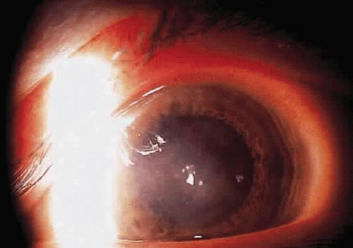 Figure 39.2 Herpes simplex virus disciform keratitis. Central corneal edema and underlying keratic endothelial precipitates. |
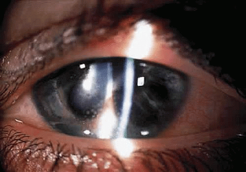 Figure 39.3 Herpes simplex virus keratouveitis. Multiple features of herpetic keratouveitis, including corneal stromal scarring, keratic endothelial precipitates, anterior synechiae, and diffuse iris atrophy. |
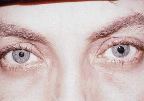 Figure 39.4 Herpes simplex virus iridocyclitis of the left eye, leading to iris sphincter atrophy and anisocoria. |
Pathophysiology
Uveitis associated with endotheliitis, disciform keratitis, and necrotizing stromal keratitis can be attributed to both immunologic reaction and live virus invasion. The pathogenesis of HSV stromal keratitis and iritis is believed to involve viral proliferation, usually in the corneal epithelium; antigenic stimulation; and host inflammatory and immune responses, although the contribution of each factor is debated.31 HSV viral particles have been recovered from the aqueous of patients with HSV iridocyclitis.31,32 Nevertheless, in a rabbit model of HSV uveitis, Oh demonstrated that live but not inactivate HSV introduced to HSV antigen-negative animals stimulated a gradual, delayed iridocyclitis, whereas either live or inactivated virus introduced to eyes that had recovered from primary disease rapidly stimulated an inflammatory response.33 These observations suggest that live virus is implicated in the iridocyclitis of primary disease, but that recurrent disease may be mediated by immunologic mechanisms not necessarily dependent on the presence of live virus. Sundmacher and Neumann-Haefelin suggested that clinical features such as elevated intraocular pressure, acute focal iritis, and endotheliitis are particularly suggestive of the presence of live HSV in the anterior chamber.32
Live virus also has been isolated from the aqueous and tears of patients with herpetic endotheliitis and stromal edema.18,20 In some cases of disciform keratitis, live virus might infect endothelial cells that demonstrate swelling and pleomorphism. It has been postulated that endothelial damage may result from direct viral cytolysis, as well as from subsequent antigen-antibody complement-mediated immunologic attack.34 Most experimental evidence, however, seems to support the theory that endothelial dysfunction and subsequent corneal swelling of disciform keratouveitis is caused by a delayed-type hypersensitivity and immunologic attack directed against endothelial cells that express herpes antigens.32,35,36
Treatment
Because the keratouveitis of epithelial HSV is mild and considered reactive, therapy is restricted to treatment of the epithelial disease either with a topical or an oral antiviral agent for 7 to 10 days, although there is only anecdotal and nonophthalmic evidence of the efficacy of oral antivirals in the treatment of active epithelial disease.37 There is pharmacokinetic evidence that acyclovir has excellent penetration into the eye and reaches therapeutic level in tear fluid.38 A topical cycloplegic agent may be used for symptomatic relief of ciliary spasm. Treatment with topical corticosteroids is discouraged because the suppression of the inflammatory response induced by these agents may lead to reduced clearance of antigen, which allows deeper spread of infection and extends the period of exposure to this stimulus, leading to destructive inflammation.39
Disciform and necrotizing stromal disease share the potential for irreversible corneal opacification, in the former because of irrevocable endothelial decompensation, and in the latter because of stromal scarring. Associated uveitis also introduces the risk of posterior and anterior synechiae, glaucoma, and cataract.
From 1994 to 1996, after several prospective, randomized, double-masked clinical trials, the HEDS group concluded that in the treatment of HSV stromal keratitis, topical corticosteroid treatment was significantly better than placebo in reducing persistence of progression of stromal inflammation and in shortening the duration of stromal keratitis.40 The HEDS group also concluded that there was no clinically significant beneficial effect of adding oral acyclovir to concomitant topical corticosteroid and trifluridine in the treatment of stromal keratitis with regard to time to treatment failure.41 Mild to moderately severe disciform keratitis without signs of live virus, such as elevated intraocular pressure or focal iris necrosis, can be treated without the use of topical or oral antiviral agents; however, because it is difficult to prove the absence of live virus, some clinicians prefer to routinely use either an oral or topical antiviral. If concurrent iridocyclitis of any significance is present, the results of another prospective, randomized HEDS trial have suggested (not statistically significant) that oral acyclovir at 400 mg five times daily administered for several weeks, along with a topical antiviral and corticosteroid drop, may enhance resolution of inflammation.42 These cases tend to be exquisitely responsive to topical corticosteroid administration. Corticosteroids need not be applied more than every 2 to 4 hours initially, and ideally, four times per day according to severity of inflammation. Topical corticosteroids should be tapered very slowly over several weeks to months as signs of inflammation resolve. Cycloplegic agents should be used to reduce discomfort caused by ciliary body spasm and prevent the formation of posterior synechiae. Elevated intraocular pressure, when secondary to trabeculitis, is often easily ameliorated with control of inflammation; however, it should be treated with appropriate intraocular pressure-lowering agents if the pressure is exceedingly high on presentation or slow to respond, even with decrease in anterior chamber inflammation.30 One must keep in mind the possibility of steroid-induced ocular hypertension and treat accordingly. Linear endotheliitis with iridocyclitis is treated in a similar fashion to stromal keratitis. In some cases, oral antiviral medication may prove beneficial in controlling inflammation and preventing a recurrence of disease.22
Severe disciform disease and necrotizing stromal disease with significant iridocyclitis can prove very difficult to treat. Because there may be live virus present, oral or topical antiviral agents should be used before onset of topical steroids. As stated previously, the HEDS group reported that frequent application (initially eight times a day) of topical corticosteroid effectively shortens the duration of stromal keratitis and reduces persistence or progression of HSV stromal keratitis.40 Oral acyclovir may enhance the resolution of herpetic iridocyclitis. Topical agents should be used to lower intraocular pressure and achieve cycloplegia. Because attacks of severe herpetic keratouveitis are often prolonged, all medicines must be tapered slowly and cautiously, with careful patient follow-up.
Regarding prevention of development of HSV stromal keratitis or iridocyclitis in patients with epithelial keratitis, the HEDS group concluded that acyclovir at 400 mg five times daily for 3 weeks, in addition to traditional treatment of epithelial disease with topical trifluridine and corticosteroid, does not prevent HSV stromal keratitis or iridocyclitis for the next year.43 However, acyclovir at 400 mg twice daily for 1 year was shown to be effective in reducing the rate of recurrent ocular and dermal HSV disease, specifically in patients with a history of stromal keratitis. Importantly, there was no rebound in the rate of HSV ocular or dermal disease 6 months after acyclovir treatment was ceased.20
Stay updated, free articles. Join our Telegram channel

Full access? Get Clinical Tree


